Información de la revista
Vol. 31. Núm. 5.
Páginas 231-248 (septiembre - octubre 2020)
Compartir
Compartir
Descargar PDF
Más opciones de artículo
Vol. 31. Núm. 5.
Páginas 231-248 (septiembre - octubre 2020)
Special article
SIXTO OBRADOR SENEC PRIZE 2019: Utility of diffusion tensor imaging as a prognostic tool in moderate to severe traumatic brain injury. Part II: Longitudinal analysis of DTI metrics and its association with patient's outcome
PREMIO SIXTO OBRADOR SENEC 2019: El uso de la secuencia Tensor de difusión como herramienta pronóstica en los pacientes con traumatismo craneoencefálico grave y moderado. Parte II: Análisis longitudinal de las características del Tensor de difusión y su relación con la evolución de los pacientes
Ana M. Castaño-Leona,
Autor para correspondencia
, Marta Cicuendezb, Blanca Navarro-Maina, Pablo M. Munarriza, Igor Paredesa, Santiago Cepedac, Amaya Hilariod, Ana Ramosd, Pedro A. Gómeza, Alfonso Lagaresa
a Servicio de Neurocirugía, Hospital Universitario 12 de Octubre, Universidad Complutense de Madrid, Madrid, Spain
b Servicio de Neurocirugía, Hospital Universitario Vall d’Hebron, Universidad de Barcelona, Barcelona, Spain
c Servicio de Neurocirugía, Hospital Universitario Río Hortega, Universidad de Valladolid, Valladolid, Spain
d Servicio de Neurorradiología, Hospital Universitario 12 de Octubre, Universidad, Complutense de Madrid, Madrid, Spain
Información del artículo
Resumen
Texto completo
Bibliografía
Descargar PDF
Estadísticas
Figuras (4)
Mostrar másMostrar menos
Tablas (9)
Table 1. Demographic, clinical and radiological characteristics of the patient cohort.
Table 2. Summary of the median and interquartile range (in brackets) of FA values. Comparison of the FA values for each of the three parts of the corpus callosum in healthy controls and patients with moderate and severe TBI using the Mann–Whitney U test.
Table 3. Summary of the median and interquartile range (in brackets) of the AD and RD values (mm2/s). Comparison of the AD and RD values for each of the three parts of the corpus callosum in healthy controls and patients with moderate and severe TBI using the Mann–Whitney U test.
Table 4. Median and interquartile range (in brackets) of the FA, AD and RD values measured in the MRI-DTI six months post-TBI. Comparison of the DTI characteristics between pairs of patient types according to the GOS-E score assigned at six months post-TBI using the Mann–Whitney U test.
Table 5. Median and interquartile range (in brackets) of the FA, AD and RD values measured in the MRI-DTI 12 months post-TBI. Comparison of the DTI characteristics between pairs of patient types according to the GOS-E score assigned at 12 months post-TBI using the Mann–Whitney U test.
Table 6. Longitudinal change in the FA value in the three parts of the corpus callosum in different time periods expressed as median absolute difference (interquartile range in brackets) and percentage of change. Identification of significant longitudinal modifications using the paired Wilcoxon T test and comparison of results between subgroups of patients according to TBI severity using the Mann–Whitney U test.
Table 7. Longitudinal change in the AD and RD values (mm2/s) in the three parts of the corpus callosum in different time periods expressed as median absolute difference (interquartile range in brackets) and percentage of change. Identification of significant longitudinal modifications using the paired Wilcoxon T test and comparison of results between subgroups of patients according to TBI severity using the Mann–Whitney U test.
Table 8. Longitudinal change in the DTI characteristics in the three parts of the corpus callosum measured from the initial DTI to the DTI at six months expressed as median absolute difference (interquartile range in brackets) and percentage of change. Identification of significant longitudinal modifications using the paired Wilcoxon T test and comparison of results between pairs of patient types according to the GOS-E score assigned at six months post-TBI using the Mann–Whitney U test.
Table 9. Longitudinal change in the DTI characteristics in the three parts of the corpus callosum measured from the initial DTI to the DTI at 12 months expressed as median absolute difference (interquartile range in brackets) and percentage of change. Identification of significant longitudinal modifications using the paired Wilcoxon T test and comparison of results between pairs of patient types according to the GOS-E score assigned at 12 months post-TBI using the Mann–Whitney U test.
Mostrar másMostrar menos
Abstract
Background and objectives

Traumatic axonal injury is the main cause of the cognitive and neuropsychological situation of patients after head trauma (TBI). Additionally, there are some evidences about the dynamic evolution of traumatic axonal injury. Although the diffusion tensor MRI (DTI) sequence is considered a useful technique for modifying the extent of the traumatic axonal injury, few studies have evaluated the longitudinal changes in the characteristics of the DTI and its relation to evolution of patients.
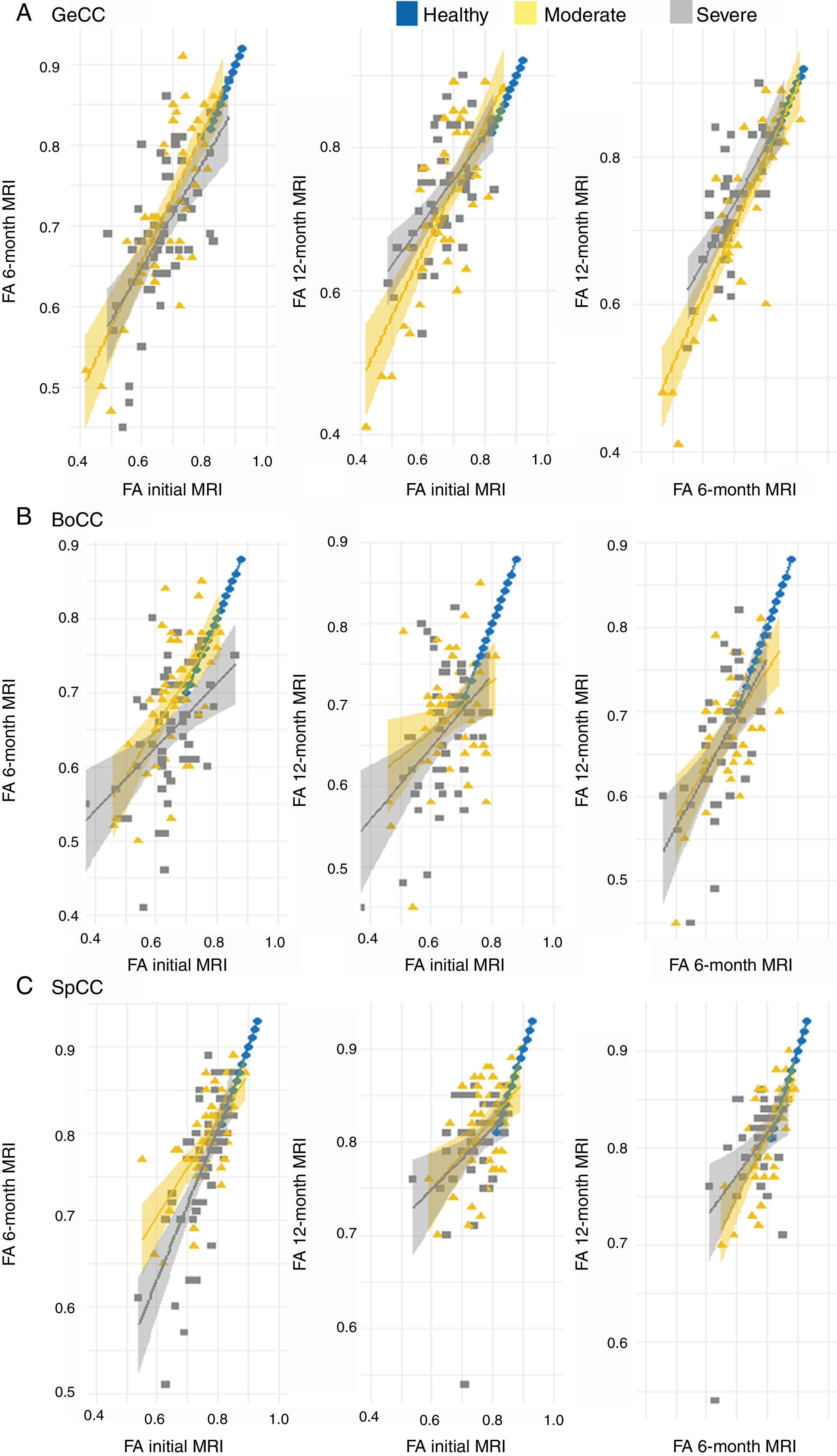
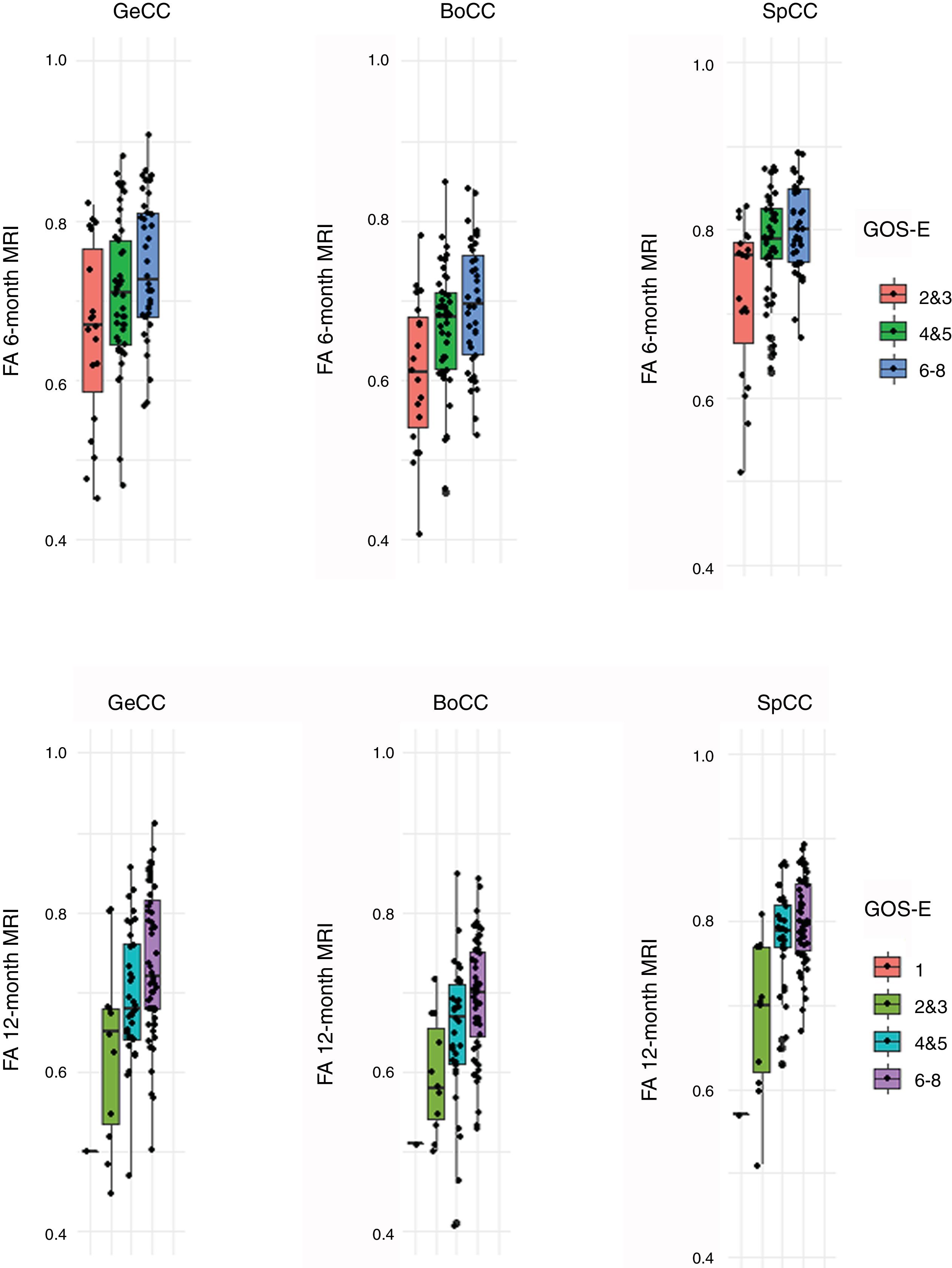
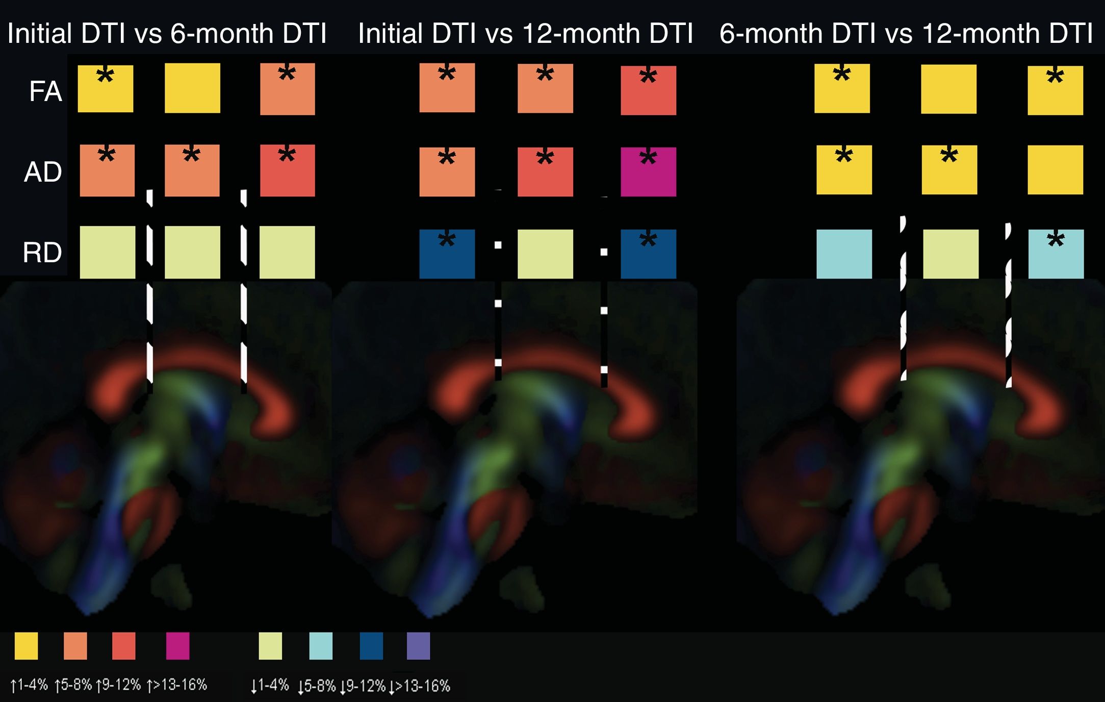
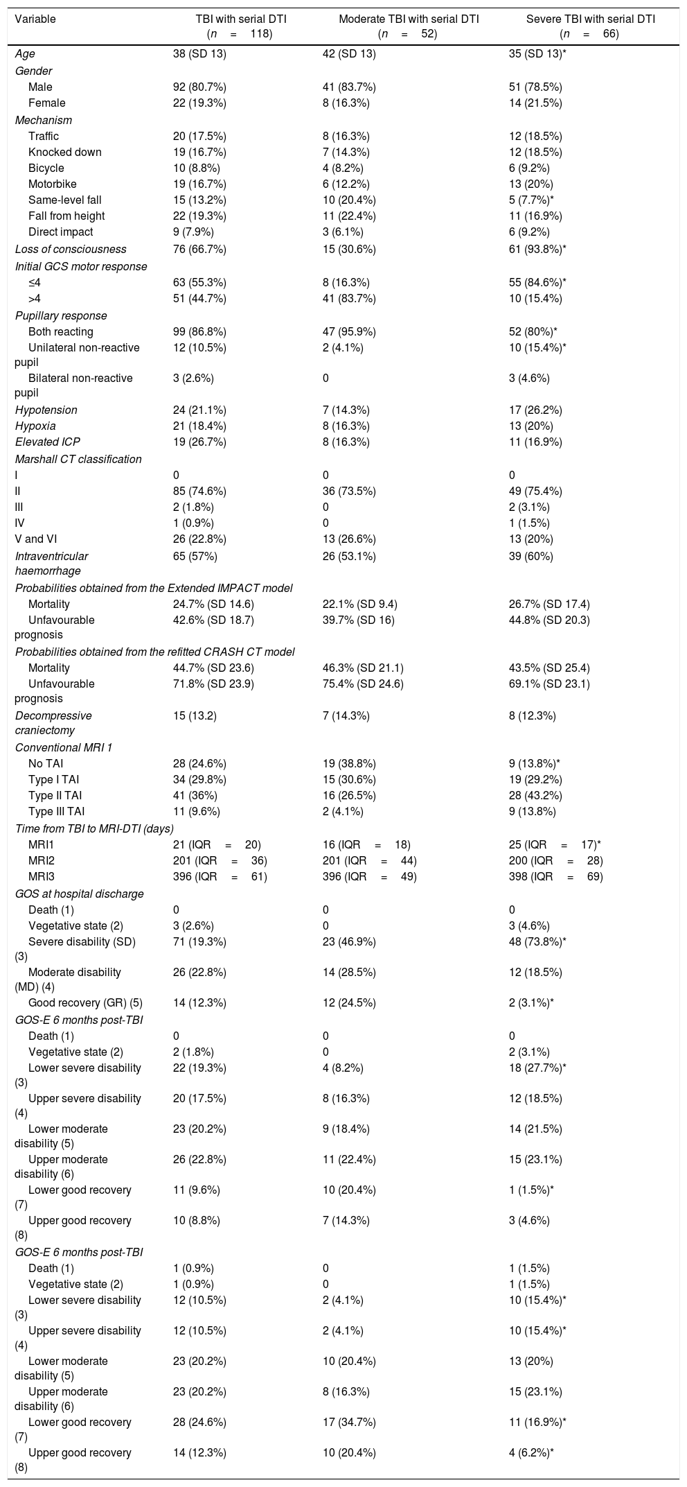
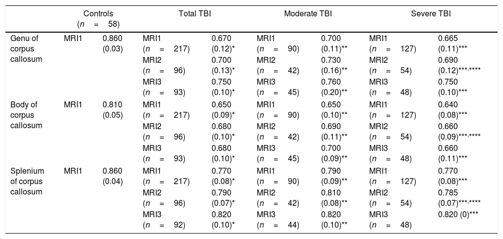
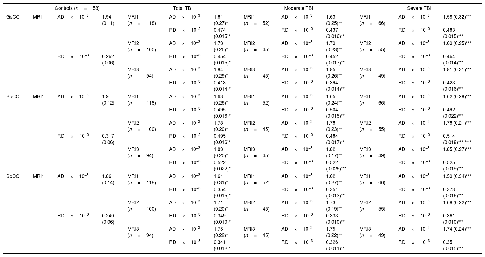
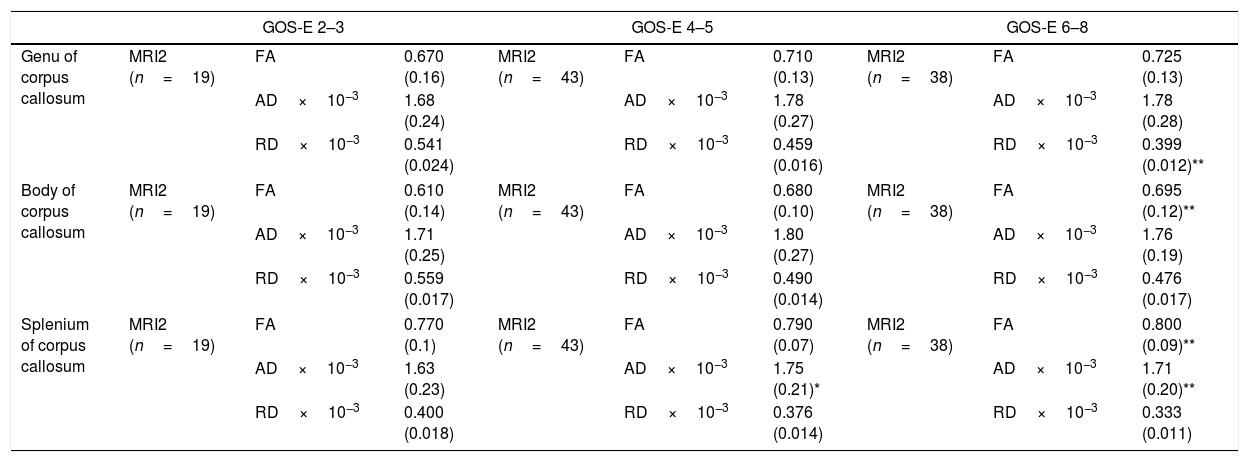
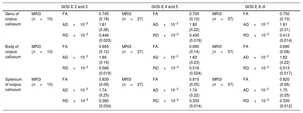
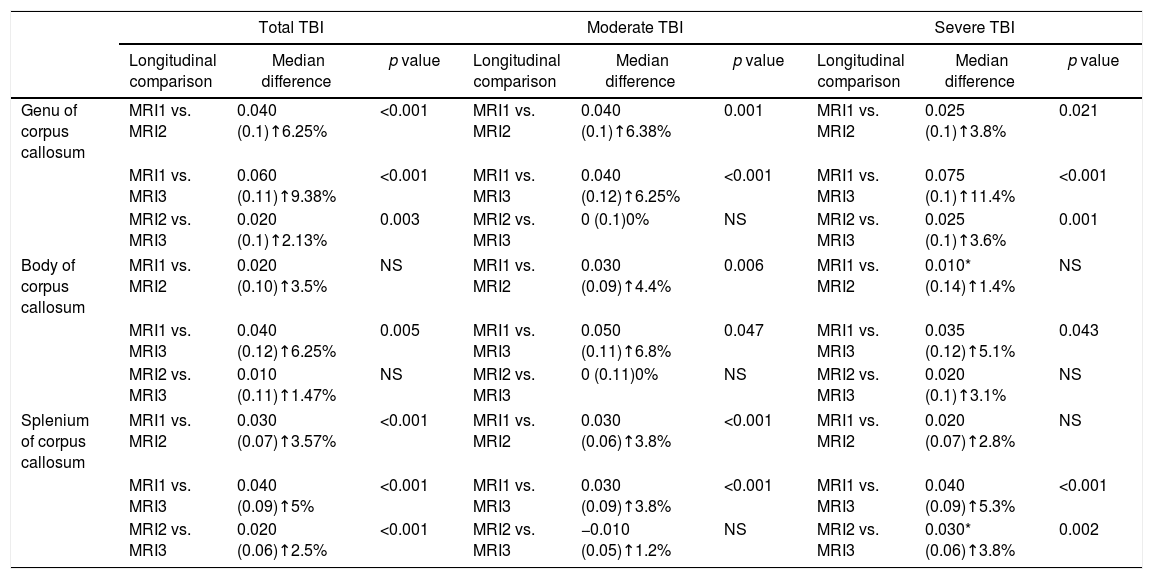
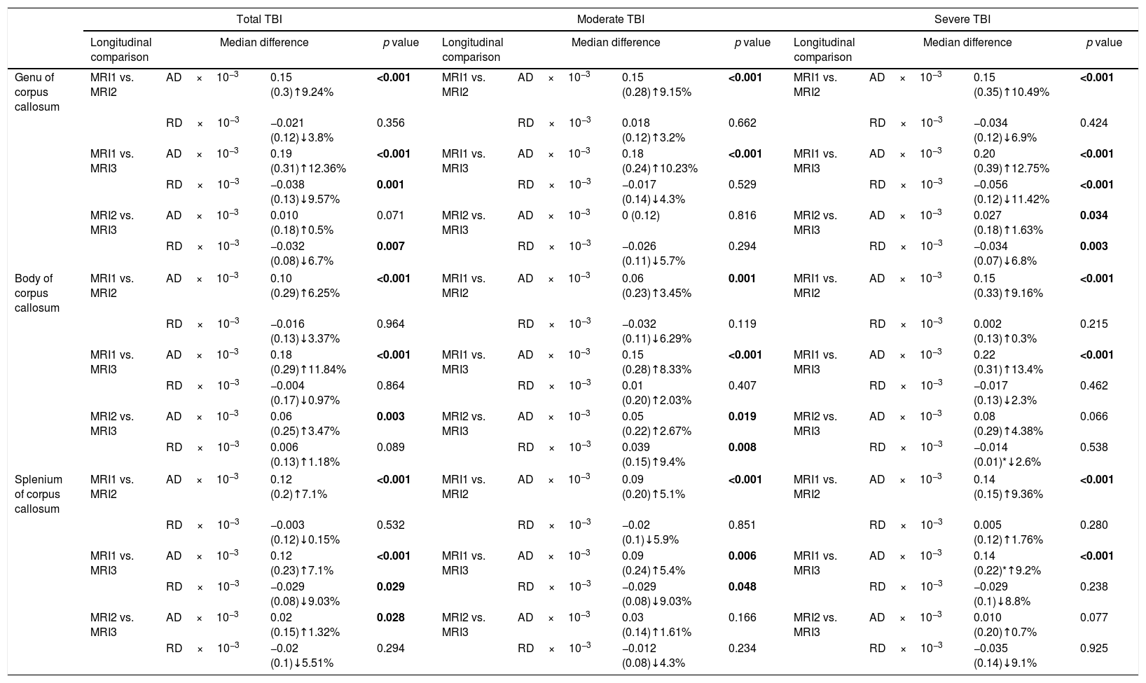
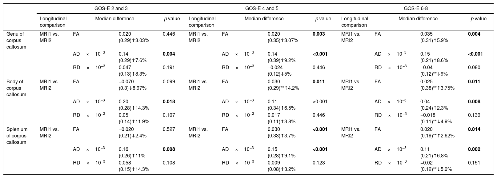
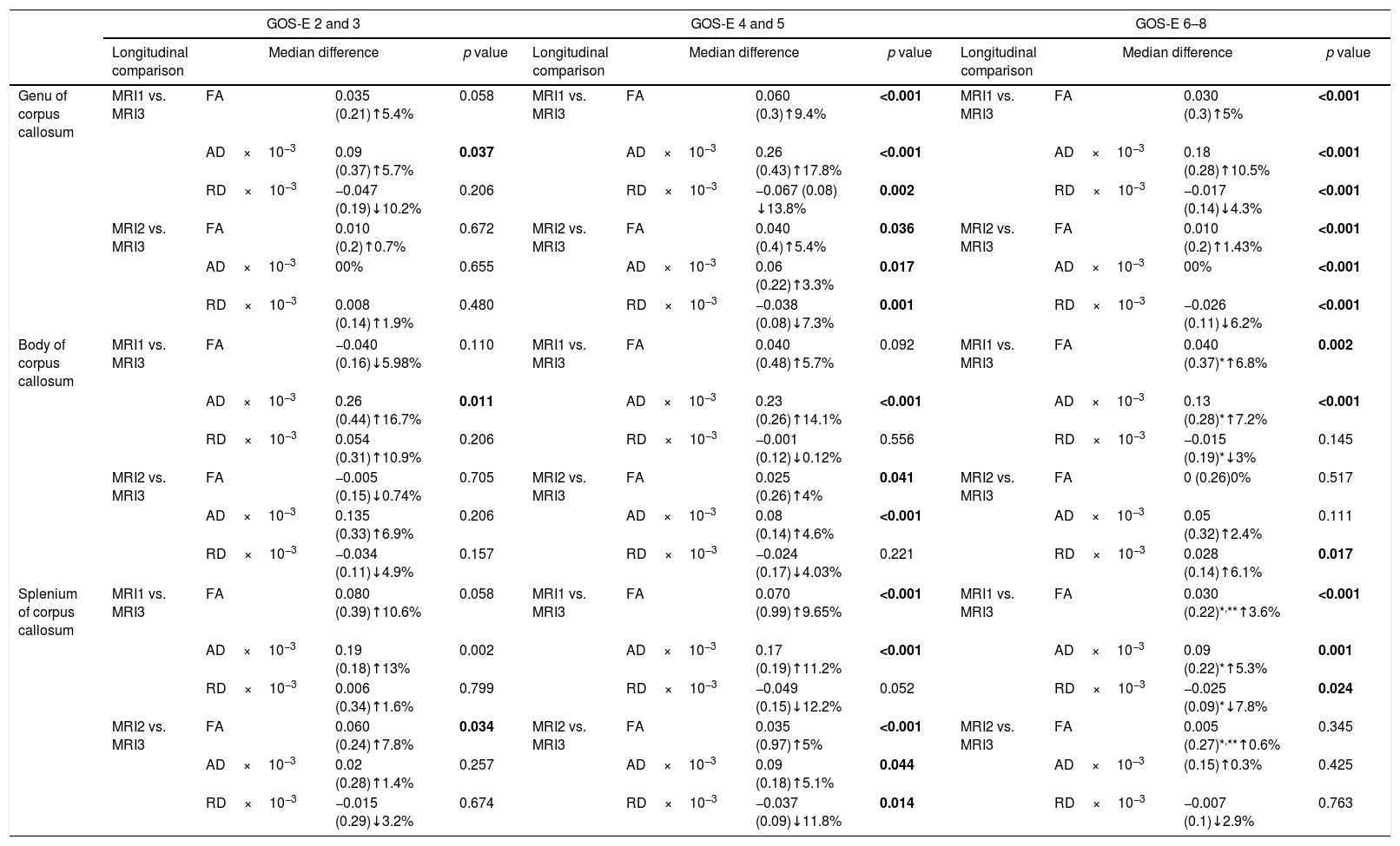
Materials and methods

We performed a prospective observational study in 118 patients with moderate to severe TBI. The study included clinical outcome assessment based on the Glasgow Outcome Scale Extended and serial DTI studies in the early subacute setting (<60 days) and 6 and 12 months after injury. Fractional anisotropy, axial and radial diffusivities were measured in the 3 portions of corpus callosum (genu, body, splenium) at each time point and compared to normalised values from an age-matched control group. Longitudinal fractional anisotropy analysis and its correlation with patient improvement was also done by non-parametric testing and ordinal regression analysis.
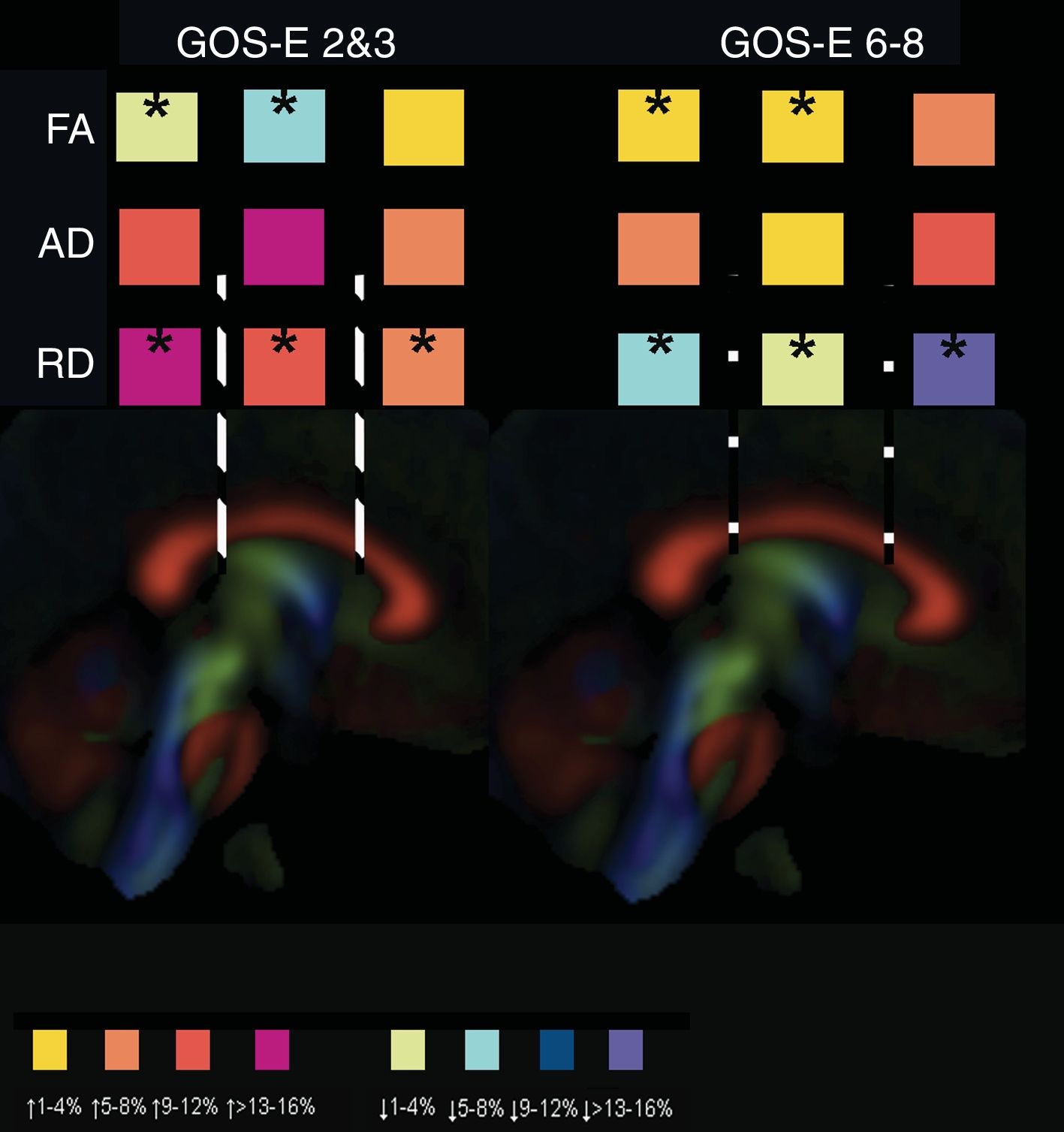
Results

Although dynamic changes in DTI characteristics have been detected in the 3 portions of corpus callosum, patients continue to show lower fractional anisotropy and axial diffusivities values and higher radial diffusivities values compared to controls at the end of the period of study. We have also found differences in the pattern of DTI metrics change between subgroups of patients according with their favourable outcome.

Conclusions

The temporal profile of the change in DTI characteristics seems to provide important information about the clinical recovery of patients after TBI.

Keywords:
Axial diffusivity
Diffusion tensor: Fractional anisotropy
Longitudinal analysis
Ordinal regression analysis
Outcome measures
Radial diffusivity
Traumatic brain injury
Resumen
Antecedentes y objetivos

La lesión axonal traumática es considerada la principal causa de las alteraciones cognitivas y neuropsicológica de los pacientes tras traumatismo craneoencefálico (TCE). Además, existen algunas evidencias sobre la evolución dinámica de la lesión axonal traumática. La secuencia de RM Tensor de difusión (DTI, diffusion tensor imaging) se considera una técnica útil para la caracterización de la lesión axonal traumática, pero son escasos los estudios que hayan evaluado los cambios longitudinales de las características del DTI y su relación con la evolución de los pacientes.

Materiales y métodos

Ciento dieciocho pacientes con TCE moderado y grave fueron estudiados mediante RM-DTI en la fase subaguda precoz (<60 días) y otros estudios sucesivos a los 6 y/o 12 meses tras TCE. Se ha medido la anisotropía fraccionada, difusión axial y radial en las 3 porciones del cuerpo calloso (rodilla, cuerpo y esplenio) y se han comparado con los valores de un grupo control. Además, se ha determinado la situación clínica de los pacientes mediante la Glasgow Outcome Scale Extended al alta hospitalaria, 6 y 12 meses tras TCE. Para el análisis longitudinal de las características del DTI y su correlación con la evolución de los pacientes se han empleado pruebas no paramétricas y un análisis de regresión ordinal.

Resultados

A pesar de haber detectado cambios dinámicos en las características del DTI en las 3 porciones del cuerpo calloso, los pacientes continuaron mostrando valores de anisotropía fraccionada y difusión axial significativamente inferiores y valores de difusión radial mayores en comparación con los controles al final del periodo de estudio. También hemos encontrado diferencias en el patrón de cambio del DTI entre subgrupos de pacientes que presentaron evolución favorable.

Conclusiones

El perfil temporal del cambio en las características del DTI parece proporcionar información importante sobre la recuperación clínica de los pacientes tras TCE.

Palabras clave:
Difusión axial
Tensor de difusión: Anisotropía fraccionada
Análisis longitudinal
Regresión ordinal
Medidas de evolución
Difusión radial
Traumatismo craneoencefálico

Artículo

Opciones para acceder a los textos completos de la publicación Neurocirugía
Socio
Socio de la Sociedad Española de Neurocirugía

Si es la primera vez que accede a la web puede obtener sus claves de acceso poniéndose en contacto con Elsevier España en suscripciones@elsevier.com o a través de su teléfono de Atención al Cliente 902 88 87 40 si llama desde territorio español o del +34 932 418 800 (de 9 a 18h., GMT + 1) si lo hace desde el extranjero.

Si ya tiene sus datos de acceso, clique aquí.

Si olvidó su clave de acceso puede recuperarla clicando aquí y seleccionando la opción "He olvidado mi contraseña".

Suscriptor
Suscriptor de la revista

Si ya tiene sus datos de acceso, clique aquí.

Si olvidó su clave de acceso puede recuperarla clicando aquí y seleccionando la opción "He olvidado mi contraseña".
Suscribirse
Suscribirse a:

Neurocirugía

Comprar
Comprar acceso al artículo

Comprando el artículo el PDF del mismo podrá ser descargado

Precio 19,34 €

Comprar ahora
Contactar
Teléfono para suscripciones e incidencias
De lunes a viernes de 9h a 18h (GMT+1) excepto los meses de julio y agosto que será de 9 a 15h
Llamadas desde España
932 415 960
Llamadas desde fuera de España
+34 932 415 960
Email
Idiomas
Neurocirugía
es en

¿Es usted profesional sanitario apto para prescribir o dispensar medicamentos?

Are you a health professional able to prescribe or dispense drugs?