El nevo de Ota y el nevo de Ito son melanocitosis dérmicas hiperpigmentarias que surgen como consecuencia de alteraciones o fallas en la migración de los melanocitos desde la cresta neural hacia la epidermis, tienen una etiopatogenia poco conocida y pueden ser congénitas o adquiridas.
Caso clínicoSe trata de un paciente con diagnóstico simultáneo de nevo de Ota y nevo de Ito desde el nacimiento que acude al Servicio de Neurocirugía del hospital Carlos Andrade Marín de la ciudad de Quito y consulta por presentar cefalea súbita de gran intensidad asociada a hemiparesia braquiocrural izquierda.
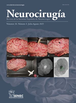
EvoluciónPor medio de exámenes complementarios es diagnosticado de dos lesiones ocupantes intracraneales extraaxiales, una parasagital frontoparietal derecha y otra localizada en polo temporal derecho. Se planificó una resección quirúrgica de la lesión parasagital cuyo diagnóstico histopatológico fue melanocitosis meníngea. La lesión de polo temporal fue derivada para tratamiento con Gamma Knife®.
ConclusiónLos tumores melanocíticos primarios son extremadamente raros, existe evidencia de su asociación con las melanocitosis dérmicas y en especial con el nevo de Ota. El caso presentado describe la coexistencia de dos melanocitosis dérmicas poco frecuentes (nevo de Ota - nevo de Ito) y un tumor melanocítico primario en el mismo paciente, un caso muy fuera de lo común.
Nevus of Ota and nevus of Ito are hyperpigmentary dermal melanocytoses which develop as a consequence of disturbances or failures during migration of melanocytes from the neural crest towards the epidermis; they have a relatively unknown aetiopathogenesis and may be congenital or acquired.
Case reportThis case involves a male patient with a simultaneous diagnosis of nevus of Ota and nevus of Ito at birth. He attended the Neurosurgery department at Carlos Andrade Marín hospital (Quito) with sudden severe headache associated with left brachio-crural hemiparesis.
ProgressInvestigations revealed two extra-axial space-occupying lesions, one parasagittal at the right frontal and parietal lobes and the other located at the right temporal lobe pole. A surgical resection was planned for the parasagittal lesion and the histopathological diagnosis was meningeal melanocytosis. The temporal pole lesion was referred for treatment with Gamma Knife®.
ConclusionPrimary melanocytic neoplasms are extremely rare. There is evidence of their association with dermal melanocytosis and, in particular, with nevus of Ota. This highly unusual case describes the coexistence of two very rare dermal melanocytoses (nevus of Ota and nevus of Ito) and a primary melanocytic neoplasms in the same patient.
Artículo
![]()
Si es la primera vez que accede a la web puede obtener sus claves de acceso poniéndose en contacto con Elsevier España en suscripciones@elsevier.com o a través de su teléfono de Atención al Cliente 902 88 87 40 si llama desde territorio español o del +34 932 418 800 (de 9 a 18h., GMT + 1) si lo hace desde el extranjero.
Si ya tiene sus datos de acceso, clique aquí.
Si olvidó su clave de acceso puede recuperarla clicando aquí y seleccionando la opción "He olvidado mi contraseña".