


A thorough understanding of cerebellum anatomy is essential in 4th ventricle approaches (more frequent in pediatric neurosurgery), avoiding relevant complications such as cerebellar mutism.
The aim of the present work is to show the feasibility of a didactic dissection of human cerebellum focusing on cerebellar peduncles and dentate nucleus (DN), which are structures at high risk during these surgical procedures.
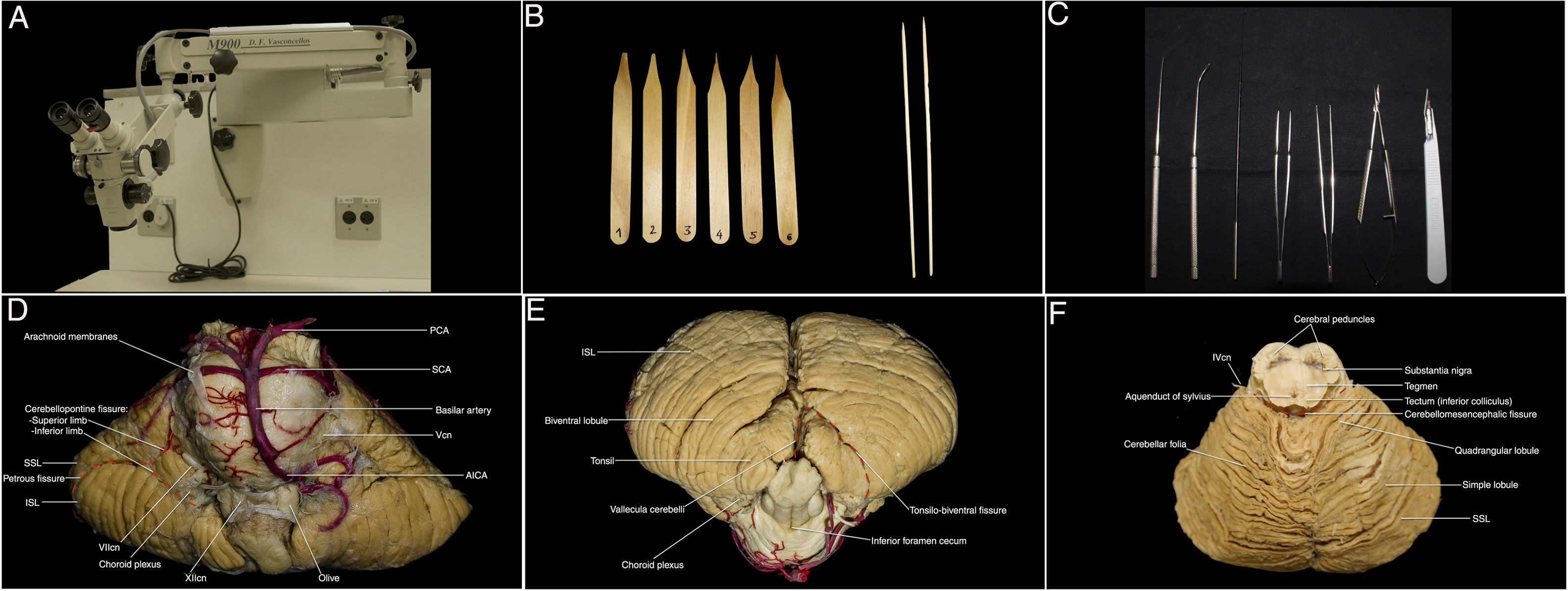
Material and methodsThe cerebellum was dissected according to the Klingler method for white matter, using standard and specific microsurgery tools. Surgical microscope magnification (×6–×40) provided by a D.F. Vasconcellos M900 was required.
A Canon EOS T7 18–55 mm digital camera was used and Adobe Lightroom Classic CC and Keynote were selected as photo enhancing software. Special methods such as LED light endoscopic transillumination were used for photographical reasons.
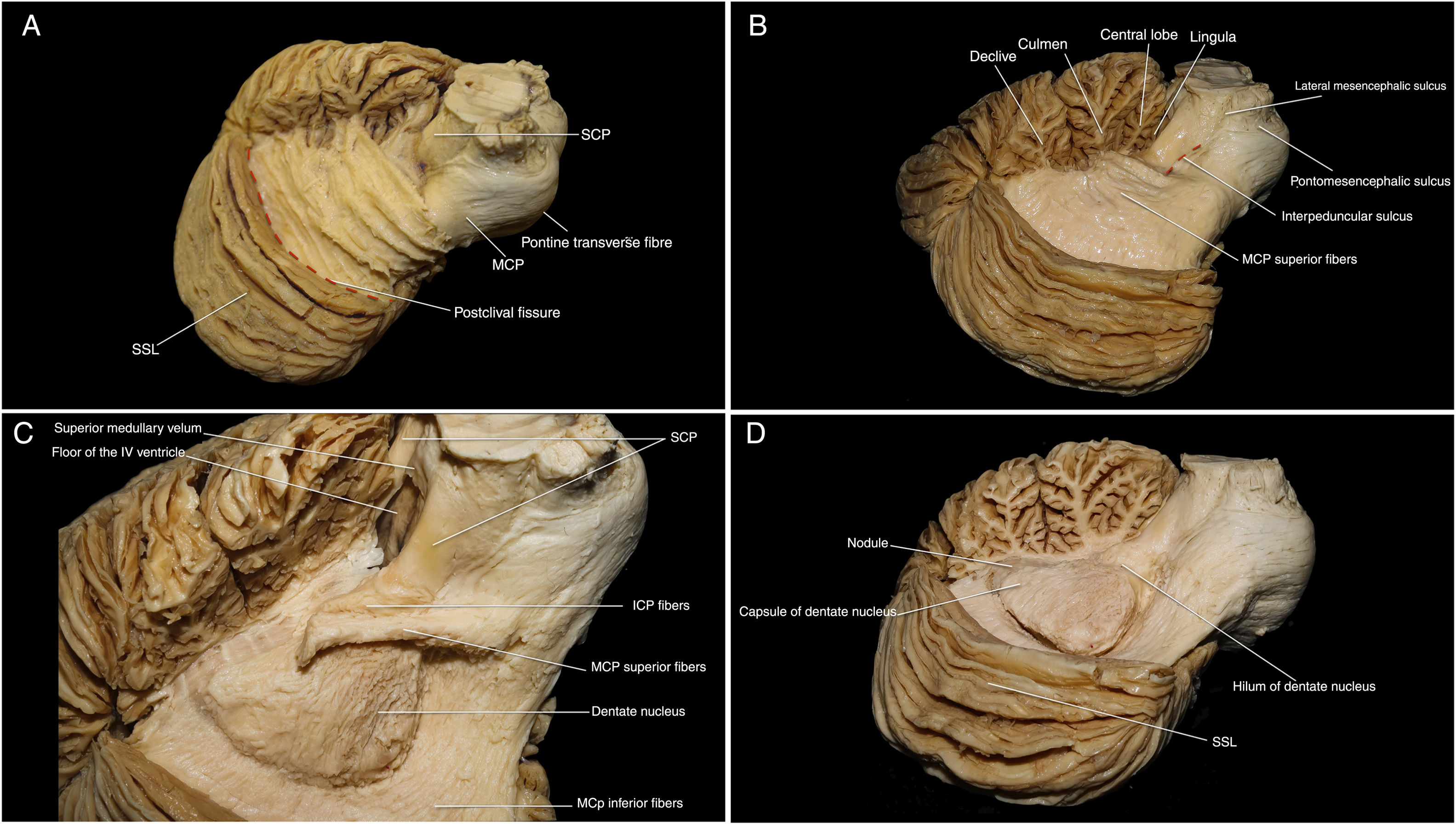
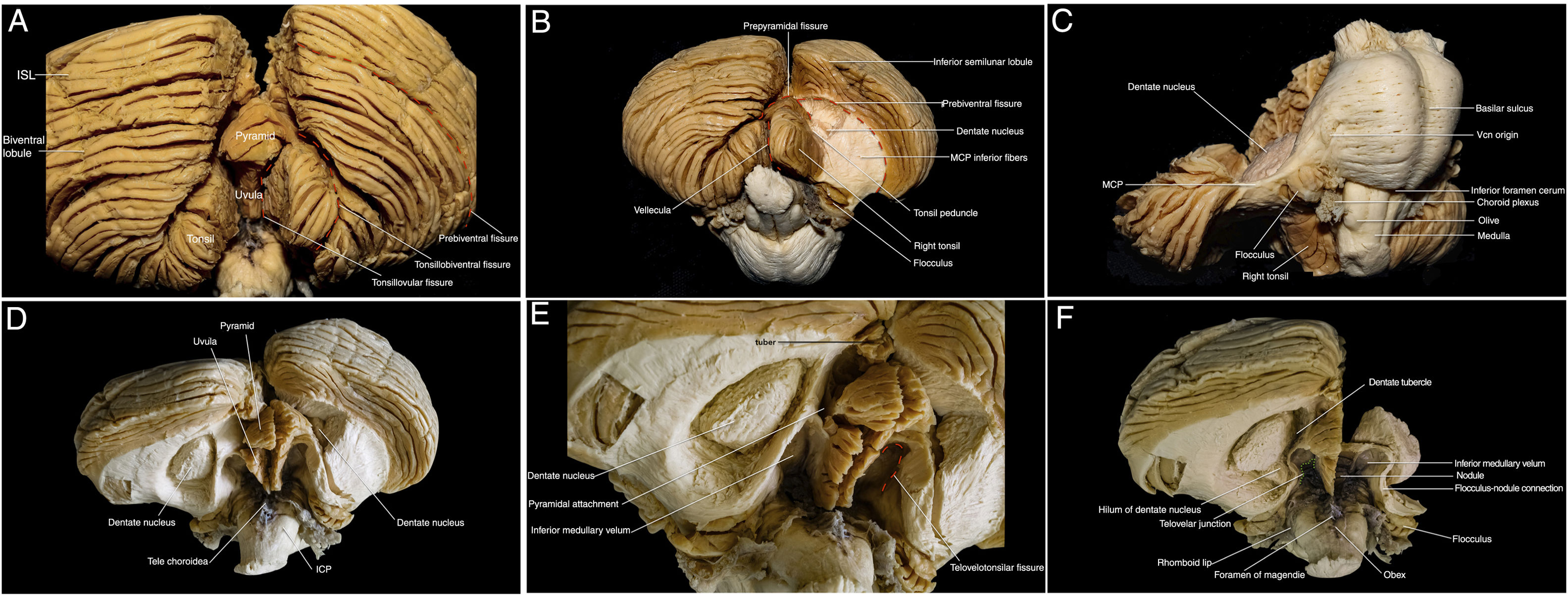
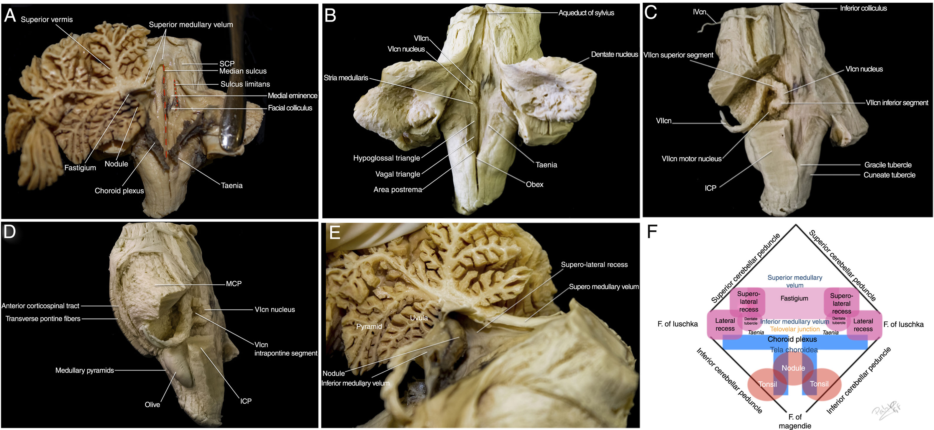
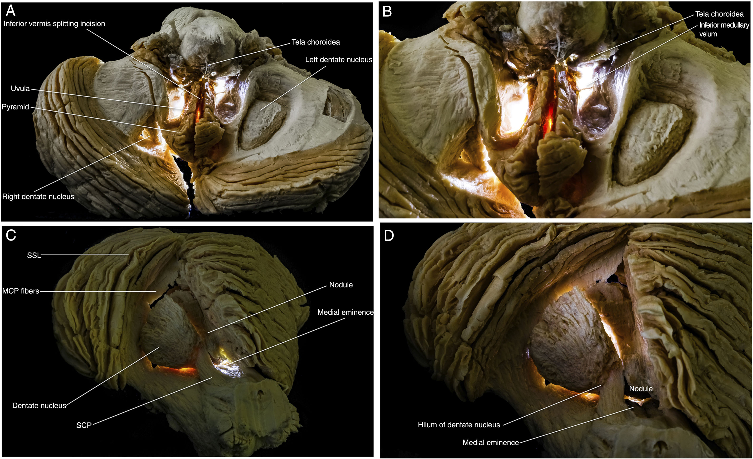
ResultsDN dissection was successfully achieved and the relations between these nucleus and the cerebellar peduncles, inferior vermis and medullary velums were described.
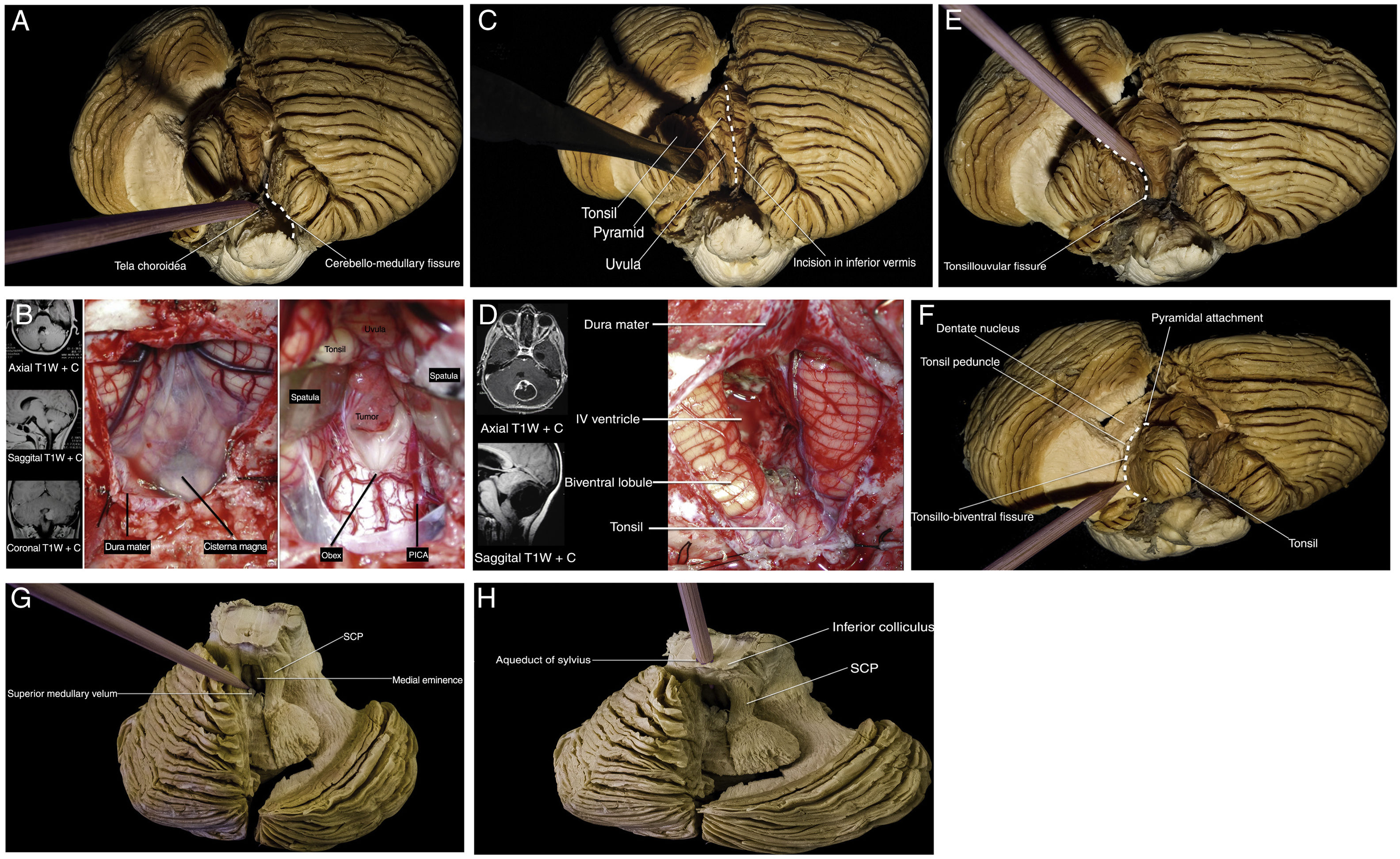
Through this three steps dissection guide (1. tentorial surface; 2. suboccipital surface; 3. 4th ventricle structures), the most relevant anatomical structures were shown and its implications in different 4th ventricle approaches were characterised.
Conclusion3 D perspective provided by real specimen anatomical dissection is critical for learning neuroanatomy.
LED transillumination was shown as a useful technique for the 4th ventricle structures photographic documentation which improves spatial recognition. This benefit can be applied for the study of the relations between the medullary velums and the rhomboid fossa foramina, which are permeable to light.
The proposed three-steps dissection guide helps to a better understanding of human cerebellum and to gain self-confidence, allowing safer practice for neurosurgeons in all stages of their career.
Un buen conocimiento anatómico del cerebelo es fundamental para llevar a cabo abordajes al IV ventrículo (frecuentes en la neurocirugía pediátrica) de forma segura, evitando secuelas como el mutismo cerebeloso.
El propósito del presente trabajo es realizar una disección didáctica del cerebelo humano centrándonos especialmente en los pedúnculos cerebelosos y en los núcleos dentados (DN); estructuras en riesgo durante estos procedimientos.
Material y métodosSe disecó el órgano, siguiendo el método de Klingler para fibras blancas, usando material de microcirugía estándar y específico, bajo un microscopio D. F. Vasconcellos M900 con aumento ×6–×40. Se utilizó una cámara Canon EOS T7 con un objetivo de 18–55 mm y se editaron las imágenes con Adobe Lightroom Classic CC y Keynote. Se emplearon métodos especiales como la iluminación endoscópica con luz LED para la obtención de algunas fotografías.
ResultadosSe logró disecar con éxito los DN del cerebelo y describir su relación con los pedúnculos cerebelosos, vermis inferior y velos medulares.
Mediante esta guía de tres pasos (1. cara tentorial; 2. cara suboccipital; 3. estructuras del IV ventrículo) se consiguió mostrar los elementos más importantes para el estudio del órgano y caracterizar sus implicaciones en los distintos abordajes al IV ventrículo.
ConclusionesLa mejor forma de completar el estudio de neuroanatomía es la disección de especímenes, ya que aporta una visión 3 D.
La transiluminación con luz LED se reveló como una herramienta útil para el registro fotográfico de estructuras del IV ventrículo, lo que mejora la visión espacial. Su principal aplicación la encontramos en los velos medulares y forámenes de la fosa romboide, ya que son permeables a la luz.
La guía de disección en tres fases propuesta en este trabajo puede ayudar a los neurocirujanos, en cualquier etapa de su formación, a comprender mejor el cerebelo, mejorando la seguridad de los procedimientos quirúrgicos y la confianza en sí mismos.
Article
![]()
If it is the first time you have accessed you can obtain your credentials by contacting Elsevier Spain in suscripciones@elsevier.com or by calling our Customer Service at902 88 87 40 if you are calling from Spain or at +34 932 418 800 (from 9 to 18h., GMT + 1) if you are calling outside of Spain.
If you already have your login data, please click here .
If you have forgotten your password you can you can recover it by clicking here and selecting the option ¿I have forgotten my password¿.


