Journal Information
Vol. 31. Issue 6.
Pages 261-267 (November - December 2020)
Visits
670
Vol. 31. Issue 6.
Pages 261-267 (November - December 2020)
Clinical research
Full text access
Postcraniectomy pain: Comparison between 2 incisions for the retrosigmoid approach. Prospective randomized study
Dolor poscraniectomía: comparación entre 2 incisiones para el abordaje retrosigmoideo. Estudio prospectivo aleatorizado
Visits
670
Emiliano Loreficea,
Corresponding author
elorefice@fleni.org.ar

Corresponding author.
, Francisco Marcó del Ponta, Sebastián J.M. Giovanninia, Sol Cavanaghb, María Teresa Goicocheab, Andrés Cervioa
a Departamento de Neurocirugía, FLENI, Buenos Aires, Argentina
b Departamento de Neurología, Clínica de cefalea, FLENI, Buenos Aires, Argentina
This item has received
Article information
Abstract
Full Text
Bibliography
Download PDF
Statistics
Figures (5)
Show moreShow less
Tables (2)
Table 1. SF-36 questionnaire items.
Table 2. Postoperative complications in the two groups.
Show moreShow less
Abstract
Background

Suboccipital lateral approach is a common practice in neurosurgery to expose the region of the cerebellopontine angle. Postcraniectomy headache (PCH) is one of the most frequent complications that diminish the quality of life of patients.

Objective

To compare postcraniectomy pain in patients operated on for vestibular neurinomas by a suboccipital lateral approach by 2 different incisions.

Material and methods

Prospective randomised research study. Follow-up of patients operated for vestibular neurinomas between July 2017 and May 2019 (n=40) by the same surgeon. One group received the classical linear incision (n=20) and another group the alternative incision in an inverted “U” (modified Dandy) (n=20). Pain intensity was evaluated by numerical scale. A minimum follow-up of 3 months was carried out. The impact on quality of life was measured by the SF-36 questionnaire short version both before and after surgery.

Results

The average age was 46.1 years. The overall PCH index was 27.5% (n=11) of the patients. The incidence of pain in the group that received the classical incision (A) was 20% (n=4) and in group B was 35% (n=7).

Conclusions

We found a higher rate of post-craniectomy headache in patients who underwent a “modified Dandy” incision. These are preliminary data of an undergoing study and we hope to obtain more representative information in the future. We recommend interdisciplinary follow up for the integral treatment of PCH.

Keywords:
Approach
Retrosigmoid
Craniectomy
Neurinoma
Postoperative pain
Resumen
Introducción

El abordaje suboccipital lateral es de práctica habitual en neurocirugía para exponer la región del ángulo pontocerebeloso. El dolor poscraniectomía (DPC) es una de las complicaciones más frecuentes que disminuyen la calidad de vida de los pacientes.

Objetivo

Comparar el DPC en pacientes operados de neurinomas vestibulares por un abordaje suboccipital lateral mediante 2 incisiones distintas.

Material y métodos

Estudio de investigación prospectivo aleatorizado. Se realizó seguimiento de un grupo de pacientes operados por neurinomas vestibulares entre julio de 2017 y mayo de 2019 (n=40) por un mismo cirujano. Un grupo recibió la incisión lineal clásica (n=20) y otro grupo la incisión alternativa en «U» invertida o «Dandy modificada» (n=20). La intensidad del dolor fue evaluada mediante escala numérica. Se realizó un seguimiento mínimo de 3 meses. El impacto en la calidad de vida se objetivó mediante cuestionario SF-36 versión corta tanto pre- como posquirúrgico.

Resultados

La edad promedio fue 46,1 años. El índice global de DPC fue del 27,5% (n=11) de los pacientes. La incidencia de cefalea en el grupo que recibió la incisión clásica (A) fue del 20% (n=4), en el grupo B fue del 35% (n=7).

Conclusión

Encontramos un mayor índice de DPC en los pacientes que recibieron una incisión tipo «Dandy modificada». Estos son datos preliminares de un estudio que continúa y esperamos obtener datos más representativos en el futuro. Recomendamos el seguimiento interdisciplinario para el tratamiento integral del DPC.

Palabras clave:
Abordaje
Retrosigmoideo
Craniectomía
Neurinoma
Dolor poscraniectomía
Full Text
Introduction

The suboccipital lateral approach (SLA) is one of the most useful in neurosurgery. It is used to treat various pathologies of the cerebellopontine angle (tumours, neurovascular decompression, vascular disease and others).1 One advantage of this approach is that it allows for excellent viewing angles and shortens operating time. However, postoperative pain at the wound site is a side effect that alters the quality of life of many patients who undergo such operations.2

At the end of the twentieth century, pain was recognised a possible direct consequence of the SLA, with the term “postcraniectomy headache” (PCH) being coined.3 This complication of the approach has a high incidence (7–50%) and a negative impact on the quality of life of patients who suffer from it.4

Although the pathophysiology of PCH is not yet certain, the most accepted theory proposes that adherences between the musculoaponeurotic and dural planes are responsible for generating the pain.5,6 Furthermore, damage to the suboccipital nerves during the surgical approach could be one of the causes.7,8

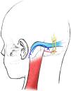
Different skin incisions and flap preparations have been proposed for this type of approach. Recently, Cohen-Gadol reported that the “modified Dandy” incision was associated with a lower rate of PCH compared to the classical retroauricular linear incision9 (Fig. 1).

Fig. 1.

Diagram of a “modified Dandy”-type retrosigmoid incision.

(0.08MB).

The objective of this work was to compare PCH in patients operated on for vestibular neurinomas via a SLA using two distinct incisions.

Materials and methodsStudy design and population

A prospective, randomised, double-blind study was conducted. The data of a group of patients who underwent surgery for the resection of vestibular neurinomas, operated on by the same senior surgeon in our institution, between July 2017 and May 2019 (n=40) was analysed.

The patients were randomly assigned to two groups (A and B) with 20 patients in each group. The patients in “group A” received a classical linear incision, while “group B” received a “modified Dandy”-type incision (Fig. 2). All received follow-up of at least three months after the surgery.

Fig. 2.

(A) Classical linear incision. (B) “Modified Dandy” incision.

(0.34MB).

Only patients with vestibular neurinomas in whom it was decided to perform a SLA were included. Those patients previously operated on via a SLA, patients with a history of chronic headache and patients with other anatomopathological diagnoses were excluded (n=8) (Fig. 3).

Fig. 3.

Diagram of patients included in the protocol.

(0.15MB).
Postcraniectomy headache study

PCH was defined according to the criteria established by the International Headache Society (IHS)10 as a secondary headache that appears within the seven days following surgery in a patient with no other causes of headache; a chronic persistent headache, such as one that persists three months or more after surgery.

All patients were assessed before and after surgery in our institution's Headache Clinic (Neurology Department). It should be noted that neither the patient nor the assessing neurologist knew the type of incision used (double-blind study).

The incidence of pain and its characteristics (oppressive or stabbing), its grading on a pain analogue scale (mild 1–5, moderate 5–8 and severe 8–10) and the use of analgesics were studied. At least three months’ follow-up was performed in all cases.

The impact of the pain on the patient's pre- and postoperative quality of life was assessed using the standardised Short Form 36 (SF-36) questionnaire (Table 1).11 This questionnaire assesses both physical health and emotional well-being. It identifies eight health dimensions: physical functioning, role limitations due to physical problems, bodily pain, general health, vitality, social functioning, role limitations due to emotional problems and mental health. In addition to these eight health scales, it includes an item that assesses the general concept of changes in perceived health status compared to one year previously. The scale goes from “0” (worst health status) to “100” (best health status) using the algorithms and indications offered in the questionnaire's scoring and interpretation manual.

Table 1.

SF-36 questionnaire items.

Meanings of scores from 0 to 100
Dimension  No. of items  “Worst” score (0)  “Best” score (100) 
Physical functioning  10  Very limited in performing all physical activities, including bathing or showering, due to health  Performs all types of physical activities, even the most vigorous, without any limitation due to health 
Role – physical  Problems with work or daily activities due to physical health  No problems with work or other daily activities due to health 
Bodily pain  Very intense pain that is extremely limiting  No pain or limitations because of it 
General health  Assesses own health as poor and believes it may get worse  Assesses own health as excellent 
Vitality  Feels tired and exhausted all the time  Feels very energetic and full of energy all the time 
Social functioning  Extreme and very frequent interference in normal social activities due to physical or emotional problems  Performs normal social activities without interference due to physical or emotional problems 
Role – emotional  Problems with work or other daily activities due to emotional problems  No problems with work or other daily activities due to emotional problems 
Mental health  Feelings of anguish and depression all the time  Feelings of happiness, tranquillity and calm all the time 
Health transition item  Believes their health is much worse than one year ago  Believes their health is much better than one year ago 

In addition to postcraniectomy headache, other complications inherent in the approach such as cerebrospinal fluid fistula and surgical site infection were assessed (Table 2).

Table 2.

Postoperative complications in the two groups.

Postop. complications  Group A (n=20)  Group B (n=20) 
Postcraniectomy headache  4 (20%)  7 (35%) 
Cerebrospinal fluid fistula  1 (5%) 
Wound infection  1 (5%) 

The research protocol was conducted in accordance with the principles established by our institution's Ethics Committee. For this reason, each patient who participated had to sign an informed consent form to be included in the protocol.

Surgical technique

A suboccipital lateral or retrosigmoid approach was used in all patients.

Position

All patients were operated on in the park bench position. The head was fixed in place using a three-pin headrest.

Incision

There were two types of incision included in the protocol.

The “modified Dandy”-type incision was performed as follows: it is an “inverted U”-shaped incision with one horizontal and one vertical branch. First a horizontal line was drawn between the root of the zygoma and the inion, representing the position of the transverse sinus at the dural level. The 7cm vertical incision is located 0.5cm posterior and parallel to the mastoid groove. The horizontal branch meets the vertical branch above the transverse sinus and its medial extension is arbitrary depending on the size of the craniectomy to be performed. Once the skin was opened, the musculocutaneous flap was repelled with an inferior pedicle (Fig. 4).

Fig. 4.

Diagram of the musculocutaneous flap with inferior pedicle.

(0.18MB).

For the classical linear incision, the mastoid apophysis and digastric groove were first marked. A vertical incision with slight lateral concavity was made 2cm medial to the mastoid apophysis, from the upper edge of the external ear to 2cm below the tip of the mastoid apophysis.12

Soft tissue dissection, craniectomy and durotomy

The craniectomy and opening of the dura were performed identically in both groups (Fig. 5).

Fig. 5.

Image showing exposure size. (A) Skin opening with exposure of the asterion. (B) Dural exposure.

(0.27MB).
Intradural time

The description of this falls outside the scope of this study.

Closure

The bone defect created was filled with bone cement (Subitón®). Polyglactin 910 00 suture material (Vicryl®) was used to close the muscle, 2–0 for subcutaneous cellular tissue and 3–0 nylon (Ethilon®) with interrupted vertical mattress sutures to close the skin.

Statistical analysis

The correlation of the qualitative variables was calculated by analysing the Phi (ϕ) coefficient to compare headaches between the two groups. The dependent T-test was used for small samples. The same test was also used to compare quality of life between the two groups based on the SF-36.

Results

There was no significant difference of distribution between genders. The mean age was 46.1 years (range: 21–73).

The overall rate of postoperative headaches across both groups was 27.5% (n=11). These were graded mild in six patients and moderate in four, while one patient experienced severe headache. The headache characteristics were similar in both groups, with stabbing-type pain in 63.6% (n=7) and oppressive pain in the rest.

Group A (n=20), in whom the classical incision was used, presented a postoperative headache incidence of 20% (n=4), these being mild in three patients and moderate in one case, with no cases of severe headache recorded.

Group B (n=20) had a postoperative headache incidence of 35% (n=7), these being mild in three cases, moderate in three cases and severe in one.

Of these, 27.2% (n=3) evolved into persistent chronic headache (three months postoperative), two in group B and one in group A.

Some 72.7% (n=8) required the use of common analgesics (ibuprofen, paracetamol, naproxen) to control the pain.

It can be inferred, with a significance level of 20%, that the patients who suffered from headache experienced a reduction in their quality of life (assessed using the SF-36), regardless of the incision used.

Comparing the incidence of headache in the two groups, a higher incidence of headache was observed in group B (ϕ coef. −0.36), without this being statistically significant.

In group A, one patient presented with a cerebrospinal fluid fistula as a complication, which resolved with the placement of a lumbar catheter, while another presented with a surgical wound infection requiring the wound to be cleaned. No fistulas or infections were recorded in group B.

Discussion

In recent times, with the use of microscopes and advances in surgical techniques, better outcomes have been attained in terms of surgical excision, nerve preservation and the morbidity and mortality of patients operated on for vestibular neurinomas.13–15 Nevertheless, this has not translated into a reduction in postoperative headache. It is for this reason that in 1980 the term “postcraniectomy headache” was coined to describe pain in the occipital region that manifests within seven days after surgery. Such headaches, together with ataxia, hypoacusis and facial paralysis, are the complications with the biggest impact on the quality of life of patients operated on for vestibular neurinomas.16,17

The presence of headache is associated with a delay in returning to work after surgery in many cases, as well as the need to continue in medical follow-up for a long period of time.18,19 People with PCH have reported more problems at work and in other daily activities in comparison with those who do not have this symptom.20 Among the different series analysed, patients with PCH state that headaches interfere with their daily activities (29–60%), sports activities (25%), work (19–38%) and social activities (8%). In 15% of cases, PCH has a negative impact on mood, while in 22% of cases the pain becomes incapacitating.21–23 They also report a higher incidence of depression and anxiety.24

Numerous factors have been proposed as causes of the pain, and they are not well-established.

With regard to the characteristics of the pain, some 73% of patients report superficial wound pain. Some authors attribute this to the musculature and soft tissues damaged during the approach.25 This has been reaffirmed by those who suggest that surgeries involving less muscle damage (because of the type of approach) have had a lower incidence of postoperative headache.26 Adherence of the musculoaponeurotic planes to the dura mater has also been suggested as a possible cause of pain. This is based on the fact that patients undergoing craniectomy have a significantly higher incidence of PCH than those who undergo craniotomy. In 2003, Schaller et al. analysed the outcomes of patients who underwent craniotomy or craniectomy via a retrosigmoid approach for the removal of vestibular schwannomas. They stated that the headaches could be due to postoperative aseptic meningitis that could be caused by the use of fibrin glue and residual bone from drilling the auditory meatus.27–29

Evidence was even found of adherence between the dura mater and the cervical musculature through a histopathological assessment in the case of one patient with persistent postoperative headache.30

On the other hand, there is a school of thought that maintains that the pain is due to suboccipital nerve lesion. In this respect, almost half of patients with pain present with a neuralgic pattern. In some cases, the pain can be reproduced on palpating the scar, and injection with local anaesthetics may be beneficial in its treatment.31–33

Beyond these two hypotheses, there is no conclusive study covering all of the aspects mentioned.

Because of all of the above, and motivated by the search for factors that might help to reduce its incidence, due to the significant impact PCH has on sufferers’ quality of life, the decision was made to conduct a prospective study on a selective group of patients to independently assess the influence of incision type on the onset of PCH and to be able to assess changes in patients’ quality of life.

In our work, after a rigorous statistical study, it was possible to infer to a significance level of 20% that patients from both groups who presented with headaches had experienced a reduction in their quality of life as assessed by the SF-36.

It is worth mentioning that the objective of this work was not to study the pathophysiology of PCH, but it should be noted that all the included surgeries consisted of a craniectomy with subsequent cranioplasty, drilling in the posterior wall of the auditory meatus and sealing with fibrin glue, regardless of the incision made.

Thus, we have sought to assess the incision's influence on postoperative pain as an independent factor and avoid including other variables that could affect the onset of pain.

The classical incision used in the retrosigmoid approach is a linear or slightly curved retroauricular incision. In 2011, Cohen-Gadol proposed an incision similar to that used by Dandy with some modifications for the retrosigmoid approach. This consists of an “inverted U”-shaped incision to avoid damaging the neurovascular bundle (occipital artery and nerve) which are often damaged in classical linear incisions. Its other advantages include that it avoids deep caudal muscle dissection towards the base of the skull, which in turn reduces adjacent adherences postoperatively. In a series of 100 patients who underwent SLA, only a single case of postoperative headache was reported, and that case presented with a subdural haematoma as a complication.

In our work, we compare the two incisions in patients operated on for the same pathology (vestibular neurinoma). The overall incidence of headache was 27.5%, which falls within the range published in the various series (7–50%). Stabbing-type headache was the most common in both groups, as was also reported in the literature.

With regard to the statistical study carried out, group A (classical incision) had a headache incidence of 20% and group B 35%, with a ϕ coefficient of −0.36, although this was not yet significant.

Group B recorded one patient with severe pain (8–10). Mild headache was the most commonly reported (n=6), with only one moderate case in both groups.

The most evident bias we encountered while conducting this study was subjective bias with regard to each individual's pain. It should also be noted that this is a prospective study that is still recruiting patients.

Conclusions

We found a higher incidence of PCH in patients who received a “modified Dandy”-type incision. In spite of this, these are preliminary data at three months, from an ongoing study, and we hope to obtain more representative data in the future. We recommend interdisciplinary follow-up for comprehensive treatment of PCH.

Limitations

These are preliminary results at three months, from a study that still needs to increase its number of patients in order to obtain more representative data.

Conflicts of interest

The authors have no conflicts of interest.

Acknowledgement

Special thanks to Dr Juan Francisco Villalonga for his thorough analysis of the structure of the work.

References
[1]
J.M. Gonzalez-Darder, V. Quilis-Quesada, E. de Oliveria.
Abordajes neuroquirúrgicos de la patología craneal y cerebral.
Elsevier, (2016),
[2]
M.B. Hanson, M.E. Glasscock, J.L. Brandes.
Medical treatment of headache after suboccipital acoustic tumor removal.
Laryngoscope, 108 (1998), pp. 1111-1114
[3]
D.A. Silverman, G.B. Hughes, S.E. Kinney, J.H. Lee.
Technical modifications of suboccipital craniectomy for prevention of postoperative headache.
Skull Base, 14 (2004), pp. 77-84
[4]
B. Schaller, A. Baumann.
Headache after removal of vestibular schwannoma via the retrosigmoid approach: a long term follow-up-study.
Otolaryngol Head Neck Surg, 128 (2003), pp. 387-395
[5]
P.J. Catalano, O. Jacobowitz, K.D. Post.
Prevention of headache after retrosigmoid removal of acoustic tumors.
Am J Otol, 17 (1996), pp. 904-908
[6]
T.J. Lovely, D.W. Lowry, P.J. Jannetta.
Functional outcome and the effect of cranioplasty after retromastoid craniectomy for microvascular decompression.
Surg Neurol, 51 (1999), pp. 191-197
[7]
J.R. Gee, Y. Ishaq, N. Vijayan.
Post craniotomy headache.
[8]
M.J. Ruckenstein, J.P. Harris, R.A. Cueva.
Pain subsequent to resection of acoustic neuromas via suboccipital and translabyrinthine approaches.
Am J Otol, 17 (1996), pp. 620-624
[9]
A.A. Cohen-Gadol.
Microvascular decompression surgery for trigeminal neuralgia and hemifacial spasm: naunces of the technique based on experiences with 100 patients and review of the literature.
Clin Neurol Neurosurg, 113 (2011), pp. 844-853
[10]
Headache Classification Committee of the International Headache Society.
The International Classification of Headache Disorders, 3rd edition (beta version).
Cephalalgia, 33 (2013), pp. 629-808
[11]
J.E. Ware Jr., C.D. Sherbourne.
The MOS 36-item short-form health survey (SF-36) (I). Conceptual framework and item selection.
Med Care, 30 (1992), pp. 473-483
[12]
R. Nader, C. Gragnaniello, S. Berta, A.J. Sabbag, M. Levy.
Retrosigmoid approach.
Neurosurgery tricks of the trade, Thieme, (2014),
[13]
A. Nanda.
Principles of posterior fossa surgery: surgical management.
Thieme, (2012),
[14]
R.L. Baldwin.
Headache prevention in retrosigmoid vestibular nerve section.
South Med J, 89 (1996), pp. 375-379
[15]
G. Andersson, L. Ekvall, A. Kinnefors, G. Nyberg, H. Rask-Andersen.
Evaluation of quality of life and symptoms after translabyrinthine acoustic neuroma surgery.
Am J Otol, 18 (1997), pp. 421-426
[16]
T. Santarius, A.R. D'Sousa, H.M. Zeitoun, G. Cruickshank, D.W. Morgan.
Audit of headache following resection of acoustic neuromas using three different techniques of suboccipital approach.
Rev Laryngol Otol Rhinol (Bord), 121 (2000), pp. 75-78
[17]
A. Nguyen, F. Girard, D. Boudreault, F. Fugere, M. Ruel, R. Moumdjian.
Scalp nerve blocks decrease the severity of pain after craniotomy.
Anesth Analg, 93 (2001), pp. 1272-1276
[18]
C.J. Schankin, C. Gall, A. Straube.
Headache syndromes after acoustic neuroma surgery and their implications for quality of life.
Cephalalgia, 29 (2009), pp. 760-771
[19]
S.A. Betchen, J. Walsh, K.D. Post.
Self-assessed quality of life after acoustic neuroma surgery.
J Neurosurg, 99 (2003), pp. 818-823
[20]
G. De Benedittis, A. Lorenzetti, M. Migliore, D. Spagnoli, F. Tiberio, R.M. Villani.
Postoperative pain in neurosurgery: a pilot study in brain surgery.
Neurosurgery, 38 (1996), pp. 466-470
[21]
D.A. Schessel, J.M. Nedzelski, D. Rowed, J.G. Feghali.
Pain after surgery for acoustic neuroma.
Otolaryngol Head Neck Surg, 107 (1992), pp. 424-429
[22]
C.A. Pedrosa, D.K. Ahern, M.J. McKenna, R.G. Ojemann, M.A. Acquadro.
Determinants and impact of headache after acoustic neuroma surgery.
Am J Otol, 15 (1994), pp. 793-797
[23]
N.M. Kane, S. Kazanas, A.R. Maw, H.B. Coakham, M.J. Torrens, M.H. Morgan, et al.
Functional outcome in patients after excision of extracanalicular acoustic neuromas using the suboccipital approach.
Ann R Coll Surg Engl, 77 (1995), pp. 210-216
[24]
A. Kaur, L. Selwa, G. Fromes, D.A. Ross.
Persistent headache after supratentorial craniotomy.
Neurosurgery, 47 (2000), pp. 633-636
[25]
P. Shirley.
Pain relief post craniotomy: a balanced approach.
Anaesthesia, 55 (2000), pp. 409
[26]
M. Thibault, F. Girard, R. Moumdjian, P. Chouinard, D. Boudreault, M. Ruel.
Craniotomy site influences postoperative pain following neurosurgical procedures: a retrospective study.
Can J Anaesth, 54 (2007), pp. 544-548
[27]
N.B. Gormley, L.N. Sekhar, D.C. Wright, D. Kamerer, D. Schessel.
Acoustic neuromas: results of current surgical management.
Neurosurgery, 41 (1997), pp. 50-60
[28]
D.A. Schessel, D.W. Rowed, J.M. Nedzelski.
Postoperative pain following excision of acoustic neuroma by the suboccipital approach: observations on possible cause and potential amelioration.
Am J Otolaryngol, 14 (1993), pp. 491-494
[29]
N.F. Quiney, R. Cooper, M.D. Stoneham, F. Walters.
Pain after craniotomy. A time for reappraisal?.
Br J Neurosurg, 10 (1996), pp. 295-299
[30]
C.G. Jackson, B.M. McGrew, J.A. Forest.
Comparison of postoperative headache after retrosigmoid approach: vestibular nerve section versus vestibular schwannoma resection.
Am J Otol, 21 (2000), pp. 412-416
[31]
H. Koperer, W. Deinsberger, A. Jodicke, D.K. Boker.
Postoperative headache after the lateral suboccipital approach: craniotomy versus craniectomy.
Minim Invasive Neurosurg, 42 (1999), pp. 175-178
[32]
K.S. Ferreira, F. Dach, J.G. Speciali.
Scar neuromas as triggers for headache after craniotomy: clinical evidence.
Arq Neuropsiquiatr, 70 (2012), pp. 206-209
[33]
H. Levo, G. Blomstedt, I. Pyykko.
Vestibular schwannoma surgery and headache.
Acta Otolaryngol Suppl, 543 (2000), pp. 23-25

Please cite this article as: Lorefice E, Marcó del Pont F, Giovannini SJM, Cavanagh S, Goicochea MT, Cervio A. Dolor poscraniectomía: comparación entre 2 incisiones para el abordaje retrosigmoideo. Estudio prospectivo aleatorizado. Neurocirugía. 2020. https://doi.org/10.1016/j.neucir.2020.01.003

Copyright © 2020. Sociedad Española de Neurocirugía
Idiomas
Neurocirugía (English edition)
Article options
Tools
es en

¿Es usted profesional sanitario apto para prescribir o dispensar medicamentos?

Are you a health professional able to prescribe or dispense drugs?