La angiografía por sustracción digital cerebral (ASD) sigue siendo el estándar de oro para el control de los restos aneurismáticos después del clipaje quirúrgico. A pesar de estar asociada a riesgos mínimos, es un procedimiento invasivo que está lejos de estar libre de iatrogenia. Además, tiene una disponibilidad limitada que puede prolongar la estancia postoperatoria del paciente. Por otro lado, la calidad de imagen de la angiografía por tomografía computarizada (ATC) ha mejorado significativamente en las últimas décadas, proporcionando una alternativa valiosa a la ASD. El objetivo de este estudio fue comparar la capacidad de la ATC y la ASD para detectar restos aneurismáticos clínicamente significativos.
MétodosDe una serie prospectiva de aneurismas tratados quirúrgicamente, aquellos con ATC y ASD postoperatorias fueron incluidos en el estudio retrospectivamente. Se realizó una reconstrucción tridimensional de la ATC utilizando el software Brainlab Elements y los resultados se compararon con los de la ASD. Además, se recogieron variables que pudieran afectar a la reconstrucción tridimensional, como el número de clips por aneurisma y el clipaje o embolización previa. En caso de existir remanente aneurismático, también se registró su tamaño.
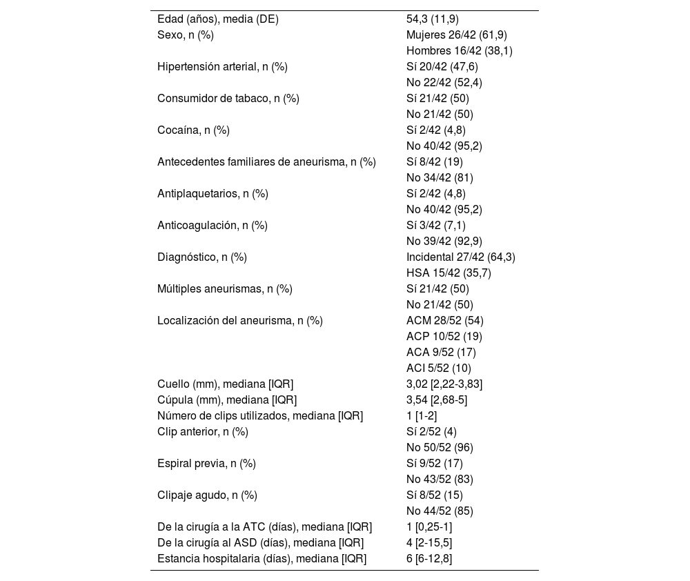
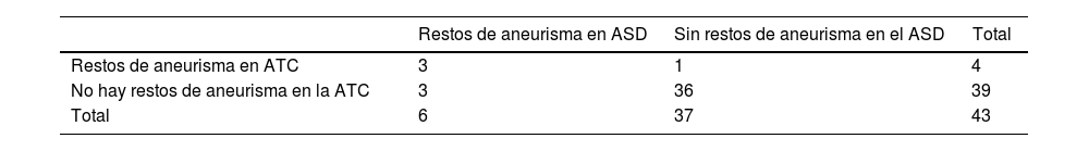
ResultadosEntre enero de 2020 y enero de 2022 se incluyeron un total de 42 pacientes en los que se pinzaron 52 aneurismas (8 de ellos rotos). La ATC presentó una sensibilidad del 50% y una especificidad del 97% en la detección de remanente aneurismático. Los casos en los que la ATC no detectó el remanente aneurismático fueron aneurismas previamente embolizados o aneurismas complejos que requirieron reconstrucción del cuello con 3 o más clips. Ninguno de los remanentes no detectados por la ATC fue lo suficientemente significativo como para justificar un retratamiento del aneurisma.
ConclusionesExcluyendo los aneurismas complejos (previamente embolizados o que requieren reconstrucción quirúrgica con 3 o más clips), las reconstrucciones tridimensionales de imágenes de ATC mostraron excelentes resultados en la detección de restos de aneurismas postoperatorios clínicamente significativos, y pueden obviar la necesidad de una ASD más invasiva y menos disponible.
Cerebral digital subtraction angiography (DSA) remains the gold standard for the control of aneurysmal remnants after surgical clipping. Despite being associated with minimal risks, it is an invasive procedure far from being iatrogenia free. Furthermore, it has limited availability which may prolong patient's postoperative stay. On the other hand, the image quality of computed tomography angiography (CTA) has improved significantly over the past decades providing a valuable alternative to DSA. The objective of this study was to compare the capacity of CTA and DSA to detect clinically significant aneurysmal remnants.
MethodsFrom a prospective series of surgically treated aneurysms, those with postoperative CTA and DSA were retrospectively included in the study. A three-dimensional reconstruction of the CTA was performed using the Brainlab Elements software and the results were compared with those of the DSA. In addition, variables that could affect the three-dimensional reconstruction were collected, such as the number of clips per aneurysm and previous clipping or embolization. In case of an aneurysm remnant, its size was also recorded.
ResultsBetween January 2020 and January 2022, a total of 42 patients in whom 52 aneurysms were clipped (8 of them ruptured) were included. CTA presented a sensitivity of 50% and a specificity of 97% in the detection of aneurysmal remnants. The cases in which CTA did not detect the aneurysmal remnant were previously embolized aneurysms or complex aneurysms that required neck reconstruction with 3 or more clips. None of the remnants undetected by CTA were significant enough to warrant retreatment of the aneurysm.
ConclusionsExcluding complex aneurysms (previously embolized or requiring surgical reconstruction with 3 or more clips), three-dimensional reconstructions of CTA images showed excellent results in detecting clinically significant postoperative aneurysm remnants and may obviate the need for a the more invasive and less available DSA.
Artículo
![]()
Si es la primera vez que accede a la web puede obtener sus claves de acceso poniéndose en contacto con Elsevier España en suscripciones@elsevier.com o a través de su teléfono de Atención al Cliente 902 88 87 40 si llama desde territorio español o del +34 932 418 800 (de 9 a 18h., GMT + 1) si lo hace desde el extranjero.
Si ya tiene sus datos de acceso, clique aquí.
Si olvidó su clave de acceso puede recuperarla clicando aquí y seleccionando la opción "He olvidado mi contraseña".