


Las malformaciones arteriovenosas son conocidas como shunts arteriovenosos que conectan el nidus, las arterias nutricias, con las venas de drenaje. Aunque no son muy comunes en el cerebro, son responsables de aproximadamente el 2% de todos los ictus. Los cambios volumétricos en los tejidos cerebrales circundantes causados por las malformaciones arteriovenosas cerebrales aún no se han reportado.
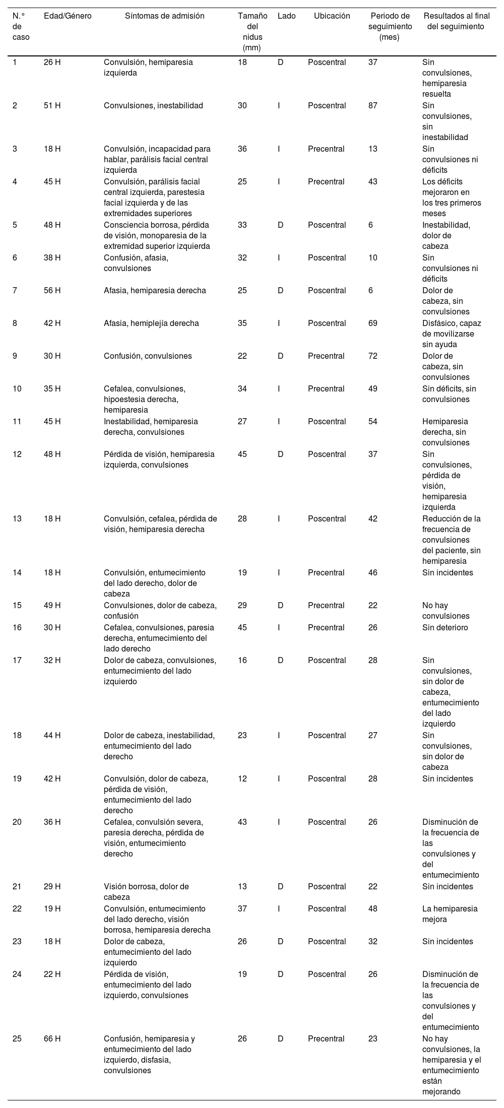
MétodosPara detectar estos cambios volumétricos se adquirieron datos de resonancia magnética (RM) de 38 controles y 25 pacientes no operados con malformaciones arteriovenosas en el giro precentral y poscentral. Los datos de RM fueron analizados con los pipelines vol2Brain, Ceres y HIPS. Los nidus de estos pacientes fueron resecados mediante disección microquirúrgica transsulcal.
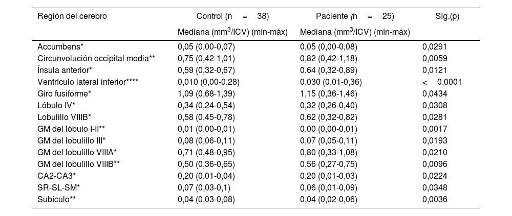
ResultadosSe realizó un análisis volumétrico integral que abarcó 135 estructuras distintas del cerebro, cerebelo e hipocampo utilizando el software Vol2brain. La comparación entre el grupo de pacientes y el grupo de control reveló diferencias volumétricas significativas. Específicamente, el grupo de pacientes exhibió volúmenes significativamente mayores en el núcleo accumbens, el giro fusiforme, el giro occipital medio, la ínsula anterior, el ventrículo lateral inferior y la sustancia gris de los lóbulos cerebelosos VIIIA y VIIIB en comparación con el grupo de control. Por el contrario, en relación con el grupo de control, el grupo de pacientes demostró volúmenes significativamente menores en la sustancia gris de los lóbulos cerebelosos IV, I-II y III, así como en las subregiones hipocampales de CA2-CA3, SR-SL-SM y el subículo.
ConclusiónLas malformaciones arteriovenosas que ocurren en el giro precentral y poscentral conducen a cambios volumétricos en estructuras distantes del sitio de la malformación, incluyendo el cerebro, el hipocampo e incluso el cerebelo. Por lo tanto, las malformaciones arteriovenosas pueden influir en los volúmenes de estructuras localizadas no solo dentro de su región cerebral inmediata, sino también en otras partes del cerebro y el cerebelo. Comprender estos cambios volumétricos puede ayudar a explicar los síntomas de los pacientes. Sin embargo, se requiere investigación adicional sobre si estos cambios volumétricos resultan del efecto de masa del nidus o se derivan de otra causa subyacente.
Arteriovenous malformations are known as arterial-venous shunts that connect nidus, the nourishing arteries, to draining veins. Although they are not very common in the brain, they are responsible for approximately 2% of all strokes. The volumetric changes in the surrounding brain tissues caused by cerebral arteriovenous malformations have not yet been reported.
MethodsTo detect these volumetric changes, MR data were acquired from 38 controls and 25 unoperated patients with arteriovenous malformations in the precentral and postcentral gyrus. MR data were analyzed with vol2Brain, Ceres and HIPS pipelines. The niduses of these patients were resected by transsulcal microsurgical dissection.
ResultsA comprehensive volumetric analysis encompassing 135 distinct brain, cerebellar, and hippocampal structures was conducted using the Vol2brain software. Comparison between the patient group and the control group revealed significant volumetric differences. Specifically, the patient group exhibited significantly larger volumes in the nucleus accumbens, fusiform gyrus, middle occipital gyrus, anterior insula, inferior lateral ventricle, and the gray matter of cerebellar lobules VIIIA and VIIIB compared with the control group. Conversely, relative to the control group, the patient group demonstrated significantly smaller volumes in the gray matter of cerebellar lobules IV, I-II, and III, as well as in the hippocampal subfields of CA2-CA3, SR-SL-SM, and the subiculum.
ConclusionArteriovenous malformations occurring in the precentral and postcentral gyrus lead to volumetric changes in structures distant from the site of the malformation, including the brain, hippocampus, and even the cerebellum. Therefore, arteriovenous malformations may influence the volumes of structures located not only within their immediate brain region but also in other parts of the cerebrum and cerebellum. Understanding these volumetric changes can aid in explaining patient symptoms. However, further research is required regarding whether these volumetric changes result from the mass effect of the nidus or stem from another underlying cause.
Artículo
![]()
Si es la primera vez que accede a la web puede obtener sus claves de acceso poniéndose en contacto con Elsevier España en suscripciones@elsevier.com o a través de su teléfono de Atención al Cliente 902 88 87 40 si llama desde territorio español o del +34 932 418 800 (de 9 a 18h., GMT + 1) si lo hace desde el extranjero.
Si ya tiene sus datos de acceso, clique aquí.
Si olvidó su clave de acceso puede recuperarla clicando aquí y seleccionando la opción "He olvidado mi contraseña".


