array:23 [
  "pii" => "S2529849622001034"
  "issn" => "25298496"
  "doi" => "10.1016/j.neucie.2022.08.003"
  "estado" => "S300"
  "fechaPublicacion" => "2023-11-01"
  "aid" => "565"
  "copyright" => "Sociedad Española de Neurocirugía"
  "copyrightAnyo" => "2022"
  "documento" => "simple-article"
  "crossmark" => 1
  "subdocumento" => "crp"
  "cita" => "Neurocirugia. 2023;34:326-8"
  "abierto" => array:3 [
    "ES" => false
    "ES2" => false
    "LATM" => false
  ]
  "gratuito" => false
  "lecturas" => array:1 [
    "total" => 0
  ]
  "Traduccion" => array:1 [
    "en" => array:19 [
      "pii" => "S113014732200080X"
      "issn" => "11301473"
      "doi" => "10.1016/j.neucir.2022.08.004"
      "estado" => "S300"
      "fechaPublicacion" => "2023-11-01"
      "aid" => "565"
      "copyright" => "Sociedad Española de Neurocirugía"
      "documento" => "simple-article"
      "crossmark" => 1
      "subdocumento" => "crp"
      "cita" => "Neurocirugia. 2023;34:326-8"
      "abierto" => array:3 [
        "ES" => false
        "ES2" => false
        "LATM" => false
      ]
      "gratuito" => false
      "lecturas" => array:1 [
        "total" => 0
      ]
      "en" => array:13 [
        "idiomaDefecto" => true
        "cabecera" => "<span class="elsevierStyleTextfn">Case report</span>"
        "titulo" => "Endoscopic third ventriculostomy limited by artery of Percheron"
        "tienePdf" => "en"
        "tieneTextoCompleto" => "en"
        "tieneResumen" => array:2 [
          0 => "en"
          1 => "es"
        ]
        "paginas" => array:1 [
          0 => array:2 [
            "paginaInicial" => "326"
            "paginaFinal" => "328"
          ]
        ]
        "titulosAlternativos" => array:1 [
          "es" => array:1 [
            "titulo" => "Ventriculostom&#237;a endosc&#243;pica limitada por arteria de Percheron"
          ]
        ]
        "contieneResumen" => array:2 [
          "en" => true
          "es" => true
        ]
        "contieneTextoCompleto" => array:1 [
          "en" => true
        ]
        "contienePdf" => array:1 [
          "en" => true
        ]
        "resumenGrafico" => array:2 [
          "original" => 0
          "multimedia" => array:7 [
            "identificador" => "fig0005"
            "etiqueta" => "Fig&#46; 1"
            "tipo" => "MULTIMEDIAFIGURA"
            "mostrarFloat" => true
            "mostrarDisplay" => false
            "figura" => array:1 [
              0 => array:4 [
                "imagen" => "gr1.jpeg"
                "Alto" => 780
                "Ancho" => 755
                "Tamanyo" => 56491
              ]
            ]
            "descripcion" => array:1 [
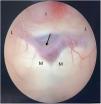
              "en" => "<p id="spar0015" class="elsevierStyleSimplePara elsevierViewall">Endoscopic vision of the third ventricle floor in which the infundibular recess &#40;I&#41; and the mammillary bodies &#40;M&#41; can be identified&#46; In the premamillary area an arterial structure is observed &#40;arrow&#41; which branches to irrigate the lateral walls of the third ventricle &#40;L&#41;&#46;</p>"
            ]
          ]
        ]
        "autores" => array:1 [
          0 => array:2 [
            "autoresLista" => "Herbert Daniel Jim&#233;nez Zapata, Adri&#225;n Fern&#225;ndez Garc&#237;a, Mercedes Carlota de Lera Alfonso, Carlos Alberto Rodr&#237;guez Arias"
            "autores" => array:4 [
              0 => array:2 [
                "nombre" => "Herbert Daniel"
                "apellidos" => "Jim&#233;nez Zapata"
              ]
              1 => array:2 [
                "nombre" => "Adri&#225;n"
                "apellidos" => "Fern&#225;ndez Garc&#237;a"
              ]
              2 => array:2 [
                "nombre" => "Mercedes Carlota"
                "apellidos" => "de Lera Alfonso"
              ]
              3 => array:2 [
                "nombre" => "Carlos Alberto"
                "apellidos" => "Rodr&#237;guez Arias"
              ]
            ]
          ]
        ]
      ]
      "idiomaDefecto" => "en"
      "Traduccion" => array:1 [
        "en" => array:9 [
          "pii" => "S2529849622001034"
          "doi" => "10.1016/j.neucie.2022.08.003"
          "estado" => "S300"
          "subdocumento" => ""
          "abierto" => array:3 [
            "ES" => false
            "ES2" => false
            "LATM" => false
          ]
          "gratuito" => false
          "lecturas" => array:1 [
            "total" => 0
          ]
          "idiomaDefecto" => "en"
          "EPUB" => "https://multimedia.elsevier.es/PublicationsMultimediaV1/item/epub/S2529849622001034?idApp=UINPBA00004B"
        ]
      ]
      "EPUB" => "https://multimedia.elsevier.es/PublicationsMultimediaV1/item/epub/S113014732200080X?idApp=UINPBA00004B"
      "url" => "/11301473/0000003400000006/v1_202311030516/S113014732200080X/v1_202311030516/en/main.assets"
    ]
  ]
  "itemAnterior" => array:19 [
    "pii" => "S2529849622001010"
    "issn" => "25298496"
    "doi" => "10.1016/j.neucie.2022.08.001"
    "estado" => "S300"
    "fechaPublicacion" => "2023-11-01"
    "aid" => "563"
    "copyright" => "Sociedad Espa&#241;ola de Neurocirug&#237;a"
    "documento" => "simple-article"
    "crossmark" => 1
    "subdocumento" => "crp"
    "cita" => "Neurocirugia. 2023;34:321-5"
    "abierto" => array:3 [
      "ES" => false
      "ES2" => false
      "LATM" => false
    ]
    "gratuito" => false
    "lecturas" => array:1 [
      "total" => 0
    ]
    "en" => array:13 [
      "idiomaDefecto" => true
      "cabecera" => "<span class="elsevierStyleTextfn">Case Report</span>"
      "titulo" => "Ventriculoperitoneal shunt migration into the pulmonary artery&#58; Case report and literature review"
      "tienePdf" => "en"
      "tieneTextoCompleto" => "en"
      "tieneResumen" => array:2 [
        0 => "en"
        1 => "es"
      ]
      "paginas" => array:1 [
        0 => array:2 [
          "paginaInicial" => "321"
          "paginaFinal" => "325"
        ]
      ]
      "titulosAlternativos" => array:1 [
        "es" => array:1 [
          "titulo" => "Cat&#233;ter de derivaci&#243;n ventr&#237;culo-peritoneal migrado a la arteria pulmonar&#58; revisi&#243;n de la literatura a prop&#243;sito de un caso"
        ]
      ]
      "contieneResumen" => array:2 [
        "en" => true
        "es" => true
      ]
      "contieneTextoCompleto" => array:1 [
        "en" => true
      ]
      "contienePdf" => array:1 [
        "en" => true
      ]
      "resumenGrafico" => array:2 [
        "original" => 0
        "multimedia" => array:7 [
          "identificador" => "fig0005"
          "etiqueta" => "Fig&#46; 1"
          "tipo" => "MULTIMEDIAFIGURA"
          "mostrarFloat" => true
          "mostrarDisplay" => false
          "figura" => array:1 [
            0 => array:4 [
              "imagen" => "gr1.jpeg"
              "Alto" => 763
              "Ancho" => 755
              "Tamanyo" => 126023
            ]
          ]
          "descripcion" => array:1 [
            "en" => "<p id="spar0025" class="elsevierStyleSimplePara elsevierViewall">3D reconstruction of cranio-cervical CT scan&#44; showing the point where distal catheter &#40;arrow heads&#41; perforates the right external jugular vein &#40;white arrows&#41;&#46;</p>"
          ]
        ]
      ]
      "autores" => array:1 [
        0 => array:2 [
          "autoresLista" => "Marta Gonz&#225;lez-Pombo, Juan Alberto Torri, Magdalena Olivares Blanco"
          "autores" => array:3 [
            0 => array:2 [
              "nombre" => "Marta"
              "apellidos" => "Gonz&#225;lez-Pombo"
            ]
            1 => array:2 [
              "nombre" => "Juan Alberto"
              "apellidos" => "Torri"
            ]
            2 => array:2 [
              "nombre" => "Magdalena"
              "apellidos" => "Olivares Blanco"
            ]
          ]
        ]
      ]
    ]
    "idiomaDefecto" => "en"
    "Traduccion" => array:1 [
      "en" => array:9 [
        "pii" => "S1130147322000781"
        "doi" => "10.1016/j.neucir.2022.08.002"
        "estado" => "S300"
        "subdocumento" => ""
        "abierto" => array:3 [
          "ES" => false
          "ES2" => false
          "LATM" => false
        ]
        "gratuito" => false
        "lecturas" => array:1 [
          "total" => 0
        ]
        "idiomaDefecto" => "en"
        "EPUB" => "https://multimedia.elsevier.es/PublicationsMultimediaV1/item/epub/S1130147322000781?idApp=UINPBA00004B"
      ]
    ]
    "EPUB" => "https://multimedia.elsevier.es/PublicationsMultimediaV1/item/epub/S2529849622001010?idApp=UINPBA00004B"
    "url" => "/25298496/0000003400000006/v2_202404171846/S2529849622001010/v2_202404171846/en/main.assets"
  ]
  "en" => array:19 [
    "idiomaDefecto" => true
    "cabecera" => "<span class="elsevierStyleTextfn">Case report</span>"
    "titulo" => "Endoscopic third ventriculostomy limited by artery of Percheron"
    "tieneTextoCompleto" => true
    "paginas" => array:1 [
      0 => array:2 [
        "paginaInicial" => "326"
        "paginaFinal" => "328"
      ]
    ]
    "autores" => array:1 [
      0 => array:4 [
        "autoresLista" => "Herbert Daniel Jim&#233;nez Zapata, Adri&#225;n Fern&#225;ndez Garc&#237;a, Mercedes Carlota de Lera Alfonso, Carlos Alberto Rodr&#237;guez Arias"
        "autores" => array:4 [
          0 => array:4 [
            "nombre" => "Herbert Daniel"
            "apellidos" => "Jim&#233;nez Zapata"
            "email" => array:1 [
              0 => "danijz88@gmail.com"
            ]
            "referencia" => array:2 [
              0 => array:2 [
                "etiqueta" => "<span class="elsevierStyleSup">a</span>"
                "identificador" => "aff0005"
              ]
              1 => array:2 [
                "etiqueta" => "<span class="elsevierStyleSup">&#42;</span>"
                "identificador" => "cor0005"
              ]
            ]
          ]
          1 => array:3 [
            "nombre" => "Adri&#225;n"
            "apellidos" => "Fern&#225;ndez Garc&#237;a"
            "referencia" => array:1 [
              0 => array:2 [
                "etiqueta" => "<span class="elsevierStyleSup">a</span>"
                "identificador" => "aff0005"
              ]
            ]
          ]
          2 => array:3 [
            "nombre" => "Mercedes Carlota"
            "apellidos" => "de Lera Alfonso"
            "referencia" => array:1 [
              0 => array:2 [
                "etiqueta" => "<span class="elsevierStyleSup">b</span>"
                "identificador" => "aff0010"
              ]
            ]
          ]
          3 => array:3 [
            "nombre" => "Carlos Alberto"
            "apellidos" => "Rodr&#237;guez Arias"
            "referencia" => array:1 [
              0 => array:2 [
                "etiqueta" => "<span class="elsevierStyleSup">a</span>"
                "identificador" => "aff0005"
              ]
            ]
          ]
        ]
        "afiliaciones" => array:2 [
          0 => array:3 [
            "entidad" => "Servicio de Neurocirug&#237;a&#44; Hospital Cl&#237;nico Universitario de Valladolid&#44; Avenida Ram&#243;n y Cajal&#44; 3&#44; 47003 Valladolid&#44; Spain"
            "etiqueta" => "a"
            "identificador" => "aff0005"
          ]
          1 => array:3 [
            "entidad" => "Servicio de Neurolog&#237;a&#44; Hospital Cl&#237;nico Universitario de Valladolid&#44; Avenida Ram&#243;n y Cajal&#44; 3&#44; 47003 Valladolid&#44; Spain"
            "etiqueta" => "b"
            "identificador" => "aff0010"
          ]
        ]
        "correspondencia" => array:1 [
          0 => array:3 [
            "identificador" => "cor0005"
            "etiqueta" => "&#8270;"
            "correspondencia" => "<span class="elsevierStyleItalic">Corresponding author</span>&#46;"
          ]
        ]
      ]
    ]
    "titulosAlternativos" => array:1 [
      "es" => array:1 [
        "titulo" => "Ventriculostom&#237;a endosc&#243;pica limitada por arteria de Percheron"
      ]
    ]
    "resumenGrafico" => array:2 [
      "original" => 0
      "multimedia" => array:7 [
        "identificador" => "fig0015"
        "etiqueta" => "Fig&#46; 3"
        "tipo" => "MULTIMEDIAFIGURA"
        "mostrarFloat" => true
        "mostrarDisplay" => false
        "figura" => array:1 [
          0 => array:4 [
            "imagen" => "gr3.jpeg"
            "Alto" => 395
            "Ancho" => 1505
            "Tamanyo" => 146697
          ]
        ]
        "descripcion" => array:1 [
          "en" => "<p id="spar0025" class="elsevierStyleSimplePara elsevierViewall">Graphic representation of Park et al&#46; classification of anatomical variations of posterior thalamoperforating arteries&#58; multiple perforating arteries arising from both P1 segments &#40;group I&#41;&#44; multiple perforating arteries in one side and a single branch in the contralateral side &#40;group II&#41;&#44; a single branch in both sides &#40;group III&#41;&#44; a single branch irrigating both the ipsilateral and contralateral sides &#40;group IV&#41;&#44; multiple branches arising in one side to irrigate it and the contralateral side also &#40;group V&#41;&#46;</p>"
        ]
      ]
    ]
    "textoCompleto" => "<span class="elsevierStyleSections"><span id="sec0005" class="elsevierStyleSection elsevierViewall"><span class="elsevierStyleSectionTitle" id="sect0025">Introduction</span><p id="par0005" class="elsevierStylePara elsevierViewall">Artery of Percheron is a rare anatomical variation of the thalamoperforating arteries&#46; Damage of this artery during neurosurgical activity can lead to fatal consequences&#46; The prevalence of artery of Percheron&#39;s infarctions is yet to be accurately defined&#44; nonetheless&#44; according to some series of cases&#44; they are deemed to represent from 0&#46;3&#37; to 2&#37; of all ischemic strokes&#46;<a class="elsevierStyleCrossRef" href="#bib0030"><span class="elsevierStyleSup">1</span></a></p><p id="par0010" class="elsevierStylePara elsevierViewall">We present the case of a patient with this anatomical variant detected during endoscopic third ventriculostomy&#46;</p></span><span id="sec0010" class="elsevierStyleSection elsevierViewall"><span class="elsevierStyleSectionTitle" id="sect0030">Case report</span><p id="par0015" class="elsevierStylePara elsevierViewall">Thirteen-year-old male was taken to the emergency department complaining about a 24<span class="elsevierStyleHsp" style=""></span>h-long headache&#46; The parents reported that&#44; during the last four months&#44; he had developed a progressive worsening of his motor skills&#44; with clumsiness and falls&#44; difficulty manipulating small objects&#44; trembling and loss of strength&#46; He had also presented a decline in his mental status affecting his concentration and memory&#46;</p><p id="par0020" class="elsevierStylePara elsevierViewall">The neurologic examination revealed lower limb hypertonia with claudication in antigravitatory maneuvers&#44; gait abnormality&#44; patellar and ankle hyper-reflexia&#44; and bilateral positive Babinski sign&#46; The rest of the examination did not reveal any abnormality&#44; with a Glasgow Coma Scale of 15&#46; The CT scan showed a remarkable obstructive hydrocephalus with a space-occupying mass at the level of the mesencephalon&#46;</p><p id="par0025" class="elsevierStylePara elsevierViewall">An endoscopic third ventriculostomy &#40;ETV&#41; was performed through a frontal burr hole following the standard technique&#46; During the procedure an artery was encountered at the level of the tuber cinereum limiting the perforation of the third ventricle floor &#40;<a class="elsevierStyleCrossRef" href="#fig0005">Fig&#46; 1</a>&#41;&#46; That artery ascended from the interpeduncular cistern finally branching into two arteries irrigating the lateral walls of the third ventricle&#46; The tumor was then biopsied &#40;anaplastic astrocytoma &#8211; grade III&#41;&#44; and&#44; due to the impossibility to perform an ETV with the proper size&#44; a ventriculoperitoneal shunt was eventually placed&#46;</p><elsevierMultimedia ident="fig0005"></elsevierMultimedia><p id="par0030" class="elsevierStylePara elsevierViewall">Postoperatively there was no complication&#44; and the magnetic resonance confirmed a lesion at the level of the mesencephalic tectum&#46; Moreover&#44; in the multiplanar reformation &#40;<a class="elsevierStyleCrossRef" href="#fig0010">Fig&#46; 2</a>&#41; it was possible to verify the presence of an artery originating from the P1 segment of the right posterior cerebral artery ascending to the premammillary area branching to irrigate both the right and left thalamus&#46;</p><elsevierMultimedia ident="fig0010"></elsevierMultimedia></span><span id="sec0015" class="elsevierStyleSection elsevierViewall"><span class="elsevierStyleSectionTitle" id="sect0035">Discussion</span><p id="par0035" class="elsevierStylePara elsevierViewall">The thalamoperforating arteries are those arising from the upper or posterior face of the P1 segment &#40;posterior thalamoperforating arteries&#41; and from the posterior communicating artery &#40;anterior thalamoperforating arteries&#41;&#44; irrigating the thalamus&#44; hypothalamus&#44; posterior limb of the internal capsule and rostral mesencephalon&#46;<a class="elsevierStyleCrossRefs" href="#bib0035"><span class="elsevierStyleSup">2&#44;3</span></a> There are several anatomic variants in the disposition of the posterior thalamoperforating arteries which&#44; following the anatomical study proposed by Park&#44; can be classified in five groups &#40;<a class="elsevierStyleCrossRef" href="#fig0015">Fig&#46; 3</a>&#41;&#46;<a class="elsevierStyleCrossRef" href="#bib0035"><span class="elsevierStyleSup">2</span></a> The fourth variant has been classically named artery of Percheron and it represents 11&#46;5&#37; of the cases&#46; The importance of this artery relates to the consequence of its occlusion&#44; either because of a stroke or iatrogenically &#40;after surgery or endovascular procedures&#41;&#44; causing bilateral infarctions of the paramedian thalamic region and rostral mesencephalon&#46; In addition&#44; in those cases in which the anterior thalamoperforating arteries are absent&#44; the occlusion of the artery of Percheron may also cause infarctions of the hypothalamus and of the anterior thalamic region&#46;<a class="elsevierStyleCrossRef" href="#bib0040"><span class="elsevierStyleSup">3</span></a></p><elsevierMultimedia ident="fig0015"></elsevierMultimedia><p id="par0040" class="elsevierStylePara elsevierViewall">Clinically&#44; artery of Percheron stroke usually cause a decline in the level of consciousness&#44; upward gaze palsy&#44; amnesia and may also cause other symptoms depending on the extension of the infarction at the level of the mesencephalon &#40;oculomotor palsy&#44; hemiplegia&#44; ataxia&#44; rubric&#47;Holmes&#8217; tremor&#41;&#46;<a class="elsevierStyleCrossRef" href="#bib0035"><span class="elsevierStyleSup">2</span></a> Hyperintensity of the pial mesencephalic surface in the interpeduncular fossa &#40;V sign&#41; as described by Lazzaro et al&#46;&#44;<a class="elsevierStyleCrossRef" href="#bib0030"><span class="elsevierStyleSup">1</span></a> is a typical radiologic finding visible in MRI&#46; There are other types of diseases that can result in bilateral thalamic lesions&#46; In the differential diagnosis of artery of Percheron occlusion&#44; we must include deep cerebral venous thrombosis&#44; top basilar syndrome and Wernickes&#39;s encephalopathy&#46;</p><p id="par0045" class="elsevierStylePara elsevierViewall">The treatment of this type of stroke is a real challenge since most of the time the use of thrombolytics &#40;such as recombinant tissue plasminogen activator&#41; is contraindicated due to the delayed in diagnosis and&#44; in addition&#44; the small diameter of artery of Percheron makes very difficult the endovascular revascularization&#46; Usually&#44; in the acute phase the patient is treated with intravenous heparin and subsequently&#44; the treatment will depend on the underlying etiology &#40;cardioembolic require oral anticoagulants whereas cryptogenic causes require antiplatelet therapy&#41;&#46;<a class="elsevierStyleCrossRef" href="#bib0045"><span class="elsevierStyleSup">4</span></a></p><p id="par0050" class="elsevierStylePara elsevierViewall">The prognosis of artery of Percheron occlusion will depend on the extent of the stroke&#46; Cases without midbrain involvement usually have a better prognosis &#40;67&#37; of patients with favorable evolution&#41;&#46; On the other hand&#44; 75&#37; of patients with midbrain or anterior thalamic involvement have unfavorable outcome&#46;<a class="elsevierStyleCrossRef" href="#bib0045"><span class="elsevierStyleSup">4</span></a></p><p id="par0055" class="elsevierStylePara elsevierViewall">ETV is a safe and effective method to treat obstructive hydrocephalus caused by aqueductal stenosis&#44; however&#44; some anatomic variations such as small prepontine space&#44; thickening of the third ventricle floor or displacement of the mammillary bodies may increase the surgical risk&#46;<a class="elsevierStyleCrossRef" href="#bib0050"><span class="elsevierStyleSup">5</span></a> To the best of our knowledge&#44; this is the first case describing an artery of Percheron antero-superiorly displaced thus impeding the safe fenestration of the third ventricle floor&#46;</p></span><span id="sec0020" class="elsevierStyleSection elsevierViewall"><span class="elsevierStyleSectionTitle" id="sect0040">Conclusion</span><p id="par0060" class="elsevierStylePara elsevierViewall">Identification of an artery of Percheron in the preoperative neuroimaging or during the operation itself stands as a keystone to prevent neurologic sequelae&#44; sometimes fatal&#44; associated to its occlusion&#46;</p></span></span>"
    "textoCompletoSecciones" => array:1 [
      "secciones" => array:9 [
        0 => array:3 [
          "identificador" => "xres2131398"
          "titulo" => "Abstract"
          "secciones" => array:1 [
            0 => array:1 [
              "identificador" => "abst0005"
            ]
          ]
        ]
        1 => array:2 [
          "identificador" => "xpalclavsec1810113"
          "titulo" => "Keywords"
        ]
        2 => array:3 [
          "identificador" => "xres2131397"
          "titulo" => "Resumen"
          "secciones" => array:1 [
            0 => array:1 [
              "identificador" => "abst0010"
            ]
          ]
        ]
        3 => array:2 [
          "identificador" => "xpalclavsec1810112"
          "titulo" => "Palabras clave"
        ]
        4 => array:2 [
          "identificador" => "sec0005"
          "titulo" => "Introduction"
        ]
        5 => array:2 [
          "identificador" => "sec0010"
          "titulo" => "Case report"
        ]
        6 => array:2 [
          "identificador" => "sec0015"
          "titulo" => "Discussion"
        ]
        7 => array:2 [
          "identificador" => "sec0020"
          "titulo" => "Conclusion"
        ]
        8 => array:1 [
          "titulo" => "References"
        ]
      ]
    ]
    "pdfFichero" => "main.pdf"
    "tienePdf" => true
    "fechaRecibido" => "2022-04-22"
    "fechaAceptado" => "2022-08-12"
    "PalabrasClave" => array:2 [
      "en" => array:1 [
        0 => array:4 [
          "clase" => "keyword"
          "titulo" => "Keywords"
          "identificador" => "xpalclavsec1810113"
          "palabras" => array:4 [
            0 => "Third ventriculostomy"
            1 => "Neuroanatomy"
            2 => "Thalamoperforating arteries"
            3 => "Stroke"
          ]
        ]
      ]
      "es" => array:1 [
        0 => array:4 [
          "clase" => "keyword"
          "titulo" => "Palabras clave"
          "identificador" => "xpalclavsec1810112"
          "palabras" => array:4 [
            0 => "Ventriculostom&#237;a"
            1 => "Neuroanatom&#237;a"
            2 => "Arterias talamoperforantes"
            3 => "Ictus"
          ]
        ]
      ]
    ]
    "tieneResumen" => true
    "resumen" => array:2 [
      "en" => array:2 [
        "titulo" => "Abstract"
        "resumen" => "<span id="abst0005" class="elsevierStyleSection elsevierViewall"><p id="spar0005" class="elsevierStyleSimplePara elsevierViewall">The irrigation of the thalamus depends mainly on the thalamoperforating arteries&#46; There are many anatomical variations in these arteries&#44; the best known being the artery of Percheron&#46; We report a case of a 13-year-old male presented with headache and decline in his mental status&#46; Imaging features showed obstructive hydrocephalus secondary to a mass at the level of the mesencephalon so an endoscopic third ventriculostomy was performed&#46; During the procedure a thalamoperforating artery was encountered at the level of the tuber cinereum limiting the perforation of the third ventricle floor&#46; The present case emphasizes the importance of knowing the anatomy of these arteries and the identification of their main variants during neurosurgical procedures&#46;</p></span>"
      ]
      "es" => array:2 [
        "titulo" => "Resumen"
        "resumen" => "<span id="abst0010" class="elsevierStyleSection elsevierViewall"><p id="spar0010" class="elsevierStyleSimplePara elsevierViewall">La irrigaci&#243;n tal&#225;mica depende principalmente de las arterias talamoperforantes&#46; Existen muchas variantes anat&#243;micas en el origen y disposici&#243;n de estas arterias siendo la m&#225;s conocida la denominada arteria de Percheron&#46; En este art&#237;culo presentamos el caso de un var&#243;n de 13 a&#241;os que acudi&#243; a urgencias por cefalea y deterioro del nivel de consciencia&#46; En las pruebas de imagen se evidenci&#243; una hidrocefalia obstructiva secundaria a una tumoraci&#243;n mesencef&#225;lica&#44; motivo por el cual se decidi&#243; realizar una ventriculostom&#237;a endosc&#243;pica&#46; Durante el procedimiento se evidenci&#243; una arteria talamoperforante a nivel del tuber cinereum que limit&#243; la fenestraci&#243;n del suelo del tercer ventr&#237;culo&#46; A partir de este caso destacamos la importancia de conocer la anatom&#237;a de estas arterias con sus posibles variantes y su identificaci&#243;n durante los procedimientos neuroquir&#250;rgicos&#46;</p></span>"
      ]
    ]
    "multimedia" => array:3 [
      0 => array:7 [
        "identificador" => "fig0005"
        "etiqueta" => "Fig&#46; 1"
        "tipo" => "MULTIMEDIAFIGURA"
        "mostrarFloat" => true
        "mostrarDisplay" => false
        "figura" => array:1 [
          0 => array:4 [
            "imagen" => "gr1.jpeg"
            "Alto" => 780
            "Ancho" => 755
            "Tamanyo" => 56491
          ]
        ]
        "descripcion" => array:1 [
          "en" => "<p id="spar0015" class="elsevierStyleSimplePara elsevierViewall">Endoscopic vision of the third ventricle floor in which the infundibular recess &#40;I&#41; and the mammillary bodies &#40;M&#41; can be identified&#46; In the premamillary area an arterial structure is observed &#40;arrow&#41; which branches to irrigate the lateral walls of the third ventricle &#40;L&#41;&#46;</p>"
        ]
      ]
      1 => array:7 [
        "identificador" => "fig0010"
        "etiqueta" => "Fig&#46; 2"
        "tipo" => "MULTIMEDIAFIGURA"
        "mostrarFloat" => true
        "mostrarDisplay" => false
        "figura" => array:1 [
          0 => array:4 [
            "imagen" => "gr2.jpeg"
            "Alto" => 495
            "Ancho" => 1505
            "Tamanyo" => 93659
          ]
        ]
        "descripcion" => array:1 [
          "en" => "<p id="spar0020" class="elsevierStyleSimplePara elsevierViewall">T2 and gadolinium enhanced-T1 weighted MRI&#46; In the axial cut &#40;left&#41; an intra-axial tumor in the mesencephalic tectum can be seen &#40;&#42;&#41;&#46; In the sagittal &#40;center&#41; and coronal &#40;right&#41; reconstructions we identify an artery &#40;arrow&#41; which arises from P1 segment of the PCA &#40;arrowhead&#41; ascending to the third ventricle&#46;</p>"
        ]
      ]
      2 => array:7 [
        "identificador" => "fig0015"
        "etiqueta" => "Fig&#46; 3"
        "tipo" => "MULTIMEDIAFIGURA"
        "mostrarFloat" => true
        "mostrarDisplay" => false
        "figura" => array:1 [
          0 => array:4 [
            "imagen" => "gr3.jpeg"
            "Alto" => 395
            "Ancho" => 1505
            "Tamanyo" => 146697
          ]
        ]
        "descripcion" => array:1 [
          "en" => "<p id="spar0025" class="elsevierStyleSimplePara elsevierViewall">Graphic representation of Park et al&#46; classification of anatomical variations of posterior thalamoperforating arteries&#58; multiple perforating arteries arising from both P1 segments &#40;group I&#41;&#44; multiple perforating arteries in one side and a single branch in the contralateral side &#40;group II&#41;&#44; a single branch in both sides &#40;group III&#41;&#44; a single branch irrigating both the ipsilateral and contralateral sides &#40;group IV&#41;&#44; multiple branches arising in one side to irrigate it and the contralateral side also &#40;group V&#41;&#46;</p>"
        ]
      ]
    ]
    "bibliografia" => array:2 [
      "titulo" => "References"
      "seccion" => array:1 [
        0 => array:2 [
          "identificador" => "bibs0015"
          "bibliografiaReferencia" => array:5 [
            0 => array:3 [
              "identificador" => "bib0030"
              "etiqueta" => "1"
              "referencia" => array:1 [
                0 => array:2 [
                  "contribucion" => array:1 [
                    0 => array:2 [
                      "titulo" => "Artery of percheron infarction&#58; imaging patterns and clinical spectrum"
                      "autores" => array:1 [
                        0 => array:2 [
                          "etal" => true
                          "autores" => array:6 [
                            0 => "N&#46;A&#46; Lazzaro"
                            1 => "B&#46; Wright"
                            2 => "M&#46; Castillo"
                            3 => "N&#46;J&#46; Fischbein"
                            4 => "C&#46;M&#46; Glastonbury"
                            5 => "P&#46;G&#46; Hildenbrand"
                          ]
                        ]
                      ]
                    ]
                  ]
                  "host" => array:1 [
                    0 => array:2 [
                      "doi" => "10.3174/ajnr.A2044"
                      "Revista" => array:6 [
                        "tituloSerie" => "AJNR Am J Neuroradiol"
                        "fecha" => "2010"
                        "volumen" => "31"
                        "paginaInicial" => "1283"
                        "paginaFinal" => "1289"
                        "link" => array:1 [
                          0 => array:2 [
                            "url" => "https://www.ncbi.nlm.nih.gov/pubmed/20299438"
                            "web" => "Medline"
                          ]
                        ]
                      ]
                    ]
                  ]
                ]
              ]
            ]
            1 => array:3 [
              "identificador" => "bib0035"
              "etiqueta" => "2"
              "referencia" => array:1 [
                0 => array:2 [
                  "contribucion" => array:1 [
                    0 => array:2 [
                      "titulo" => "Morphological characteristics of the thalamoperforating arteries"
                      "autores" => array:1 [
                        0 => array:2 [
                          "etal" => false
                          "autores" => array:6 [
                            0 => "S&#46;Q&#46; Park"
                            1 => "H&#46;-G&#46; Bae"
                            2 => "S&#46;-M&#46; Yoon"
                            3 => "J&#46;-J&#46; Shim"
                            4 => "I&#46;-G&#46; Yun"
                            5 => "S&#46;-K&#46; Choi"
                          ]
                        ]
                      ]
                    ]
                  ]
                  "host" => array:1 [
                    0 => array:2 [
                      "doi" => "10.3340/jkns.2010.47.1.36"
                      "Revista" => array:6 [
                        "tituloSerie" => "J Korean Neurosurg Soc"
                        "fecha" => "2010"
                        "volumen" => "47"
                        "paginaInicial" => "36"
                        "paginaFinal" => "41"
                        "link" => array:1 [
                          0 => array:2 [
                            "url" => "https://www.ncbi.nlm.nih.gov/pubmed/20157376"
                            "web" => "Medline"
                          ]
                        ]
                      ]
                    ]
                  ]
                ]
              ]
            ]
            2 => array:3 [
              "identificador" => "bib0040"
              "etiqueta" => "3"
              "referencia" => array:1 [
                0 => array:2 [
                  "contribucion" => array:1 [
                    0 => array:2 [
                      "titulo" => "The artery of Percheron revisited&#58; a cadaveric anatomical study"
                      "autores" => array:1 [
                        0 => array:2 [
                          "etal" => false
                          "autores" => array:4 [
                            0 => "H&#46; Kocaeli"
                            1 => "S&#46; Yilmazlar"
                            2 => "T&#46; Kuytu"
                            3 => "E&#46; Korfali"
                          ]
                        ]
                      ]
                    ]
                  ]
                  "host" => array:1 [
                    0 => array:2 [
                      "doi" => "10.1007/s00701-012-1548-1"
                      "Revista" => array:6 [
                        "tituloSerie" => "Acta Neurochir &#40;Wien&#41;"
                        "fecha" => "2013"
                        "volumen" => "155"
                        "paginaInicial" => "533"
                        "paginaFinal" => "539"
                        "link" => array:1 [
                          0 => array:2 [
                            "url" => "https://www.ncbi.nlm.nih.gov/pubmed/23139104"
                            "web" => "Medline"
                          ]
                        ]
                      ]
                    ]
                  ]
                ]
              ]
            ]
            3 => array:3 [
              "identificador" => "bib0045"
              "etiqueta" => "4"
              "referencia" => array:1 [
                0 => array:2 [
                  "contribucion" => array:1 [
                    0 => array:2 [
                      "titulo" => "Artery of Percheron infarction&#58; a short review"
                      "autores" => array:1 [
                        0 => array:2 [
                          "etal" => false
                          "autores" => array:5 [
                            0 => "A&#46; Kichloo"
                            1 => "S&#46;M&#46; Jamal"
                            2 => "E&#46;A&#46; Zain"
                            3 => "F&#46; Wani"
                            4 => "N&#46; Vipparala"
                          ]
                        ]
                      ]
                    ]
                  ]
                  "host" => array:1 [
                    0 => array:1 [
                      "Revista" => array:3 [
                        "tituloSerie" => "J Investig Med High Impact Case Rep"
                        "fecha" => "2019"
                        "volumen" => "7"
                      ]
                    ]
                  ]
                ]
              ]
            ]
            4 => array:3 [
              "identificador" => "bib0050"
              "etiqueta" => "5"
              "referencia" => array:1 [
                0 => array:2 [
                  "contribucion" => array:1 [
                    0 => array:2 [
                      "titulo" => "Anatomic variations of the floor of the third ventricle&#58; an endoscopic study"
                      "autores" => array:1 [
                        0 => array:2 [
                          "etal" => false
                          "autores" => array:5 [
                            0 => "M&#46;E&#46; Sughrue"
                            1 => "J&#46; Chiou"
                            2 => "J&#46;D&#46; Burks"
                            3 => "P&#46;A&#46; Bonney"
                            4 => "C&#46; Teo"
                          ]
                        ]
                      ]
                    ]
                  ]
                  "host" => array:1 [
                    0 => array:2 [
                      "doi" => "10.1016/j.wneu.2016.02.037"
                      "Revista" => array:6 [
                        "tituloSerie" => "World Neurosurg"
                        "fecha" => "2016"
                        "volumen" => "90"
                        "paginaInicial" => "211"
                        "paginaFinal" => "227"
                        "link" => array:1 [
                          0 => array:2 [
                            "url" => "https://www.ncbi.nlm.nih.gov/pubmed/26906894"
                            "web" => "Medline"
                          ]
                        ]
                      ]
                    ]
                  ]
                ]
              ]
            ]
          ]
        ]
      ]
    ]
  ]
  "idiomaDefecto" => "en"
  "url" => "/25298496/0000003400000006/v2_202404171846/S2529849622001034/v2_202404171846/en/main.assets"
  "Apartado" => array:4 [
    "identificador" => "92441"
    "tipo" => "SECCION"
    "en" => array:2 [
      "titulo" => "Case Reports"
      "idiomaDefecto" => true
    ]
    "idiomaDefecto" => "en"
  ]
  "PDF" => "https://static.elsevier.es/multimedia/25298496/0000003400000006/v2_202404171846/S2529849622001034/v2_202404171846/en/main.pdf?idApp=UINPBA00004B&text.app=https://revistaneurocirugia.com/"
  "EPUB" => "https://multimedia.elsevier.es/PublicationsMultimediaV1/item/epub/S2529849622001034?idApp=UINPBA00004B"
]
Share
Journal Information
Vol. 34. Issue 6.
Pages 326-328 (November - December 2023)
Share
Share
Download PDF
More article options
Visits
22
Vol. 34. Issue 6.
Pages 326-328 (November - December 2023)
Case report
Endoscopic third ventriculostomy limited by artery of Percheron
Ventriculostomía endoscópica limitada por arteria de Percheron
Visits
22
Herbert Daniel Jiménez Zapataa,
Corresponding author
danijz88@gmail.com

Corresponding author.
, Adrián Fernández Garcíaa, Mercedes Carlota de Lera Alfonsob, Carlos Alberto Rodríguez Ariasa
a Servicio de Neurocirugía, Hospital Clínico Universitario de Valladolid, Avenida Ramón y Cajal, 3, 47003 Valladolid, Spain
b Servicio de Neurología, Hospital Clínico Universitario de Valladolid, Avenida Ramón y Cajal, 3, 47003 Valladolid, Spain
This item has received
Article information
Abstract
Full Text
Bibliography
Download PDF
Statistics
Figures (3)
Show moreShow less
Abstract

The irrigation of the thalamus depends mainly on the thalamoperforating arteries. There are many anatomical variations in these arteries, the best known being the artery of Percheron. We report a case of a 13-year-old male presented with headache and decline in his mental status. Imaging features showed obstructive hydrocephalus secondary to a mass at the level of the mesencephalon so an endoscopic third ventriculostomy was performed. During the procedure a thalamoperforating artery was encountered at the level of the tuber cinereum limiting the perforation of the third ventricle floor. The present case emphasizes the importance of knowing the anatomy of these arteries and the identification of their main variants during neurosurgical procedures.

Keywords:
Third ventriculostomy
Neuroanatomy
Thalamoperforating arteries
Stroke
Resumen

La irrigación talámica depende principalmente de las arterias talamoperforantes. Existen muchas variantes anatómicas en el origen y disposición de estas arterias siendo la más conocida la denominada arteria de Percheron. En este artículo presentamos el caso de un varón de 13 años que acudió a urgencias por cefalea y deterioro del nivel de consciencia. En las pruebas de imagen se evidenció una hidrocefalia obstructiva secundaria a una tumoración mesencefálica, motivo por el cual se decidió realizar una ventriculostomía endoscópica. Durante el procedimiento se evidenció una arteria talamoperforante a nivel del tuber cinereum que limitó la fenestración del suelo del tercer ventrículo. A partir de este caso destacamos la importancia de conocer la anatomía de estas arterias con sus posibles variantes y su identificación durante los procedimientos neuroquirúrgicos.

Palabras clave:
Ventriculostomía
Neuroanatomía
Arterias talamoperforantes
Ictus

Article

These are the options to access the full texts of the publication Neurocirugía (English edition)
Member
Member of the Sociedad Española de Neurocirugía

If it is the first time you have accessed you can obtain your credentials by contacting Elsevier Spain in suscripciones@elsevier.com or by calling our Customer Service at902 88 87 40 if you are calling from Spain or at +34 932 418 800 (from 9 to 18h., GMT + 1) if you are calling outside of Spain.

If you already have your login data, please click here .

If you have forgotten your password you can you can recover it by clicking here and selecting the option ¿I have forgotten my password¿.

Subscriber
Subscriber

If you already have your login data, please click here .

If you have forgotten your password you can you can recover it by clicking here and selecting the option “I have forgotten my password”
Subscribe
Subscribe to

Neurocirugía (English edition)

Purchase
Purchase article

Purchasing article the PDF version will be downloaded

Price 19.34 €

Purchase now
Contact
Phone for subscriptions and reporting of errors
From Monday to Friday from 9 a.m. to 6 p.m. (GMT + 1) except for the months of July and August which will be from 9 a.m. to 3 p.m.
Calls from Spain
932 415 960
Calls from outside Spain
+34 932 415 960
Email
Idiomas
Neurocirugía (English edition)
Article options
Tools
es en

¿Es usted profesional sanitario apto para prescribir o dispensar medicamentos?

Are you a health professional able to prescribe or dispense drugs?