Normal perfusion pressure breakthrough (NPPB) phenomenon is a major life-threatening complication that restricts the treatment of complex intracranial arteriovenous malformations. The aim of the study it to develop a rat model mimicking NPPB phenomenon that enables the evaluation of any therapy to prevent such complication.
MethodsTwenty Wistar male rats were randomly assigned to either a study or a control group. Study animals underwent an end-to-side left external jugular vein-common carotid artery anastomosis and ligation of bilateral external carotid arteries. Control animals only underwent ligation of bilateral external carotid arteries. All animals were sacrificed sixty days after the procedure. Hemodynamic parameters [mean arterial pressure (MAP), intracranial pressure (ICP) and cerebral perfusion pressure (CPP)], blood–brain barrier (BBB) permeability (measured by fluorescein staining) and histological features were then compared between both groups.
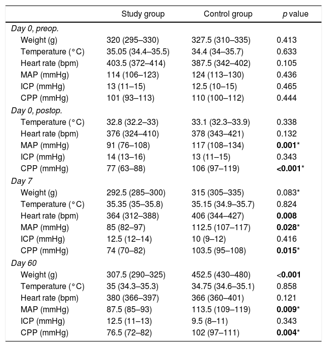
ResultsA significant decrease in MAP and CPP was confirmed in the study group. An increase in ICP was also observed. A significant decrease in MAP and CPP was also present in the study group when comparing preoperative values with those recorded on days 0 (postoperative), 7 and 60. Fluorescein staining findings were consistent with signs of BBB disruption in study animals. Histological analysis demonstrated an increased number of pyknotic neurons in the ipsilateral hemisphere of rat brains included in the study group.
ConclusionThese results confirm that this model mimics a vascular steal state with chronic cerebral hypoperfusion comparable to patients with AVMs behavior and disruption of the BBB after fistula closure comparable to NPPB phenomenon disorders.
El síndrome de restablecimiento de la presión de perfusión cerebral (PPC) normal es una complicación grave, que supone un riesgo vital y limita el tratamiento de malformaciones arteriovenosas complejas. El objetivo de este estudio es desarrollar un modelo animal remedando dicho síndrome que permita evaluar terapias para su prevención.
MétodosVeinte ratas macho Wistar fueron asignadas aleatoriamente a un grupo estudio o control. Los animales de estudio se sometieron a una anastomosis término-lateral entre la vena yugular externa y la arteria carótida común izquierdas y ligadura bilateral de las arterias carótidas externas. Los animales control se sometieron a la ligadura bilateral de las arterias carótidas externas. Todos los animales se sacrificaron 60 días después. Se compararon parámetros hemodinámicos (presión arterial media [PAM], presión intracraneal [PIC] y PPC), permeabilidad de la barrera hemato-encefálica (BHE) y características histológicas entre ambos grupos.
ResultadosEl grupo estudio mostró un descenso significativo de la PAM y la PPC, así como un aumento de la PIC respecto al grupo control. Al comparar los valores preoperatorios con aquéllos registrados los días 0 (postoperatorio), 7 y 60 en el grupo estudio, también se confirmó un descenso significativo de la PAM y la PPC. La disrupción de la BHE fue constatada únicamente en el grupo estudio mediante la extravasación de fluoresceína sódica. El análisis histológico demostró mayor número de neuronas picnóticas en el hemisferio ipsilateral a la anastomosis de los animales estudio.
ConclusiónLos resultados descritos apuntan a un modelo que remeda el estado de robo vascular con hipoperfusión cerebral crónica comparable al que sufren los pacientes con malformaciones arteriovenosas, así como la disrupción de la BHE tras el cierre de la anastomosis, comparable al acontecido en el síndrome de restablecimiento de la PPC normal tras la exclusión de la malformación.
Article

If it is the first time you have accessed you can obtain your credentials by contacting Elsevier Spain in suscripciones@elsevier.com or by calling our Customer Service at902 88 87 40 if you are calling from Spain or at +34 932 418 800 (from 9 to 18h., GMT + 1) if you are calling outside of Spain.
If you already have your login data, please click here .
If you have forgotten your password you can you can recover it by clicking here and selecting the option ¿I have forgotten my password¿.


