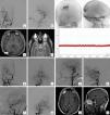
A 36-year-old male presented to the Emergency Department with clinical symptoms of blurred vision of progressive onset of two years of evolution. The ophthalmological examination revealed the existence of bilateral papilledema. Using cranial computed tomography and magnetic resonance imaging, the presence of a right occipital pial arteriovenous malformation was certified. Arteriographically, pial arterial contributions dependent on the right middle cerebral artery and the right posterior cerebral artery were identified. Venous drainage was located at the level of the superior sagittal sinus. An associated right transverse sinus stenosis was also identified. The existence of secondary intracranial hypertension was corroborated by monitoring with an intracranial pressure sensor. An interventional procedure was carried out consisting of embolization of the arterial supplies of the lesion using Onyx®. The clinical-radiological findings after the procedure were favorable: the papilledema disappeared and complete exclusion of the malformation was achieved. A new intracranial pressure measurement showed resolution of intracranial hypertension. Subsequent regulated radiological controls showed complete exclusion of the malformation up to 5 years later.
Varón de 36 años de edad que acudió a servicio de Urgencias ante cuadro clínico de visión borrosa de instauración progresiva de dos años de evolución. El examen oftalmológico reveló la existencia de edema de papila bilateral. Mediante tomografía computerizada y resonancia magnética craneales se certificó la presencia de una malformación arteriovenosa pial occipital derecha. Arteriográficamente se identificaron aportes arteriales piales dependientes de la arteria cerebral media derecha y de la arteria cerebral posterior derecha. El drenaje venoso fue localizado a nivel del seno sagital superior. Se identificó así mismo una estenosis del seno trasverso derecho asociada. La existencia de hipertensión intracraneal secundaria fue corroborada mediante monitorización con sensor de presión intracraneal. Se llevó a cabo procedimiento intervencionista consistente en embolización de los aportes arteriales de la lesión mediante Onyx®. Los hallazgos clínico-radiológicos tras el procedimiento resultaron favorables: desapareció el edema de papila y se logró la exclusión completa de la malformación. Una nueva medida de presión intracraneal mostró la resolución de la hipertensión intracraneal. Los controles radiológicos reglados posteriores mostraron exclusión completa de la malformación hasta 5 años después.
Article

If it is the first time you have accessed you can obtain your credentials by contacting Elsevier Spain in suscripciones@elsevier.com or by calling our Customer Service at902 88 87 40 if you are calling from Spain or at +34 932 418 800 (from 9 to 18h., GMT + 1) if you are calling outside of Spain.
If you already have your login data, please click here .
If you have forgotten your password you can you can recover it by clicking here and selecting the option ¿I have forgotten my password¿.


