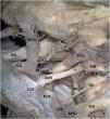
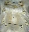
This study investigates the mobilization of cranial nerves in the upper clival region to improve surgical approaches. Cadaveric specimens (n = 20) were dissected to examine the oculomotor, trochlear, and abducens nerves. Dissection techniques focused on the nerves' intradural course and their relationship to surrounding structures.
MethodsPre-dissection revealed the nerves' entry points into the clival dura and their proximity to each other. Measurements were taken to quantify these distances. Following intradural dissection, measurements were again obtained to assess the degree of nerve mobilization.
ResultsDissection showed that the abducens nerve takes three folds during its course: at the dural foramen, towards the posterior cavernous sinus, and lastly within the cavernous sinus. The trochlear nerve enters the dura and makes two bends before entering the cavernous sinus. The oculomotor nerve enters the cavernous sinus directly and runs parallel to the trochlear nerve. Importantly, intradural dissection increased the space between the abducens nerves (by 4.21 mm) and between the oculomotor and trochlear nerves (by 3.09 mm on average). This indicates that nerve mobilization can create wider surgical corridors for approaching lesions in the upper clivus region.
ConclusionsThis study provides a detailed anatomical analysis of the oculomotor, trochlear, and abducens nerves in the upper clivus. The cadaveric dissections and measurements demonstrate the feasibility of mobilizing these nerves to achieve wider surgical corridors. This information can be valuable for surgeons planning endoscopic or microscopic approaches to lesions in the upper clivus region.
Este estudio investiga la movilización de los nervios craneales en la región clival superior para mejorar los abordajes quirúrgicos. Se diseccionaron ejemplares de cadáveres (n = 20) para examinar los nervios oculomotor, troclear y abducens. Las técnicas de disección se centraron en el trayecto intradural de los nervios y su relación con las estructuras circundantes.
MétodosLa pre-disección reveló los puntos de entrada de los nervios en la dura clival y su proximidad entre sí. Se tomaron medidas para cuantificar estas distancias. Tras la disección intradural, se volvieron a obtener medidas para evaluar el grado de movilización nerviosa.
ResultadosLa disección mostró que el nervio abducens da tres pliegues durante su recorrido: en el foramen dural, hacia el seno cavernoso posterior y por último dentro del seno cavernoso. El nervio troclear ingresa en la dura y realiza dos curvas antes de entrar en el seno cavernoso. El nervio oculomotor entra directamente en el seno cavernoso y corre paralelo al nervio troclear. Es importante destacar que la disección intradural aumentó el espacio entre los nervios abducens (en 4,21 mm) y entre los nervios oculomotor y troclear (en 3,09 mm de promedio). Esto indica que la movilización nerviosa puede crear corredores quirúrgicos más amplios para abordar lesiones en la región del clivus superior.
ConclusionesEste estudio proporciona un análisis anatómico detallado de los nervios oculomotor, troclear y abducens en el clivus superior. Las disecciones y mediciones de cadáveres demuestran la viabilidad de movilizar estos nervios para lograr corredores quirúrgicos más amplios. Esta información puede ser valiosa para los cirujanos que planean abordajes endoscópicos o microscópicos de lesiones en la región del clivus superior.
Article

If it is the first time you have accessed you can obtain your credentials by contacting Elsevier Spain in suscripciones@elsevier.com or by calling our Customer Service at902 88 87 40 if you are calling from Spain or at +34 932 418 800 (from 9 to 18h., GMT + 1) if you are calling outside of Spain.
If you already have your login data, please click here .
If you have forgotten your password you can you can recover it by clicking here and selecting the option ¿I have forgotten my password¿.


