


In spontaneous subarachnoid haemorrhage (SAH) accurate determination of the bleeding source is paramount to guide treatment. Traditionally, the bleeding pattern has been used to predict the aneurysm location. Here, we have tested a software-based tool, which quantifies the volume of intracranial blood and stratifies it according to the regional distribution, to predict the location of the ruptured aneurysm.
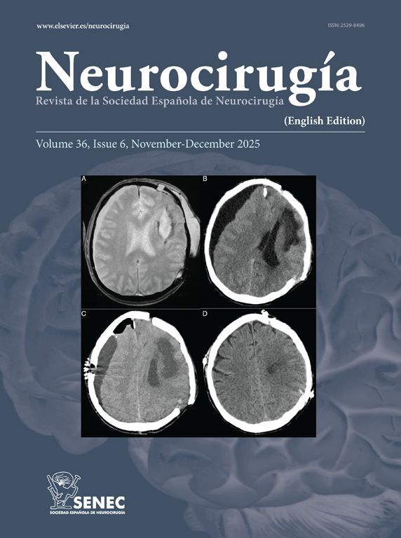
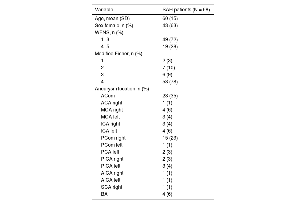
MethodsA consecutive series of SAH patients admitted to a single tertiary centre between 2012–2018, within 72 h of onset, harbouring a single intracranial aneurysm. A semi-automatized method of blood quantification, based on the relative density increase, was applied to initial non-contrast CTs. Five regions were used to define the bleeding patterns and to correlate them with aneurysm location: perimesencephalic, interhemispheric, right/left hemisphere and intraventricular.
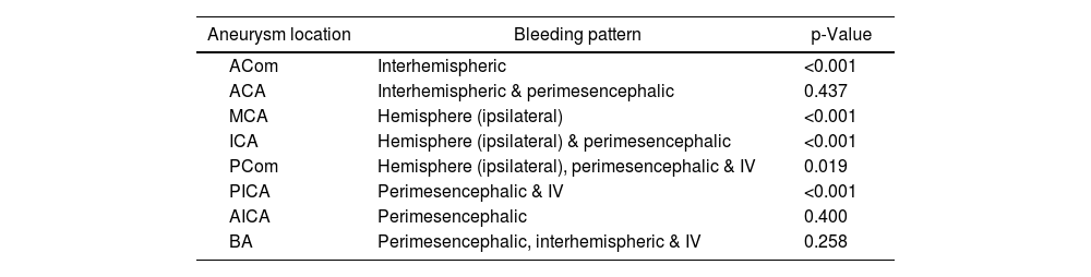
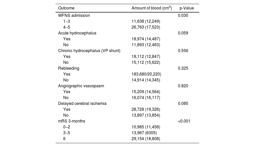
Results68 patients were included for analysis. There was a strong association between the distribution of blood and the aneurysm location (p < 0.001). In particular: ACom and interhemispheric fissure (p < 0.001), MCA and ipsilateral hemisphere (p < 0.001), ICA and ipsilateral hemisphere and perimesencephalic cisterns (p < 0.001), PCom and hemispheric, perimesencephalic and intraventricular (p = 0.019), and PICA and perimesencephalic and intraventricular (p < 0.001). The internal diagnostic value was high (AUROC ≥ 0.900) for these locations.
ConclusionRegional automatised volumetry seems a reliable and objective tool to quantify and describe the distribution of blood within the subarachnoid spaces. This tool accurately predicts the location of the ruptured aneurysm; its use may be prospectively considered in the emergency setting when speed and simplicity are attained.
En la hemorragia subaracnoidea espontánea (HAS), la determinación fehaciente del punto de sangrado es fundamental para guiar el tratamiento. De forma tradicional, la determinación de la localización del aneurisma se basa en el patrón de sangrado. En el presente estudio, evaluamos una herramienta computarizada, capaz de cuantificar el volumen de sangre intracraneal y de estratificarlo según la distribución regional, con el objetivo de predecir la localización del aneurisma roto.
MétodosUna serie consecutiva de pacientes ingresados en nuestro centro terciario durante el periodo 2012–2018, con hemorragia subaracnoidea de debut en < 72 h y con aneurisma único. Se aplicó el método cuantitativo semiautomatizado a las tomografías computarizadas basales de dichos pacientes. La detección del sistema se basa en el aumento relativo de densidad atribuido a la presencia de sangre. Se contemplaron cinco regiones para definir los patrones de sangrado y para su posterior correlación con la localización del aneurisma: perimesencefálico, interhemisférico, hemisférico derecho/izquierdo e intraventricular.
ResultadosEl análisis incluyó 68 pacientes. Se detectó una fuerte asociación entre el patrón de sangrado y la localización del aneurisma (p < 0,001). En concreto: ACom con la distribución interhemisférica (p < 0,001); ACM con la hemisférica ipsilateral (p < 0,001); ACI con la hemisférica ipsilateral y perimesencefálica (p < 0,001); ACoP la hemisférica ipsilateral, perimesencefálica e intraventricular (p = 0,019); ACPI con la perimesencefálica e intraventricular (p < 0,001). El valor diagnóstico interno del modelo fue elevado para estas localizaciones (AUROC ≥ 0.900).
ConclusionesLa volumetría regional automatizada parece una herramienta fiable y objetiva para cuantificar y describir la distribución de sangre en los espacios subaracnoideos. Este método es capaz de predecir de forma precisa la localización del aneurisma roto; su aplicación prospectiva es en la medicina de urgencias, una vez asegurada la agilidad y simplicidad del funcionamiento.
Article
![]()
If it is the first time you have accessed you can obtain your credentials by contacting Elsevier Spain in suscripciones@elsevier.com or by calling our Customer Service at902 88 87 40 if you are calling from Spain or at +34 932 418 800 (from 9 to 18h., GMT + 1) if you are calling outside of Spain.
If you already have your login data, please click here .
If you have forgotten your password you can you can recover it by clicking here and selecting the option ¿I have forgotten my password¿.


