Multiple lesion glioblastoma (M-GBM) represent a group of GBM patients in which there exist multiple foci of tumor enhancement. The prognosis is poorer than that of single-lesion GBM patients, but this actually is a controversial data. Is unknown whether multifocality has a genetic and molecular basis. Our specific aim is to identify the molecular characteristics of M-GBM by performing a comprehensive multidimensional analysis.
MethodsThe surgical, radiological and clinical outcomes of patients that underwent surgery for GBM at our institution for 2 years have been retrospectively reviewed. We compared the overall survival (OS), progression free survival and extent of resection (EOR) between M-GBM tumors (type I) and S-GBM (single contrast-enhancing lesion, type II).
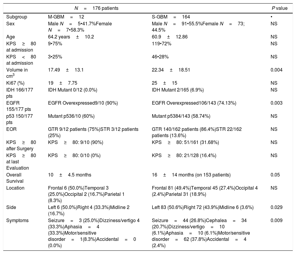
ResultsA total of 177 patients were included in the final cohort, 12 patients had M-GBM and 165 patients had S-GBM. Although patients with M-GBM had higher tumor volumes and midline location, the EOR was not different between both type of lesions. Higher percentage of tumors with EGFR overexpression was detected in M-GBM. PFS and OS was significantly shorter in M-GBM.
ConclusionsConsidering no differences in EOR, patients with M-GBM showed shorter PFS and OS in comparison with S-GBM. Evidences about the M-GBM origin as a multifocal lesion because its molecular profile are suggested.
El glioblastoma multiforme multifocal (M-GBM) representa un grupo de pacientes con GBM en el que existen múltiples focos de mejora tumoral. El pronóstico es peor que el de los pacientes con GBM de lesión única, pero en realidad es un dato controvertido. Se desconoce si la multifocalidad tiene una base genèc)tica y molecular. Nuestro objetivo específico es identificar las características moleculares de M-GBM mediante la realización de un análisis multidimensional integral.
Mèc)todosLos resultados quirúrgicos, radiológicos y clínicos de los pacientes que se sometieron a cirugía para GBM en nuestra institución durante 2 años han sido revisados retrospectivamente. Comparamos la supervivencia general (SG), la supervivencia libre de progresión y el grado de resección (EOR) entre los tumores M-GBM (tipo I) y S-GBM (lesión única que mejora el contraste, tipo II).
ResultadosUn total de 177 pacientes fueron incluidos en la cohorte final, 12 pacientes tenían M-GBM y 165 pacientes tenían S-GBM. Aunque los pacientes con M-GBM tenían mayores volúmenes tumorales y ubicación en la línea media, el EOR no fue diferente entre ambos tipos de lesiones. Se detectó un mayor porcentaje de tumores con sobreexpresión de EGFR en M-GBM. PFS y OS fue significativamente más corto en M-GBM.
ConclusionesTeniendo en cuenta que no hay diferencias en EOR, los pacientes con M-GBM mostraron PFS y OS más cortos en comparación con S-GBM. Se sugieren evidencias sobre el origen de M-GBM como una lesión multifocal porque se sugiere su perfil molecular.
Article

If it is the first time you have accessed you can obtain your credentials by contacting Elsevier Spain in suscripciones@elsevier.com or by calling our Customer Service at902 88 87 40 if you are calling from Spain or at +34 932 418 800 (from 9 to 18h., GMT + 1) if you are calling outside of Spain.
If you already have your login data, please click here .
If you have forgotten your password you can you can recover it by clicking here and selecting the option ¿I have forgotten my password¿.


