Share
array:24 [
"pii" => "S2529849624000029"
"issn" => "25298496"
"doi" => "10.1016/j.neucie.2023.10.001"
"estado" => "S300"
"fechaPublicacion" => "2024-03-01"
"aid" => "593"
"copyright" => "Sociedad Española de Neurocirugía"
"copyrightAnyo" => "2023"
"documento" => "article"
"crossmark" => 1
"subdocumento" => "fla"
"cita" => "Neurocirugia. 2024;35:87-94"
"abierto" => array:3 [
"ES" => false
"ES2" => false
"LATM" => false
]
"gratuito" => false
"lecturas" => array:1 [
"total" => 0
]
"Traduccion" => array:1 [
"es" => array:19 [
"pii" => "S1130147323003895"
"issn" => "11301473"
"doi" => "10.1016/j.neucir.2023.10.002"
"estado" => "S300"
"fechaPublicacion" => "2024-03-01"
"aid" => "593"
"copyright" => "Sociedad Española de Neurocirugía"
"documento" => "article"
"crossmark" => 1
"subdocumento" => "fla"
"cita" => "Neurocirugia. 2024;35:87-94"
"abierto" => array:3 [
"ES" => false
"ES2" => false
"LATM" => false
]
"gratuito" => false
"lecturas" => array:1 [
"total" => 0
]
"es" => array:13 [
"idiomaDefecto" => true
"cabecera" => "<span class="elsevierStyleTextfn">Investigación clínica</span>"
"titulo" => "Ependimomas de fosa posterior de tipo lateral en la edad pediátrica"
"tienePdf" => "es"
"tieneTextoCompleto" => "es"
"tieneResumen" => array:2 [
0 => "es"
1 => "en"
]
"paginas" => array:1 [
0 => array:2 [
"paginaInicial" => "87"
"paginaFinal" => "94"
]
]
"titulosAlternativos" => array:1 [
"en" => array:1 [
"titulo" => "Lateral-type posterior fossa ependymomas in pediatric population"
]
]
"contieneResumen" => array:2 [
"es" => true
"en" => true
]
"contieneTextoCompleto" => array:1 [
"es" => true
]
"contienePdf" => array:1 [
"es" => true
]
"resumenGrafico" => array:2 [
"original" => 0
"multimedia" => array:7 [
"identificador" => "fig0005"
"etiqueta" => "Figura 1"
"tipo" => "MULTIMEDIAFIGURA"
"mostrarFloat" => true
"mostrarDisplay" => false
"figura" => array:1 [
0 => array:4 [
"imagen" => "gr1.jpeg"
"Alto" => 2879
"Ancho" => 2713
"Tamanyo" => 503331
]
]
"descripcion" => array:1 [
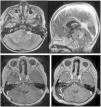
"es" => "<p id="spar0045" class="elsevierStyleSimplePara elsevierViewall">Ependimoma de ángulo pontocerebeloso: imágenes pre- y postoperatorias.</p> <p id="spar0050" class="elsevierStyleSimplePara elsevierViewall"><span class="elsevierStyleItalic">Superior izquierda</span>: Imagen preoperatoria RM axial T2. Aspecto característico de ependimoma de fosa posterior de tipo lateral que ocupa el ángulo pontocerebeloso, engloba las estructuras neurovasculares y comprime y rota el tronco en sentido opuesto al tumor. <span class="elsevierStyleItalic">Superior derecha</span>: Imagen preoperatoria RM sagital T1 con contraste que muestra la extensión de la lesión desde el acueducto de Silvio hasta el agujero magno. <span class="elsevierStyleItalic">Inferior izquierda:</span> Imagen postoperatoria de RM axial T1 con contraste que muestra una resección casi completa, con resto en la cara lateral del bulbo y en relación con la salida del nervio facial derecho. <span class="elsevierStyleItalic">Inferior derecha</span>: Imagen tras la segunda intervención, RM axial T1 con contraste que muestra una resección completa.</p>"
]
]
]
"autores" => array:1 [
0 => array:2 [
"autoresLista" => "Pablo Miranda-Lloret, Estela Plaza-Ramírez, Juan Antonio Simal-Julián, Giovanni Pancucci, Adela Cañete, Alejandro Montoya-Filardi, Gemma Llavador"
"autores" => array:7 [
0 => array:2 [
"nombre" => "Pablo"
"apellidos" => "Miranda-Lloret"
]
1 => array:2 [
"nombre" => "Estela"
"apellidos" => "Plaza-Ramírez"
]
2 => array:2 [
"nombre" => "Juan Antonio"
"apellidos" => "Simal-Julián"
]
3 => array:2 [
"nombre" => "Giovanni"
"apellidos" => "Pancucci"
]
4 => array:2 [
"nombre" => "Adela"
"apellidos" => "Cañete"
]
5 => array:2 [
"nombre" => "Alejandro"
"apellidos" => "Montoya-Filardi"
]
6 => array:2 [
"nombre" => "Gemma"
"apellidos" => "Llavador"
]
]
]
]
]
"idiomaDefecto" => "es"
"Traduccion" => array:1 [
"en" => array:9 [
"pii" => "S2529849624000029"
"doi" => "10.1016/j.neucie.2023.10.001"
"estado" => "S300"
"subdocumento" => ""
"abierto" => array:3 [
"ES" => false
"ES2" => false
"LATM" => false
]
"gratuito" => false
"lecturas" => array:1 [
"total" => 0
]
"idiomaDefecto" => "en"
"EPUB" => "https://multimedia.elsevier.es/PublicationsMultimediaV1/item/epub/S2529849624000029?idApp=UINPBA00004B"
]
]
"EPUB" => "https://multimedia.elsevier.es/PublicationsMultimediaV1/item/epub/S1130147323003895?idApp=UINPBA00004B"
"url" => "/11301473/0000003500000002/v1_202403060452/S1130147323003895/v1_202403060452/es/main.assets"
]
]
"itemSiguiente" => array:19 [
"pii" => "S2529849624000042"
"issn" => "25298496"
"doi" => "10.1016/j.neucie.2023.10.003"
"estado" => "S300"
"fechaPublicacion" => "2024-03-01"
"aid" => "592"
"copyright" => "Sociedad Española de Neurocirugía"
"documento" => "article"
"crossmark" => 1
"subdocumento" => "fla"
"cita" => "Neurocirugia. 2024;35:95-112"
"abierto" => array:3 [
"ES" => false
"ES2" => false
"LATM" => false
]
"gratuito" => false
"lecturas" => array:1 [
"total" => 0
]
"en" => array:12 [
"idiomaDefecto" => true
"cabecera" => "<span class="elsevierStyleTextfn">Special article</span>"
"titulo" => "A new standardized nomenclature in neurosurgery: Criteria and quantitative and qualitative evaluation indicators of medical procedures"
"tienePdf" => "en"
"tieneTextoCompleto" => "en"
"tieneResumen" => array:2 [
0 => "en"
1 => "es"
]
"paginas" => array:1 [
0 => array:2 [
"paginaInicial" => "95"
"paginaFinal" => "112"
]
]
"titulosAlternativos" => array:1 [
"es" => array:1 [
"titulo" => "Nuevo nomenclátor estandarizado de Neurocirugía: criterios e indicadores cuantitativos y cualitativos de baremación de actos médicos"
]
]
"contieneResumen" => array:2 [
"en" => true
"es" => true
]
"contieneTextoCompleto" => array:1 [
"en" => true
]
"contienePdf" => array:1 [
"en" => true
]
"autores" => array:1 [
0 => array:2 [
"autoresLista" => "Ángel Horcajadas Almansa, Luis Ley Urzaiz, Roberto Garcia Leal, Francisco González Llanos, Mónica Lara Almunia, Ruben Martinez Laez, Jose M. Torres Campa, Idoia Zaspe Cenoz, Jesús Lafuente Baraza"
"autores" => array:9 [
0 => array:2 [
"nombre" => "Ángel"
"apellidos" => "Horcajadas Almansa"
]
1 => array:2 [
"nombre" => "Luis"
"apellidos" => "Ley Urzaiz"
]
2 => array:2 [
"nombre" => "Roberto"
"apellidos" => "Garcia Leal"
]
3 => array:2 [
"nombre" => "Francisco"
"apellidos" => "González Llanos"
]
4 => array:2 [
"nombre" => "Mónica"
"apellidos" => "Lara Almunia"
]
5 => array:2 [
"nombre" => "Ruben"
"apellidos" => "Martinez Laez"
]
6 => array:2 [
"nombre" => "Jose M."
"apellidos" => "Torres Campa"
]
7 => array:2 [
"nombre" => "Idoia"
"apellidos" => "Zaspe Cenoz"
]
8 => array:2 [
"nombre" => "Jesús"
"apellidos" => "Lafuente Baraza"
]
]
]
]
]
"idiomaDefecto" => "en"
"Traduccion" => array:1 [
"es" => array:9 [
"pii" => "S1130147323003883"
"doi" => "10.1016/j.neucir.2023.10.001"
"estado" => "S300"
"subdocumento" => ""
"abierto" => array:3 [
"ES" => false
"ES2" => false
"LATM" => false
]
"gratuito" => false
"lecturas" => array:1 [
"total" => 0
]
"idiomaDefecto" => "es"
"EPUB" => "https://multimedia.elsevier.es/PublicationsMultimediaV1/item/epub/S1130147323003883?idApp=UINPBA00004B"
]
]
"EPUB" => "https://multimedia.elsevier.es/PublicationsMultimediaV1/item/epub/S2529849624000042?idApp=UINPBA00004B"
"url" => "/25298496/0000003500000002/v1_202403091432/S2529849624000042/v1_202403091432/en/main.assets"
]
"itemAnterior" => array:18 [
"pii" => "S2529849623000424"
"issn" => "25298496"
"doi" => "10.1016/j.neucie.2023.09.001"
"estado" => "S300"
"fechaPublicacion" => "2024-03-01"
"aid" => "591"
"copyright" => "Sociedad Española de Neurocirugía"
"documento" => "article"
"crossmark" => 1
"subdocumento" => "fla"
"cita" => "Neurocirugia. 2024;35:79-86"
"abierto" => array:3 [
"ES" => false
"ES2" => false
"LATM" => false
]
"gratuito" => false
"lecturas" => array:1 [
"total" => 0
]
"en" => array:12 [
"idiomaDefecto" => true
"cabecera" => "<span class="elsevierStyleTextfn">Clinical Research</span>"
"titulo" => "Intracerebral hemorrhage after deep brain stimulation surgery guided with microelectrode recording: analysis of 297 procedures"
"tienePdf" => "en"
"tieneTextoCompleto" => "en"
"tieneResumen" => array:2 [
0 => "en"
1 => "es"
]
"paginas" => array:1 [
0 => array:2 [
"paginaInicial" => "79"
"paginaFinal" => "86"
]
]
"titulosAlternativos" => array:1 [
"es" => array:1 [
"titulo" => "Hemorragia intracerebral después de cirugía de estimulación cerebral profunda guiada por microrregistro: un análisis de 297 procedimientos"
]
]
"contieneResumen" => array:2 [
"en" => true
"es" => true
]
"contieneTextoCompleto" => array:1 [
"en" => true
]
"contienePdf" => array:1 [
"en" => true
]
"autores" => array:1 [
0 => array:2 [
"autoresLista" => "Beatriz Dos Santos, Rui Vaz, Ana Cristina Braga, Manuel Rito, Diana Lucas, Clara Chamadoira"
"autores" => array:6 [
0 => array:2 [
"nombre" => "Beatriz"
"apellidos" => "Dos Santos"
]
1 => array:2 [
"nombre" => "Rui"
"apellidos" => "Vaz"
]
2 => array:2 [
"nombre" => "Ana Cristina"
"apellidos" => "Braga"
]
3 => array:2 [
"nombre" => "Manuel"
"apellidos" => "Rito"
]
4 => array:2 [
"nombre" => "Diana"
"apellidos" => "Lucas"
]
5 => array:2 [
"nombre" => "Clara"
"apellidos" => "Chamadoira"
]
]
]
]
]
"idiomaDefecto" => "en"
"EPUB" => "https://multimedia.elsevier.es/PublicationsMultimediaV1/item/epub/S2529849623000424?idApp=UINPBA00004B"
"url" => "/25298496/0000003500000002/v1_202403091432/S2529849623000424/v1_202403091432/en/main.assets"
]
"en" => array:19 [
"idiomaDefecto" => true
"cabecera" => "<span class="elsevierStyleTextfn">Clinical research</span>"
"titulo" => "Lateral-type posterior fossa ependymomas in pediatric population"
"tieneTextoCompleto" => true
"paginas" => array:1 [
0 => array:2 [
"paginaInicial" => "87"
"paginaFinal" => "94"
]
]
"autores" => array:1 [
0 => array:4 [
"autoresLista" => "Pablo Miranda-Lloret, Estela Plaza-Ramírez, Juan Antonio Simal-Julián, Giovanni Pancucci, Adela Cañete, Alejandro Montoya-Filardi, Gemma Llavador"
"autores" => array:7 [
0 => array:4 [
"nombre" => "Pablo"
"apellidos" => "Miranda-Lloret"
"email" => array:1 [
0 => "Miranda_pab@gva.es"
]
"referencia" => array:1 [
0 => array:2 [
"etiqueta" => "<span class="elsevierStyleSup">*</span>"
"identificador" => "cor0005"
]
]
]
1 => array:2 [
"nombre" => "Estela"
"apellidos" => "Plaza-Ramírez"
]
2 => array:2 [
"nombre" => "Juan Antonio"
"apellidos" => "Simal-Julián"
]
3 => array:2 [
"nombre" => "Giovanni"
"apellidos" => "Pancucci"
]
4 => array:2 [
"nombre" => "Adela"
"apellidos" => "Cañete"
]
5 => array:2 [
"nombre" => "Alejandro"
"apellidos" => "Montoya-Filardi"
]
6 => array:2 [
"nombre" => "Gemma"
"apellidos" => "Llavador"
]
]
"afiliaciones" => array:1 [
0 => array:2 [
"entidad" => "Servicios de Neurocirugía, Oncología Pediátrica, Radiología Pediátrica y Anatomía Patológica, Hospital Universitario y Politécnico La Fe, Valencia, Spain"
"identificador" => "aff0005"
]
]
"correspondencia" => array:1 [
0 => array:3 [
"identificador" => "cor0005"
"etiqueta" => "⁎"
"correspondencia" => "<span class="elsevierStyleItalic">Corresponding author</span>."
]
]
]
]
"titulosAlternativos" => array:1 [
"es" => array:1 [
"titulo" => "Ependimomas de fosa posterior de tipo lateral en la edad pediátrica"
]
]
"resumenGrafico" => array:2 [
"original" => 0
"multimedia" => array:8 [
"identificador" => "fig0005"
"etiqueta" => "Figure 1"
"tipo" => "MULTIMEDIAFIGURA"
"mostrarFloat" => true
"mostrarDisplay" => false
"figura" => array:1 [
0 => array:4 [
"imagen" => "gr1.jpeg"
"Alto" => 2879
"Ancho" => 2713
"Tamanyo" => 503973
]
]
"detalles" => array:1 [
0 => array:3 [
"identificador" => "at0005"
"detalle" => "Figure "
"rol" => "short"
]
]
"descripcion" => array:1 [
"en" => "<p id="spar0005" class="elsevierStyleSimplePara elsevierViewall">Cerebellopontine angle ependymoma: pre- and postoperative images.</p> <p id="spar0010" class="elsevierStyleSimplePara elsevierViewall"><span class="elsevierStyleItalic">Top left</span>: preoperative axial T2-weighted MRI image. Characteristic appearance of a lateral posterior fossa ependymoma occupying the cerebellopontine angle, encompassing the neurovascular structures and compressing and rotating the brainstem in the opposite direction to the tumour. <span class="elsevierStyleItalic">Top right</span>: preoperative sagittal T1-weighted contrast-enhanced MRI image showing the extension of the lesion from the cerebral aqueduct to the foramen magnum. <span class="elsevierStyleItalic">Bottom left:</span> postoperative axial T1-weighted contrast-enhanced MRI image showing near-total resection, with residual tumour on the lateral aspect of the medulla and in relation to the exit point of the right facial nerve. <span class="elsevierStyleItalic">Bottom right:</span> image after the second operation, axial T1-weighted contrast-enhanced MRI showing total resection.</p>"
]
]
]
"textoCompleto" => "<span class="elsevierStyleSections"><span id="sec0005" class="elsevierStyleSection elsevierViewall"><span class="elsevierStyleSectionTitle" id="sect0065">Introduction</span><p id="par0005" class="elsevierStylePara elsevierViewall">Ependymomas are rosette- and pseudorosette-forming glial tumours with a relatively low incidence. The new WHO classification uses anatomical, histological and molecular criteria to properly classify these lesions.<a class="elsevierStyleCrossRef" href="#bib0005"><span class="elsevierStyleSup">1</span></a> Although they can occur throughout the nervous system, the relative incidence of ependymomas in the posterior fossa is significantly higher in the paediatric population. Posterior fossa ependymomas can be classified into subtypes A and B depending on the methylation pattern and the presence of mutations in EZHIP and H3K27. While the new WHO classification provides for the staging of these lesions at grades II and III, a higher histological grade does not necessarily mean a poorer prognosis. In the latest edition of the WHO tumour classification, the term <span class="elsevierStyleItalic">anaplastic</span> ependymoma has been withdrawn, as it is also not considered to provide prognostic information.</p><p id="par0010" class="elsevierStylePara elsevierViewall">Prior to this molecular classification, there were reports in the literature from paediatric neurosurgeons of a subtype of posterior fossa ependymomas whose treatment was particularly problematic. This subtype has been referred to by different authors as cerebellopontine angle ependymoma or lateral posterior fossa ependymoma.<a class="elsevierStyleCrossRefs" href="#bib0010"><span class="elsevierStyleSup">2–5</span></a> These lesions characteristically present as large neoplasms affecting young children, with a marked tendency to involve vital structures of the posterior fossa such as the vertebral, basilar and posterior inferior cerebellar arteries or the lower and middle cranial nerve roots. They are independent of the floor of the fourth ventricle and they appear to originate from the lateral aspect of the medulla and the lateral recess of the fourth ventricle. From this origin, as they grow they characteristically displace and rotate the medulla and pons in the opposite direction to the tumour. Surgically, these are very difficult lesions that require significant anatomical and technical knowledge.<a class="elsevierStyleCrossRefs" href="#bib0015"><span class="elsevierStyleSup">3,6</span></a> Although the morbidity associated with these procedures is not negligible, the possibility of total resection is the most important therapeutic prognostic factor and conventional treatment is still maximal safe resection followed by focal radiotherapy.<a class="elsevierStyleCrossRefs" href="#bib0035"><span class="elsevierStyleSup">7–14</span></a> More recent studies have shown that this subtype of ependymoma corresponds to type A posterior fossa ependymoma,<a class="elsevierStyleCrossRefs" href="#bib0025"><span class="elsevierStyleSup">5,15</span></a> although new subtypes and classifications are continuously being described and developed.<a class="elsevierStyleCrossRefs" href="#bib0080"><span class="elsevierStyleSup">16,17</span></a></p><p id="par0015" class="elsevierStylePara elsevierViewall">In light of the above, the aim of this study is to review our series of lateral posterior fossa ependymomas in the paediatric population, compare our results with those in the literature and assess whether or not histological grade has any significant implications for surgical treatment.</p></span><span id="sec0010" class="elsevierStyleSection elsevierViewall"><span class="elsevierStyleSectionTitle" id="sect0070">Material and methods</span><p id="par0020" class="elsevierStylePara elsevierViewall">We designed a retrospective, descriptive case series study. From our hospital's pathology records, 30 paediatric patients who had undergone surgery for ependymomas in the last 10 years were identified. Of these, seven patients had cerebellopontine angle or lateral posterior fossa ependymomas according to the criteria described by Sanford and Boop.<a class="elsevierStyleCrossRef" href="#bib0015"><span class="elsevierStyleSup">3</span></a> In each case, the clinical, radiological, histological and therapeutic variables at diagnosis and throughout follow-up necessary for the study were collected. The product of the three diameters of the tumour divided by two was used as an approximation to calculate the tumour volume.<a class="elsevierStyleCrossRef" href="#bib0090"><span class="elsevierStyleSup">18</span></a> The Evans index<a class="elsevierStyleCrossRef" href="#bib0095"><span class="elsevierStyleSup">19</span></a> was used to assess enlarged ventricular size. To determine the extent of tumour resection, pre- and postoperative MRI were compared and the degree of resection was classified as total (no visible residual lesion), near-total (when the residual tumour volume was less than 5% of the initial tumour volume) or partial (when the residual tumour volume was greater than 5% of the initial tumour volume). The House-Brackmann scale<a class="elsevierStyleCrossRef" href="#bib0100"><span class="elsevierStyleSup">20</span></a> was used to classify the severity of postoperative facial nerve involvement, and major involvement was considered if the score was 3 or more. The postoperative onset of hemiparesis, meningitis, major cranial nerve deficits and any other potentially fatal circumstance were considered to be major morbidity.<a class="elsevierStyleCrossRef" href="#bib0015"><span class="elsevierStyleSup">3</span></a> The need for tracheostomy or gastrostomy in these patients was assessed in a multidisciplinary manner by a team of paediatric intensive medicine specialists, paediatric surgeons, ear, nose and throat (ENT) specialists and paediatricians. Patients were assessed on an outpatient basis at three-monthly intervals for the first year and every six months thereafter. Patient quality of life at follow-up was determined according to the Lansky scale.<a class="elsevierStyleCrossRef" href="#bib0105"><span class="elsevierStyleSup">21</span></a> The patients received supplementary oncological treatment following the recommendations subsequently published by the European Association of Neuro-Oncology (EANO), also participating in the SIOP (International Society of Paediatric Oncology) study for ependymomas in paediatric patients since 2015.<a class="elsevierStyleCrossRefs" href="#bib0055"><span class="elsevierStyleSup">11,22</span></a></p><p id="par0025" class="elsevierStylePara elsevierViewall">Data for continuous variables are shown with reference to their mean and standard deviation. Student's <span class="elsevierStyleItalic">t</span>-test was used to compare independent sample means. Bivariate associations were explored with the Chi-square test and Fisher's exact test. Survival studies are shown as Kaplan-Meier curves. The level of statistical significance was set at 0.05. SPSS version 21 (IBM, New York, USA) was used to support the statistical analysis.</p></span><span id="sec0015" class="elsevierStyleSection elsevierViewall"><span class="elsevierStyleSectionTitle" id="sect0075">Results</span><p id="par0030" class="elsevierStylePara elsevierViewall">The series comprises a total of seven patients, three boys and four girls. The main demographic and radiological characteristics are shown in <a class="elsevierStyleCrossRef" href="#tbl0005">Table 1</a>. The mean age at diagnosis was less than four years old (mean 45 months). The mean volume of the lesions exceeded 60 cm<span class="elsevierStyleSup">3</span> and they were often tumours with diameters close to 5 cm. Virtually all patients had intracranial hypertension related to the associated hydrocephalus, except for one female patient who had abnormal gait and torticollis. Six patients showed an increase in ventricular size with an Evans index above 0.3, and in four of them it was considered necessary to implant a ventricular drain preoperatively. None of the patients had spinal tumour dissemination at diagnosis or during the course of their disease. However, one of our patients developed tumour recurrence after an apparently total resection.</p><elsevierMultimedia ident="tbl0005"></elsevierMultimedia><span id="sec0020" class="elsevierStyleSection elsevierViewall"><span class="elsevierStyleSectionTitle" id="sect0080">Surgical approach</span><p id="par0035" class="elsevierStylePara elsevierViewall">The most important factors relating to treatment and follow-up are summarised in <a class="elsevierStyleCrossRef" href="#tbl0010">Table 2</a>. The surgical approach used consisted of a combination of a midline suboccipital approach followed by a lateral suboccipital approach for resection of the cisternal compartment and cranial nerves, according to the technique described by Sanford and Boop.<a class="elsevierStyleCrossRef" href="#bib0015"><span class="elsevierStyleSup">3</span></a> In three cases, more than one surgical procedure was necessary to achieve optimal resection of the lesion: one female patient developed tumour haemorrhage with deterioration to coma and required urgent surgery for posterior fossa decompression and partial tumour resection, followed by resective surgery; another female patient required two lengthy operations due to the close relationship of the tumour to the lower cranial nerves and the posterior inferior vertebral and cerebellar arteries; the third case is described in more detail as an example of the series.</p><elsevierMultimedia ident="tbl0010"></elsevierMultimedia><p id="par0040" class="elsevierStylePara elsevierViewall"><span class="elsevierStyleItalic">Case number 3</span> (<a class="elsevierStyleCrossRef" href="#fig0005">Fig. 1</a>). This was a four-year-old female patient who consulted with headache and vomiting. Examination revealed bilateral papilloedema in the fundus of the eye and right sixth cranial nerve palsy. Brain CT images showed obstructive hydrocephalus in relation to a posterior fossa tumour and it was decided to urgently implant an external ventricular drain (EVD), which resolved the symptoms and the neurological examination returned to normal. Brain MRI confirmed a probable posterior fossa ependymoma of right lateral topography measuring 4 × 5 × 4.5 cm in diameter. Planned surgery was performed using an extended suboccipital approach on the right side, accessing first the component related to the fourth ventricle and subsequently over the cerebellopontine angle, where a near-total resection was achieved, leaving a millimetre-sized remnant adjacent to the exit of the right facial nerve, where manipulation produced tonic discharges on neuromonitoring. After surgery, the patient had House-Brackmann 2 facial paralysis, which resolved completely within a few days after surgery. The ventricular drain was removed early. However, progressive hydrocephalus developed, which was successfully managed by endoscopic ventriculocisternostomy of the floor of the third ventricle. The histological diagnosis was grade II ependymoma. The patient received adjuvant focal radiotherapy with a total dose of 59.4 Gy, and was included in the SIOP stratum 2 trial. Post-radiotherapy imaging showed the persistence of a 5 mm-diameter tumour lesion on the lateral aspect of the pons. It was decided to explore this lesion surgically and total resection was achieved without incident in relation to neuromonitoring and without complications in the clinical outcome. The patient is living a normal life, free of disease and deficits, and she has now been under follow-up for over eight years.</p><elsevierMultimedia ident="fig0005"></elsevierMultimedia></span><span id="sec0025" class="elsevierStyleSection elsevierViewall"><span class="elsevierStyleSectionTitle" id="sect0085">Extent of resection. Management of hydrocephalus. Adjuvant radiotherapy</span><p id="par0045" class="elsevierStylePara elsevierViewall">In the series overall, postoperatively, four patients required ventriculoperitoneal (VP) shunt implantation, in two cases endoscopic ventriculostomy was successfully employed, and one female patient did not develop postoperative hydrocephalus. Patients who developed early hydrocephalus were managed with the use of VP CSF shunts with anti-siphon devices, while the two patients who developed subacute hydrocephalus were treated with endoscopic ventriculostomy of the floor of the third ventricle, due to the associated high Endoscopic Third Ventriculostomy Success Score (ETVSS)<a class="elsevierStyleCrossRef" href="#bib0115"><span class="elsevierStyleSup">23</span></a> and the fact that reliable clinical follow-up was possible. Total resection was achieved in six cases, and in the remaining case it was not achieved despite a second operation: this was a female patient diagnosed with an anaplastic ependymoma in whom an infiltration of the lateral aspect of the medulla was observed intraoperatively and was considered not resectable; this residual lesion was difficult to identify on postoperative imaging and disappeared after adjuvant proton therapy. Adjuvant radiotherapy was administered by photons in three patients (54.9 Gy) until the availability of proton therapy, which was used in the last four patients.</p></span><span id="sec0030" class="elsevierStyleSection elsevierViewall"><span class="elsevierStyleSectionTitle" id="sect0090">Pathology assessment of quality of life</span><p id="par0050" class="elsevierStylePara elsevierViewall">Four patients were diagnosed with grade II ependymoma and three with grade III ependymoma. However, the patient who developed tumour progression after an apparently total resection developed first grade progression and then medullary infiltration that eventually led to death. This was the only fatal case observed in the series after a mean follow-up of 58 months (<a class="elsevierStyleCrossRef" href="#fig0010">Fig. 2</a>). This patient also suffered a progressive deterioration of his general condition and at the last consultation his Lansky scale score was 50. The remaining patients had normal functional status (four cases) or a moderate limitation for play (two patients, Lansky 70 at the last outpatient assessment). Notably, five of our patients developed major postoperative morbidity requiring tracheostomy and/or gastrostomy in the postoperative period in relation to lower cranial nerve dysfunction and despite a documented favourable neurophysiological record at the end of the intervention in all cases. For all of these patients, decannulation from the tracheostomy was possible and they were able to resume normal oral feeding six months after the end of treatment. The two patients with a Lansky score of 70 had mild motor deficits and diplopia, as well as symptomatic shunt overdrainage in the process of up-regulation at the last check-up.</p><elsevierMultimedia ident="fig0010"></elsevierMultimedia><p id="par0055" class="elsevierStylePara elsevierViewall">When we explored the relationship between histological grade and the behaviour of these tumours in our series (<a class="elsevierStyleCrossRef" href="#tbl0015">Table 3</a>), we found that high-grade tumours were detected as larger lesions at younger ages, required more time to achieve appropriate surgical resection and were associated with a higher morbidity rate in the early postoperative period. However, these associations did not show statistical significance in any of the cases.</p><elsevierMultimedia ident="tbl0015"></elsevierMultimedia></span></span><span id="sec0035" class="elsevierStyleSection elsevierViewall"><span class="elsevierStyleSectionTitle" id="sect0095">Discussion</span><p id="par0060" class="elsevierStylePara elsevierViewall">Brain and central nervous system tumours are the leading cause of oncological disease in the paediatric population, with an incidence of 5.74 cases per 100,000 population under 15 years of age. However, ependymomas are a relatively rare type of tumour, accounting for about 5% of primary tumours of the nervous system at this age.<a class="elsevierStyleCrossRef" href="#bib0120"><span class="elsevierStyleSup">24</span></a> Overall survival rates for these tumours exceed 70% after total resection and targeted adjuvant therapy.<a class="elsevierStyleCrossRefs" href="#bib0035"><span class="elsevierStyleSup">7,11,25</span></a> However, there are subtypes of ependymomas whose prognosis is significantly worse.</p><p id="par0065" class="elsevierStylePara elsevierViewall">With regards to posterior fossa ependymomas, type A have the worst prognosis. Within this subgroup, new genetic characterisations are able to predict even more aggressive behaviour,<a class="elsevierStyleCrossRefs" href="#bib0080"><span class="elsevierStyleSup">16,17,26</span></a> although radiological and anatomical correlation with these new subtypes has not shown a significant association to date<a class="elsevierStyleCrossRef" href="#bib0075"><span class="elsevierStyleSup">15</span></a>.</p><p id="par0070" class="elsevierStylePara elsevierViewall">Genetic studies have identified lateral ependymomas of the posterior fossa as type A ependymomas.</p><p id="par0075" class="elsevierStylePara elsevierViewall">In our series, these cerebellopontine angle ependymomas accounted for 23% of paediatric posterior fossa ependymomas treated in the last 10 years, a figure similar to the initially described series<a class="elsevierStyleCrossRefs" href="#bib0010"><span class="elsevierStyleSup">2,4</span></a> but lower than more recent ones, where the relative incidence is as high as 40% of posterior fossa ependymomas.<a class="elsevierStyleCrossRefs" href="#bib0015"><span class="elsevierStyleSup">3,5</span></a> It is possible that the progressive recognition of this disease in imaging studies will enable more precise classification and explain these differences. Nonetheless, as this is a series with a small number of patients, the variability observed may represent an expected deviation in relation to the heterogeneity of the samples.</p><p id="par0080" class="elsevierStylePara elsevierViewall">The most common form of presentation of these lesions is intracranial hypertension associated with hydrocephalus, and to a lesser extent cranial nerve dysfunction or abnormal gait. In our series, one female patient developed an intratumoural haemorrhage on the eve of planned surgery, requiring urgent decompressive posterior fossa craniectomy due to deterioration in her level of consciousness to coma, with this being an exceptional occurrence. As a rule, patients with cerebellopontine angle ependymomas will require ventricular drainage as part of perioperative management. Although many of them will require a permanent CSF shunt postoperatively,<a class="elsevierStyleCrossRefs" href="#bib0010"><span class="elsevierStyleSup">2,3</span></a> the ETVSS<a class="elsevierStyleCrossRef" href="#bib0115"><span class="elsevierStyleSup">23</span></a> in these cases is high. In our series, two of the six patients with post-surgical CSF dysfunction were treated by endoscopic ventriculostomy, which was effective and long-lasting: this treatment would also avoid the potential risk of the tumour spreading through the shunt, although in our series we found no cases of spread.</p><p id="par0085" class="elsevierStylePara elsevierViewall">Achieving total resection in these lesions is key to improving the oncological prognosis for these patients,<a class="elsevierStyleCrossRefs" href="#bib0035"><span class="elsevierStyleSup">7,8,10,11</span></a><span class="elsevierStyleSup">,</span><a class="elsevierStyleCrossRefs" href="#bib0065"><span class="elsevierStyleSup">13,22</span></a> but the rates of radical resection are highly variable: for some authors total resection is not feasible<a class="elsevierStyleCrossRef" href="#bib0020"><span class="elsevierStyleSup">4</span></a> or is only possible in a minority of cases,<a class="elsevierStyleCrossRefs" href="#bib0025"><span class="elsevierStyleSup">5,27</span></a> while the more experienced group of Sanford and Boop achieve total resection in virtually all cases<a class="elsevierStyleCrossRef" href="#bib0015"><span class="elsevierStyleSup">3</span></a>. In our series, total resection was achieved in six out of seven patients: in one female patient infiltration of the lateral aspect of the pons by the tumour was noted and radical resection was not considered appropriate, analogous to invasion of the floor of the fourth ventricle in midline ependymomas.</p><p id="par0090" class="elsevierStylePara elsevierViewall">The morbidity rate associated with surgery for these lesions is very significant. In the 1997 Sanford et al. series on 11 patients with cerebellopontine angle ependymomas,<a class="elsevierStyleCrossRef" href="#bib0010"><span class="elsevierStyleSup">2</span></a> one patient died within 48 h after surgery, four had postoperative hemiparesis and nine required tracheostomy or gastrostomy. The same author in 2009, in the largest published series,<a class="elsevierStyleCrossRef" href="#bib0015"><span class="elsevierStyleSup">3</span></a> reported that out of a total of 45 patients, 13 required a gastrostomy and tracheostomy, for a period of less than one year in all cases. According to the authors, the experience gained made it possible for them to reduce the morbidity associated with the surgeries without abandoning radical resection, achieving an overall survival rate of 64%. In our series, five patients were managed with a tracheostomy and gastrostomy, also on a temporary basis, for a period of less than six months after the end of treatment. Our current survival rate is 85%, but the median follow-up time is still less than five years.</p><p id="par0095" class="elsevierStylePara elsevierViewall">Last of all, the measurement of quality of life in these patients is of enormous importance. Although specific dedicated scales exist, they often have limited validation and applicability in day-to-day clinical practice.<a class="elsevierStyleCrossRef" href="#bib0140"><span class="elsevierStyleSup">28</span></a> Simpler scales such as Lansky's<a class="elsevierStyleCrossRef" href="#bib0105"><span class="elsevierStyleSup">21</span></a> can be applied routinely and provide us with a first rough idea of this aspect. In our series, most of the patients had normal functional status at the last clinic assessment, while two showed mild or moderate functional limitation. The development of posterior fossa syndrome or dense cerebellar mutism is unusual in these patients and we did not observe this complication in our series. In young patients, however, it is possible that certain symptoms of irritability and dysphoria are not so much reactive to the fact of having cancer, but rather a sign of cerebellar dysfunction,<a class="elsevierStyleCrossRef" href="#bib0145"><span class="elsevierStyleSup">29</span></a> and uphold the need to maximise surgical precautions and assess the need for potentially harmful adjuvant treatments on a personalised basis in these patients.<a class="elsevierStyleCrossRefs" href="#bib0055"><span class="elsevierStyleSup">11,22,30</span></a></p></span><span id="sec0040" class="elsevierStyleSection elsevierViewall"><span class="elsevierStyleSectionTitle" id="sect0100">Conclusions</span><p id="par0100" class="elsevierStylePara elsevierViewall">Total resection of lateral posterior fossa ependymomas is the only factor on which neurosurgeons can act to significantly improve the prognosis of these patients, making it possible to achieve long-term survival rates of over 60%.<a class="elsevierStyleCrossRef" href="#bib0015"><span class="elsevierStyleSup">3</span></a> In our experience, surgery-associated morbidity rates are high, especially in relation to lower cranial nerve dysfunction. Although 70% of patients will require a gastrostomy and tracheostomy in postoperative management, the norm is that it will only be required on a temporary basis for less than six months. Histological classification into grade II and III does not seem to have any prognostic implication in the literature, nor did we observe it in our series. Continuing advances in the genetic and molecular characterisation of these tumours open the door to the future discovery of treatment targets that will improve the prognosis of our patients.</p></span><span id="sec0045" class="elsevierStyleSection elsevierViewall"><span class="elsevierStyleSectionTitle" id="sect0105">Funding</span><p id="par0105" class="elsevierStylePara elsevierViewall">This study received no specific funding from public, private or non-profit organisations.</p></span><span id="sec0050" class="elsevierStyleSection elsevierViewall"><span class="elsevierStyleSectionTitle" id="sect0110">Conflicts of interest</span><p id="par0110" class="elsevierStylePara elsevierViewall">The authors declare that they have no conflicts of interest.</p></span></span>"
"textoCompletoSecciones" => array:1 [
"secciones" => array:12 [
0 => array:3 [
"identificador" => "xres2105013"
"titulo" => "Abstract"
"secciones" => array:4 [
0 => array:2 [
"identificador" => "abst0005"
"titulo" => "Background"
]
1 => array:2 [
"identificador" => "abst0010"
"titulo" => "Methods"
]
2 => array:2 [
"identificador" => "abst0015"
"titulo" => "Results"
]
3 => array:2 [
"identificador" => "abst0020"
"titulo" => "Conclusions"
]
]
]
1 => array:2 [
"identificador" => "xpalclavsec1793817"
"titulo" => "Keywords"
]
2 => array:3 [
"identificador" => "xres2105012"
"titulo" => "Resumen"
"secciones" => array:4 [
0 => array:2 [
"identificador" => "abst0025"
"titulo" => "Antecedentes y objetivos"
]
1 => array:2 [
"identificador" => "abst0030"
"titulo" => "Materiales y métodos"
]
2 => array:2 [
"identificador" => "abst0035"
"titulo" => "Resultados"
]
3 => array:2 [
"identificador" => "abst0040"
"titulo" => "Conclusiones"
]
]
]
3 => array:2 [
"identificador" => "xpalclavsec1793818"
"titulo" => "Palabras clave"
]
4 => array:2 [
"identificador" => "sec0005"
"titulo" => "Introduction"
]
5 => array:2 [
"identificador" => "sec0010"
"titulo" => "Material and methods"
]
6 => array:3 [
"identificador" => "sec0015"
"titulo" => "Results"
"secciones" => array:3 [
0 => array:2 [
"identificador" => "sec0020"
"titulo" => "Surgical approach"
]
1 => array:2 [
"identificador" => "sec0025"
"titulo" => "Extent of resection. Management of hydrocephalus. Adjuvant radiotherapy"
]
2 => array:2 [
"identificador" => "sec0030"
"titulo" => "Pathology assessment of quality of life"
]
]
]
7 => array:2 [
"identificador" => "sec0035"
"titulo" => "Discussion"
]
8 => array:2 [
"identificador" => "sec0040"
"titulo" => "Conclusions"
]
9 => array:2 [
"identificador" => "sec0045"
"titulo" => "Funding"
]
10 => array:2 [
"identificador" => "sec0050"
"titulo" => "Conflicts of interest"
]
11 => array:1 [
"titulo" => "References"
]
]
]
"pdfFichero" => "main.pdf"
"tienePdf" => true
"fechaRecibido" => "2023-07-19"
"fechaAceptado" => "2023-10-03"
"PalabrasClave" => array:2 [
"en" => array:1 [
0 => array:4 [
"clase" => "keyword"
"titulo" => "Keywords"
"identificador" => "xpalclavsec1793817"
"palabras" => array:6 [
0 => "Ependymoma"
1 => "Posterior fossa"
2 => "Lateral-type"
3 => "Pontocerebellar"
4 => "Morbidity"
5 => "Survival"
]
]
]
"es" => array:1 [
0 => array:4 [
"clase" => "keyword"
"titulo" => "Palabras clave"
"identificador" => "xpalclavsec1793818"
"palabras" => array:5 [
0 => "Ependimoma"
1 => "Fosa posterior"
2 => "Ángulo pontocerebeloso"
3 => "Morbilidad"
4 => "Supervivencia"
]
]
]
]
"tieneResumen" => true
"resumen" => array:2 [
"en" => array:3 [
"titulo" => "Abstract"
"resumen" => "<span id="abst0005" class="elsevierStyleSection elsevierViewall"><span class="elsevierStyleSectionTitle" id="sect0010">Background</span><p id="spar0035" class="elsevierStyleSimplePara elsevierViewall">Lateral-type posterior fossa ependymomas are a well-defined subtype of tumours both clinically and pathologically, with a poor prognosis. Their incidence is low and surgical management is challenging. The objective of the present work is to review our series of lateral-tye posterior fossa ependymomas and compare our results with those of previous series.</p></span> <span id="abst0010" class="elsevierStyleSection elsevierViewall"><span class="elsevierStyleSectionTitle" id="sect0015">Methods</span><p id="spar0040" class="elsevierStyleSimplePara elsevierViewall">Among 30 cases of ependymoma operated in our paediatric department in the last ten years, we identified seven cases of lateral-type posterior fossa ependymomas. We then performed a retrospective, descriptive study.</p></span> <span id="abst0015" class="elsevierStyleSection elsevierViewall"><span class="elsevierStyleSectionTitle" id="sect0020">Results</span><p id="spar0045" class="elsevierStyleSimplePara elsevierViewall">Mean age of our patients was 3.75 years. 6 cases presented with hydrocephalus. Mean tumour volume at diagnosis was 61 cc. A complete resection was achieved in six cases and a near-total resection in one patient. 5 patients transiently required a gastrostomy and a tracheostomy. Mean follow-up was 58 months. One case progressed along this period and eventually died. 4 cases of hydrocephalus required a ventriculoperitoneal CSF shunt and two were managed with a third ventriculostomy. At last follow-up 4 patients carried a normal life and two displayed a mild restriction according to Lansky´s scale.</p></span> <span id="abst0020" class="elsevierStyleSection elsevierViewall"><span class="elsevierStyleSectionTitle" id="sect0025">Conclusions</span><p id="spar0050" class="elsevierStyleSimplePara elsevierViewall">The aim of surgical treatment in lateral-type posterior fossa ependymomas is complete resection. Neurological deficits associated to lower cranial nerve dysfunction are common but transient. Deeper genetic characterization of these tumours may identify risk factors that guide stratification of adjuvant therapies.</p></span>"
"secciones" => array:4 [
0 => array:2 [
"identificador" => "abst0005"
"titulo" => "Background"
]
1 => array:2 [
"identificador" => "abst0010"
"titulo" => "Methods"
]
2 => array:2 [
"identificador" => "abst0015"
"titulo" => "Results"
]
3 => array:2 [
"identificador" => "abst0020"
"titulo" => "Conclusions"
]
]
]
"es" => array:3 [
"titulo" => "Resumen"
"resumen" => "<span id="abst0025" class="elsevierStyleSection elsevierViewall"><span class="elsevierStyleSectionTitle" id="sect0035">Antecedentes y objetivos</span><p id="spar0055" class="elsevierStyleSimplePara elsevierViewall">Los ependimomas de fosa posterior de tipo lateral son un subtipo clínico e histológico característico, con un pronóstico poco favorable. Su incidencia es baja y su manejo quirúrgico es particularmente complejo. El objetivo del presente trabajo es revisar nuestra serie de ependimomas de fosa posterior de tipo lateral y contrastar nuestros resultados con la literatura disponible.</p></span> <span id="abst0030" class="elsevierStyleSection elsevierViewall"><span class="elsevierStyleSectionTitle" id="sect0040">Materiales y métodos</span><p id="spar0060" class="elsevierStyleSimplePara elsevierViewall">Sobre una muestra de 30 ependimomas intervenidos en neurocirugía pediátrica en los últimos diez años, se identifican 7 casos de ependimomas de tipo lateral de la fosa posterior. Sobre esta serie de casos se realiza un estudio descriptivo retrospectivo.</p></span> <span id="abst0035" class="elsevierStyleSection elsevierViewall"><span class="elsevierStyleSectionTitle" id="sect0045">Resultados</span><p id="spar0065" class="elsevierStyleSimplePara elsevierViewall">La edad media de nuestros pacientes al diagnóstico fue de 3,75 años. 6 se presentaron con hidrocefalia. El volumen tumoral medio al diagnóstico fue de 61cc. En 6 casos se llevó a cabo una resección completa y en un caso una resección casi completa. 5 pacientes precisaron de forma transitoria una traqueostomía y una gastrostomía. La media de seguimiento fue de 58 meses. Durante este tiempo se produjo un caso de recidiva que posteriormente evolucionó a exitus. 4 casos de hidrocefalia posquirúrgica precisaron una derivación ventriculoperitoneal de LCR y 2 casos fueron manejados con ventriculostomía endoscópica. En la última revisión en consulta en 4 pacientes llevaban una vida normal y 2 mostraban una restricción leve de la actividad de acuerdo a la escala de Lansky.</p></span> <span id="abst0040" class="elsevierStyleSection elsevierViewall"><span class="elsevierStyleSectionTitle" id="sect0050">Conclusiones</span><p id="spar0070" class="elsevierStyleSimplePara elsevierViewall">El objetivo del tratamiento quirúrgico de los ependimomas de tipo lateral de fosa posterior es la resección completa. Los déficits asociados a la disfunción de las pares bajos en nuestra serie fueron muy frecuentes pero transitorios. La progresiva caracterización molecular de estos tumores puede identificar diferentes grupos de riesgo sobre los que dirigir de forma adecuada la intensidad de los tratamientos adyuvantes.</p></span>"
"secciones" => array:4 [
0 => array:2 [
"identificador" => "abst0025"
"titulo" => "Antecedentes y objetivos"
]
1 => array:2 [
"identificador" => "abst0030"
"titulo" => "Materiales y métodos"
]
2 => array:2 [
"identificador" => "abst0035"
"titulo" => "Resultados"
]
3 => array:2 [
"identificador" => "abst0040"
"titulo" => "Conclusiones"
]
]
]
]
"multimedia" => array:5 [
0 => array:8 [
"identificador" => "fig0005"
"etiqueta" => "Figure 1"
"tipo" => "MULTIMEDIAFIGURA"
"mostrarFloat" => true
"mostrarDisplay" => false
"figura" => array:1 [
0 => array:4 [
"imagen" => "gr1.jpeg"
"Alto" => 2879
"Ancho" => 2713
"Tamanyo" => 503973
]
]
"detalles" => array:1 [
0 => array:3 [
"identificador" => "at0005"
"detalle" => "Figure "
"rol" => "short"
]
]
"descripcion" => array:1 [
"en" => "<p id="spar0005" class="elsevierStyleSimplePara elsevierViewall">Cerebellopontine angle ependymoma: pre- and postoperative images.</p> <p id="spar0010" class="elsevierStyleSimplePara elsevierViewall"><span class="elsevierStyleItalic">Top left</span>: preoperative axial T2-weighted MRI image. Characteristic appearance of a lateral posterior fossa ependymoma occupying the cerebellopontine angle, encompassing the neurovascular structures and compressing and rotating the brainstem in the opposite direction to the tumour. <span class="elsevierStyleItalic">Top right</span>: preoperative sagittal T1-weighted contrast-enhanced MRI image showing the extension of the lesion from the cerebral aqueduct to the foramen magnum. <span class="elsevierStyleItalic">Bottom left:</span> postoperative axial T1-weighted contrast-enhanced MRI image showing near-total resection, with residual tumour on the lateral aspect of the medulla and in relation to the exit point of the right facial nerve. <span class="elsevierStyleItalic">Bottom right:</span> image after the second operation, axial T1-weighted contrast-enhanced MRI showing total resection.</p>"
]
]
1 => array:8 [
"identificador" => "fig0010"
"etiqueta" => "Figure 2"
"tipo" => "MULTIMEDIAFIGURA"
"mostrarFloat" => true
"mostrarDisplay" => false
"figura" => array:1 [
0 => array:4 [
"imagen" => "gr2.jpeg"
"Alto" => 1872
"Ancho" => 2500
"Tamanyo" => 109560
]
]
"detalles" => array:1 [
0 => array:3 [
"identificador" => "at0010"
"detalle" => "Figure "
"rol" => "short"
]
]
"descripcion" => array:1 [
"en" => "<p id="spar0015" class="elsevierStyleSimplePara elsevierViewall">Overall survival and progression-free survival.</p>"
]
]
2 => array:8 [
"identificador" => "tbl0005"
"etiqueta" => "Table 1"
"tipo" => "MULTIMEDIATABLA"
"mostrarFloat" => true
"mostrarDisplay" => false
"detalles" => array:1 [
0 => array:3 [
"identificador" => "at0015"
"detalle" => "Table "
"rol" => "short"
]
]
"tabla" => array:1 [
"tablatextoimagen" => array:1 [
0 => array:2 [
"tabla" => array:1 [
0 => """
<table border="0" frame="\n
\t\t\t\t\tvoid\n
\t\t\t\t" class=""><tbody title="tbody"><tr title="table-row"><td class="td-with-role" title="\n
\t\t\t\t\ttable-entry\n
\t\t\t\t ; entry_with_role_rowhead " align="left" valign="\n
\t\t\t\t\ttop\n
\t\t\t\t"><span class="elsevierStyleItalic">Gender</span> \t\t\t\t\t\t\n
\t\t\t\t</td><td class="td" title="\n
\t\t\t\t\ttable-entry\n
\t\t\t\t " align="left" valign="\n
\t\t\t\t\ttop\n
\t\t\t\t"> \t\t\t\t\t\t\n
\t\t\t\t</td></tr><tr title="table-row"><td class="td-with-role" title="\n
\t\t\t\t\ttable-entry\n
\t\t\t\t ; entry_with_role_rowhead " align="left" valign="\n
\t\t\t\t\ttop\n
\t\t\t\t"><span class="elsevierStyleHsp" style=""></span>Boys \t\t\t\t\t\t\n
\t\t\t\t</td><td class="td" title="\n
\t\t\t\t\ttable-entry\n
\t\t\t\t " align="left" valign="\n
\t\t\t\t\ttop\n
\t\t\t\t">3 (42.86%) \t\t\t\t\t\t\n
\t\t\t\t</td></tr><tr title="table-row"><td class="td-with-role" title="\n
\t\t\t\t\ttable-entry\n
\t\t\t\t ; entry_with_role_rowhead " align="left" valign="\n
\t\t\t\t\ttop\n
\t\t\t\t"><span class="elsevierStyleHsp" style=""></span>Girls \t\t\t\t\t\t\n
\t\t\t\t</td><td class="td" title="\n
\t\t\t\t\ttable-entry\n
\t\t\t\t " align="left" valign="\n
\t\t\t\t\ttop\n
\t\t\t\t">4 (57.14%) \t\t\t\t\t\t\n
\t\t\t\t</td></tr><tr title="table-row"><td class="td-with-role" title="\n
\t\t\t\t\ttable-entry\n
\t\t\t\t ; entry_with_role_rowhead " align="left" valign="\n
\t\t\t\t\ttop\n
\t\t\t\t"><span class="elsevierStyleItalic">Age at diagnosis (months)</span> \t\t\t\t\t\t\n
\t\t\t\t</td><td class="td" title="\n
\t\t\t\t\ttable-entry\n
\t\t\t\t " align="left" valign="\n
\t\t\t\t\ttop\n
\t\t\t\t">45 ± 23 \t\t\t\t\t\t\n
\t\t\t\t</td></tr><tr title="table-row"><td class="td-with-role" title="\n
\t\t\t\t\ttable-entry\n
\t\t\t\t ; entry_with_role_rowhead " align="left" valign="\n
\t\t\t\t\ttop\n
\t\t\t\t"><span class="elsevierStyleItalic">Affected side</span> \t\t\t\t\t\t\n
\t\t\t\t</td><td class="td" title="\n
\t\t\t\t\ttable-entry\n
\t\t\t\t " align="left" valign="\n
\t\t\t\t\ttop\n
\t\t\t\t"> \t\t\t\t\t\t\n
\t\t\t\t</td></tr><tr title="table-row"><td class="td-with-role" title="\n
\t\t\t\t\ttable-entry\n
\t\t\t\t ; entry_with_role_rowhead " align="left" valign="\n
\t\t\t\t\ttop\n
\t\t\t\t"><span class="elsevierStyleHsp" style=""></span>Left \t\t\t\t\t\t\n
\t\t\t\t</td><td class="td" title="\n
\t\t\t\t\ttable-entry\n
\t\t\t\t " align="left" valign="\n
\t\t\t\t\ttop\n
\t\t\t\t">3 (42.86%) \t\t\t\t\t\t\n
\t\t\t\t</td></tr><tr title="table-row"><td class="td-with-role" title="\n
\t\t\t\t\ttable-entry\n
\t\t\t\t ; entry_with_role_rowhead " align="left" valign="\n
\t\t\t\t\ttop\n
\t\t\t\t"><span class="elsevierStyleHsp" style=""></span>Right \t\t\t\t\t\t\n
\t\t\t\t</td><td class="td" title="\n
\t\t\t\t\ttable-entry\n
\t\t\t\t " align="left" valign="\n
\t\t\t\t\ttop\n
\t\t\t\t">4 (57.14%) \t\t\t\t\t\t\n
\t\t\t\t</td></tr><tr title="table-row"><td class="td-with-role" title="\n
\t\t\t\t\ttable-entry\n
\t\t\t\t ; entry_with_role_rowhead " align="left" valign="\n
\t\t\t\t\ttop\n
\t\t\t\t"><span class="elsevierStyleItalic">Mean lesion size</span><span class="elsevierStyleItalic">(diameter in mm)</span><span class="elsevierStyleItalic">(lateral-lateral, superior-inferior, anterior-posterior)</span> \t\t\t\t\t\t\n
\t\t\t\t</td><td class="td" title="\n
\t\t\t\t\ttable-entry\n
\t\t\t\t " align="left" valign="\n
\t\t\t\t\ttop\n
\t\t\t\t">42.71 – 58.57 – 46.57 \t\t\t\t\t\t\n
\t\t\t\t</td></tr><tr title="table-row"><td class="td-with-role" title="\n
\t\t\t\t\ttable-entry\n
\t\t\t\t ; entry_with_role_rowhead " align="left" valign="\n
\t\t\t\t\ttop\n
\t\t\t\t"><span class="elsevierStyleItalic">Mean tumour volume (cm</span><span class="elsevierStyleSup">3</span><span class="elsevierStyleItalic">)</span> \t\t\t\t\t\t\n
\t\t\t\t</td><td class="td" title="\n
\t\t\t\t\ttable-entry\n
\t\t\t\t " align="left" valign="\n
\t\t\t\t\ttop\n
\t\t\t\t">61.32 ± 32.39 \t\t\t\t\t\t\n
\t\t\t\t</td></tr><tr title="table-row"><td class="td-with-role" title="\n
\t\t\t\t\ttable-entry\n
\t\t\t\t ; entry_with_role_rowhead " align="left" valign="\n
\t\t\t\t\ttop\n
\t\t\t\t"><span class="elsevierStyleItalic">Hydrocephalus</span> \t\t\t\t\t\t\n
\t\t\t\t</td><td class="td" title="\n
\t\t\t\t\ttable-entry\n
\t\t\t\t " align="left" valign="\n
\t\t\t\t\ttop\n
\t\t\t\t">6 (85.71%) \t\t\t\t\t\t\n
\t\t\t\t</td></tr><tr title="table-row"><td class="td-with-role" title="\n
\t\t\t\t\ttable-entry\n
\t\t\t\t ; entry_with_role_rowhead " align="left" valign="\n
\t\t\t\t\ttop\n
\t\t\t\t"><span class="elsevierStyleItalic">Clinical signs at diagnosis</span> \t\t\t\t\t\t\n
\t\t\t\t</td><td class="td" title="\n
\t\t\t\t\ttable-entry\n
\t\t\t\t " align="left" valign="\n
\t\t\t\t\ttop\n
\t\t\t\t"> \t\t\t\t\t\t\n
\t\t\t\t</td></tr><tr title="table-row"><td class="td-with-role" title="\n
\t\t\t\t\ttable-entry\n
\t\t\t\t ; entry_with_role_rowhead " align="left" valign="\n
\t\t\t\t\ttop\n
\t\t\t\t"><span class="elsevierStyleHsp" style=""></span>Hydrocephalus \t\t\t\t\t\t\n
\t\t\t\t</td><td class="td" title="\n
\t\t\t\t\ttable-entry\n
\t\t\t\t " align="left" valign="\n
\t\t\t\t\ttop\n
\t\t\t\t">6 (85.71%) \t\t\t\t\t\t\n
\t\t\t\t</td></tr><tr title="table-row"><td class="td-with-role" title="\n
\t\t\t\t\ttable-entry\n
\t\t\t\t ; entry_with_role_rowhead " align="left" valign="\n
\t\t\t\t\ttop\n
\t\t\t\t"><span class="elsevierStyleHsp" style=""></span>Abnormal gait \t\t\t\t\t\t\n
\t\t\t\t</td><td class="td" title="\n
\t\t\t\t\ttable-entry\n
\t\t\t\t " align="left" valign="\n
\t\t\t\t\ttop\n
\t\t\t\t">1 (14.29%) \t\t\t\t\t\t\n
\t\t\t\t</td></tr><tr title="table-row"><td class="td-with-role" title="\n
\t\t\t\t\ttable-entry\n
\t\t\t\t ; entry_with_role_rowhead " align="left" valign="\n
\t\t\t\t\ttop\n
\t\t\t\t"><span class="elsevierStyleItalic">Tumour spread</span> \t\t\t\t\t\t\n
\t\t\t\t</td><td class="td" title="\n
\t\t\t\t\ttable-entry\n
\t\t\t\t " align="left" valign="\n
\t\t\t\t\ttop\n
\t\t\t\t">0 \t\t\t\t\t\t\n
\t\t\t\t</td></tr></tbody></table>
"""
]
"imagenFichero" => array:1 [
0 => "xTab3482250.png"
]
]
]
]
"descripcion" => array:1 [
"en" => "<p id="spar0020" class="elsevierStyleSimplePara elsevierViewall">Demographic and radiological characteristics.</p>"
]
]
3 => array:8 [
"identificador" => "tbl0010"
"etiqueta" => "Table 2"
"tipo" => "MULTIMEDIATABLA"
"mostrarFloat" => true
"mostrarDisplay" => false
"detalles" => array:1 [
0 => array:3 [
"identificador" => "at0020"
"detalle" => "Table "
"rol" => "short"
]
]
"tabla" => array:1 [
"tablatextoimagen" => array:1 [
0 => array:2 [
"tabla" => array:1 [
0 => """
<table border="0" frame="\n
\t\t\t\t\tvoid\n
\t\t\t\t" class=""><tbody title="tbody"><tr title="table-row"><td class="td-with-role" title="\n
\t\t\t\t\ttable-entry\n
\t\t\t\t ; entry_with_role_rowhead " align="left" valign="\n
\t\t\t\t\ttop\n
\t\t\t\t"><span class="elsevierStyleItalic">Preoperative EVD requirement</span> \t\t\t\t\t\t\n
\t\t\t\t</td><td class="td" title="\n
\t\t\t\t\ttable-entry\n
\t\t\t\t " align="left" valign="\n
\t\t\t\t\ttop\n
\t\t\t\t">4 (57.14%) \t\t\t\t\t\t\n
\t\t\t\t</td></tr><tr title="table-row"><td class="td-with-role" title="\n
\t\t\t\t\ttable-entry\n
\t\t\t\t ; entry_with_role_rowhead " align="left" valign="\n
\t\t\t\t\ttop\n
\t\t\t\t"><span class="elsevierStyleItalic">Postoperative hydrocephalus</span> \t\t\t\t\t\t\n
\t\t\t\t</td><td class="td" title="\n
\t\t\t\t\ttable-entry\n
\t\t\t\t " align="left" valign="\n
\t\t\t\t\ttop\n
\t\t\t\t"> \t\t\t\t\t\t\n
\t\t\t\t</td></tr><tr title="table-row"><td class="td-with-role" title="\n
\t\t\t\t\ttable-entry\n
\t\t\t\t ; entry_with_role_rowhead " align="left" valign="\n
\t\t\t\t\ttop\n
\t\t\t\t"><span class="elsevierStyleHsp" style=""></span>No hydrocephalus \t\t\t\t\t\t\n
\t\t\t\t</td><td class="td" title="\n
\t\t\t\t\ttable-entry\n
\t\t\t\t " align="left" valign="\n
\t\t\t\t\ttop\n
\t\t\t\t">1 (14.29%) \t\t\t\t\t\t\n
\t\t\t\t</td></tr><tr title="table-row"><td class="td-with-role" title="\n
\t\t\t\t\ttable-entry\n
\t\t\t\t ; entry_with_role_rowhead " align="left" valign="\n
\t\t\t\t\ttop\n
\t\t\t\t"><span class="elsevierStyleHsp" style=""></span>VP shunt \t\t\t\t\t\t\n
\t\t\t\t</td><td class="td" title="\n
\t\t\t\t\ttable-entry\n
\t\t\t\t " align="left" valign="\n
\t\t\t\t\ttop\n
\t\t\t\t">4 (57.14%) \t\t\t\t\t\t\n
\t\t\t\t</td></tr><tr title="table-row"><td class="td-with-role" title="\n
\t\t\t\t\ttable-entry\n
\t\t\t\t ; entry_with_role_rowhead " align="left" valign="\n
\t\t\t\t\ttop\n
\t\t\t\t"><span class="elsevierStyleHsp" style=""></span>Ventriculostomy \t\t\t\t\t\t\n
\t\t\t\t</td><td class="td" title="\n
\t\t\t\t\ttable-entry\n
\t\t\t\t " align="left" valign="\n
\t\t\t\t\ttop\n
\t\t\t\t">2 (28.57%) \t\t\t\t\t\t\n
\t\t\t\t</td></tr><tr title="table-row"><td class="td-with-role" title="\n
\t\t\t\t\ttable-entry\n
\t\t\t\t ; entry_with_role_rowhead " align="left" valign="\n
\t\t\t\t\ttop\n
\t\t\t\t"><span class="elsevierStyleItalic">Need for 2nd tumour resection surgery</span> \t\t\t\t\t\t\n
\t\t\t\t</td><td class="td" title="\n
\t\t\t\t\ttable-entry\n
\t\t\t\t " align="left" valign="\n
\t\t\t\t\ttop\n
\t\t\t\t">3 (42.86%) \t\t\t\t\t\t\n
\t\t\t\t</td></tr><tr title="table-row"><td class="td-with-role" title="\n
\t\t\t\t\ttable-entry\n
\t\t\t\t ; entry_with_role_rowhead " align="left" valign="\n
\t\t\t\t\ttop\n
\t\t\t\t"><span class="elsevierStyleItalic">Total tumour resection time (hours)</span> \t\t\t\t\t\t\n
\t\t\t\t</td><td class="td" title="\n
\t\t\t\t\ttable-entry\n
\t\t\t\t " align="left" valign="\n
\t\t\t\t\ttop\n
\t\t\t\t">11.4 ± 3.28 \t\t\t\t\t\t\n
\t\t\t\t</td></tr><tr title="table-row"><td class="td-with-role" title="\n
\t\t\t\t\ttable-entry\n
\t\t\t\t ; entry_with_role_rowhead " align="left" valign="\n
\t\t\t\t\ttop\n
\t\t\t\t"><span class="elsevierStyleItalic">Final extent of tumour resection</span> \t\t\t\t\t\t\n
\t\t\t\t</td><td class="td" title="\n
\t\t\t\t\ttable-entry\n
\t\t\t\t " align="left" valign="\n
\t\t\t\t\ttop\n
\t\t\t\t"> \t\t\t\t\t\t\n
\t\t\t\t</td></tr><tr title="table-row"><td class="td-with-role" title="\n
\t\t\t\t\ttable-entry\n
\t\t\t\t ; entry_with_role_rowhead " align="left" valign="\n
\t\t\t\t\ttop\n
\t\t\t\t"><span class="elsevierStyleHsp" style=""></span>Total \t\t\t\t\t\t\n
\t\t\t\t</td><td class="td" title="\n
\t\t\t\t\ttable-entry\n
\t\t\t\t " align="left" valign="\n
\t\t\t\t\ttop\n
\t\t\t\t">6 (85.71%) \t\t\t\t\t\t\n
\t\t\t\t</td></tr><tr title="table-row"><td class="td-with-role" title="\n
\t\t\t\t\ttable-entry\n
\t\t\t\t ; entry_with_role_rowhead " align="left" valign="\n
\t\t\t\t\ttop\n
\t\t\t\t"><span class="elsevierStyleHsp" style=""></span>Near-total \t\t\t\t\t\t\n
\t\t\t\t</td><td class="td" title="\n
\t\t\t\t\ttable-entry\n
\t\t\t\t " align="left" valign="\n
\t\t\t\t\ttop\n
\t\t\t\t">1 (14.29%) \t\t\t\t\t\t\n
\t\t\t\t</td></tr><tr title="table-row"><td class="td-with-role" title="\n
\t\t\t\t\ttable-entry\n
\t\t\t\t ; entry_with_role_rowhead " align="left" valign="\n
\t\t\t\t\ttop\n
\t\t\t\t"><span class="elsevierStyleItalic">Histological diagnosis</span> \t\t\t\t\t\t\n
\t\t\t\t</td><td class="td" title="\n
\t\t\t\t\ttable-entry\n
\t\t\t\t " align="left" valign="\n
\t\t\t\t\ttop\n
\t\t\t\t"> \t\t\t\t\t\t\n
\t\t\t\t</td></tr><tr title="table-row"><td class="td-with-role" title="\n
\t\t\t\t\ttable-entry\n
\t\t\t\t ; entry_with_role_rowhead " align="left" valign="\n
\t\t\t\t\ttop\n
\t\t\t\t"><span class="elsevierStyleHsp" style=""></span>Grade II \t\t\t\t\t\t\n
\t\t\t\t</td><td class="td" title="\n
\t\t\t\t\ttable-entry\n
\t\t\t\t " align="left" valign="\n
\t\t\t\t\ttop\n
\t\t\t\t">4 (57.14%) \t\t\t\t\t\t\n
\t\t\t\t</td></tr><tr title="table-row"><td class="td-with-role" title="\n
\t\t\t\t\ttable-entry\n
\t\t\t\t ; entry_with_role_rowhead " align="left" valign="\n
\t\t\t\t\ttop\n
\t\t\t\t"><span class="elsevierStyleHsp" style=""></span>Grade III \t\t\t\t\t\t\n
\t\t\t\t</td><td class="td" title="\n
\t\t\t\t\ttable-entry\n
\t\t\t\t " align="left" valign="\n
\t\t\t\t\ttop\n
\t\t\t\t">3 (42.86%) \t\t\t\t\t\t\n
\t\t\t\t</td></tr><tr title="table-row"><td class="td-with-role" title="\n
\t\t\t\t\ttable-entry\n
\t\t\t\t ; entry_with_role_rowhead " align="left" valign="\n
\t\t\t\t\ttop\n
\t\t\t\t"><span class="elsevierStyleItalic">Major post-surgical morbidity</span> \t\t\t\t\t\t\n
\t\t\t\t</td><td class="td" title="\n
\t\t\t\t\ttable-entry\n
\t\t\t\t " align="left" valign="\n
\t\t\t\t\ttop\n
\t\t\t\t">5 (71.43%) \t\t\t\t\t\t\n
\t\t\t\t</td></tr><tr title="table-row"><td class="td-with-role" title="\n
\t\t\t\t\ttable-entry\n
\t\t\t\t ; entry_with_role_rowhead " align="left" valign="\n
\t\t\t\t\ttop\n
\t\t\t\t"><span class="elsevierStyleItalic">Morbidity longer than 6 months</span> \t\t\t\t\t\t\n
\t\t\t\t</td><td class="td" title="\n
\t\t\t\t\ttable-entry\n
\t\t\t\t " align="left" valign="\n
\t\t\t\t\ttop\n
\t\t\t\t">0 \t\t\t\t\t\t\n
\t\t\t\t</td></tr><tr title="table-row"><td class="td-with-role" title="\n
\t\t\t\t\ttable-entry\n
\t\t\t\t ; entry_with_role_rowhead " align="left" valign="\n
\t\t\t\t\ttop\n
\t\t\t\t"><span class="elsevierStyleItalic">Tumour progression</span> \t\t\t\t\t\t\n
\t\t\t\t</td><td class="td" title="\n
\t\t\t\t\ttable-entry\n
\t\t\t\t " align="left" valign="\n
\t\t\t\t\ttop\n
\t\t\t\t">1 (14.29%) \t\t\t\t\t\t\n
\t\t\t\t</td></tr><tr title="table-row"><td class="td-with-role" title="\n
\t\t\t\t\ttable-entry\n
\t\t\t\t ; entry_with_role_rowhead " align="left" valign="\n
\t\t\t\t\ttop\n
\t\t\t\t"><span class="elsevierStyleItalic">Mean follow-up time (months)</span> \t\t\t\t\t\t\n
\t\t\t\t</td><td class="td" title="\n
\t\t\t\t\ttable-entry\n
\t\t\t\t " align="left" valign="\n
\t\t\t\t\ttop\n
\t\t\t\t">58.14 ± 49.69 \t\t\t\t\t\t\n
\t\t\t\t</td></tr><tr title="table-row"><td class="td-with-role" title="\n
\t\t\t\t\ttable-entry\n
\t\t\t\t ; entry_with_role_rowhead " align="left" valign="\n
\t\t\t\t\ttop\n
\t\t\t\t"><span class="elsevierStyleItalic">Latest score on the Lansky scale</span> \t\t\t\t\t\t\n
\t\t\t\t</td><td class="td" title="\n
\t\t\t\t\ttable-entry\n
\t\t\t\t " align="left" valign="\n
\t\t\t\t\ttop\n
\t\t\t\t"> \t\t\t\t\t\t\n
\t\t\t\t</td></tr><tr title="table-row"><td class="td-with-role" title="\n
\t\t\t\t\ttable-entry\n
\t\t\t\t ; entry_with_role_rowhead " align="left" valign="\n
\t\t\t\t\ttop\n
\t\t\t\t"><span class="elsevierStyleHsp" style=""></span>50 \t\t\t\t\t\t\n
\t\t\t\t</td><td class="td" title="\n
\t\t\t\t\ttable-entry\n
\t\t\t\t " align="left" valign="\n
\t\t\t\t\ttop\n
\t\t\t\t">1 (14.29%) \t\t\t\t\t\t\n
\t\t\t\t</td></tr><tr title="table-row"><td class="td-with-role" title="\n
\t\t\t\t\ttable-entry\n
\t\t\t\t ; entry_with_role_rowhead " align="left" valign="\n
\t\t\t\t\ttop\n
\t\t\t\t"><span class="elsevierStyleHsp" style=""></span>70 \t\t\t\t\t\t\n
\t\t\t\t</td><td class="td" title="\n
\t\t\t\t\ttable-entry\n
\t\t\t\t " align="left" valign="\n
\t\t\t\t\ttop\n
\t\t\t\t">2 (28.57%) \t\t\t\t\t\t\n
\t\t\t\t</td></tr><tr title="table-row"><td class="td-with-role" title="\n
\t\t\t\t\ttable-entry\n
\t\t\t\t ; entry_with_role_rowhead " align="left" valign="\n
\t\t\t\t\ttop\n
\t\t\t\t"><span class="elsevierStyleHsp" style=""></span>90 \t\t\t\t\t\t\n
\t\t\t\t</td><td class="td" title="\n
\t\t\t\t\ttable-entry\n
\t\t\t\t " align="left" valign="\n
\t\t\t\t\ttop\n
\t\t\t\t">1 (14.29%) \t\t\t\t\t\t\n
\t\t\t\t</td></tr><tr title="table-row"><td class="td-with-role" title="\n
\t\t\t\t\ttable-entry\n
\t\t\t\t ; entry_with_role_rowhead " align="left" valign="\n
\t\t\t\t\ttop\n
\t\t\t\t"><span class="elsevierStyleHsp" style=""></span>100 \t\t\t\t\t\t\n
\t\t\t\t</td><td class="td" title="\n
\t\t\t\t\ttable-entry\n
\t\t\t\t " align="left" valign="\n
\t\t\t\t\ttop\n
\t\t\t\t">3 (42.86%) \t\t\t\t\t\t\n
\t\t\t\t</td></tr></tbody></table>
"""
]
"imagenFichero" => array:1 [
0 => "xTab3482252.png"
]
]
]
]
"descripcion" => array:1 [
"en" => "<p id="spar0025" class="elsevierStyleSimplePara elsevierViewall">Factors related to treatment and follow-up.</p>"
]
]
4 => array:8 [
"identificador" => "tbl0015"
"etiqueta" => "Table 3"
"tipo" => "MULTIMEDIATABLA"
"mostrarFloat" => true
"mostrarDisplay" => false
"detalles" => array:1 [
0 => array:3 [
"identificador" => "at0025"
"detalle" => "Table "
"rol" => "short"
]
]
"tabla" => array:1 [
"tablatextoimagen" => array:1 [
0 => array:2 [
"tabla" => array:1 [
0 => """
<table border="0" frame="\n
\t\t\t\t\tvoid\n
\t\t\t\t" class=""><thead title="thead"><tr title="table-row"><th class="td" title="\n
\t\t\t\t\ttable-head\n
\t\t\t\t " align="left" valign="\n
\t\t\t\t\ttop\n
\t\t\t\t" scope="col" style="border-bottom: 2px solid black"> \t\t\t\t\t\t\n
\t\t\t\t\t\t</th><th class="td" title="\n
\t\t\t\t\ttable-head\n
\t\t\t\t " align="left" valign="\n
\t\t\t\t\ttop\n
\t\t\t\t" scope="col" style="border-bottom: 2px solid black">Mean(±standard deviation) \t\t\t\t\t\t\n
\t\t\t\t\t\t</th><th class="td" title="\n
\t\t\t\t\ttable-head\n
\t\t\t\t " align="left" valign="\n
\t\t\t\t\ttop\n
\t\t\t\t" scope="col" style="border-bottom: 2px solid black">Grade II ependymoma \t\t\t\t\t\t\n
\t\t\t\t\t\t</th><th class="td" title="\n
\t\t\t\t\ttable-head\n
\t\t\t\t " align="left" valign="\n
\t\t\t\t\ttop\n
\t\t\t\t" scope="col" style="border-bottom: 2px solid black">Grade III ependymoma \t\t\t\t\t\t\n
\t\t\t\t\t\t</th><th class="td" title="\n
\t\t\t\t\ttable-head\n
\t\t\t\t " align="left" valign="\n
\t\t\t\t\ttop\n
\t\t\t\t" scope="col" style="border-bottom: 2px solid black">Statistical analysis \t\t\t\t\t\t\n
\t\t\t\t\t\t</th><th class="td" title="\n
\t\t\t\t\ttable-head\n
\t\t\t\t " align="left" valign="\n
\t\t\t\t\ttop\n
\t\t\t\t" scope="col" style="border-bottom: 2px solid black">p-value \t\t\t\t\t\t\n
\t\t\t\t\t\t</th></tr></thead><tbody title="tbody"><tr title="table-row"><td class="td-with-role" title="\n
\t\t\t\t\ttable-entry\n
\t\t\t\t ; entry_with_role_rowhead " align="left" valign="\n
\t\t\t\t\ttop\n
\t\t\t\t"><span class="elsevierStyleItalic">Age (months)</span> \t\t\t\t\t\t\n
\t\t\t\t</td><td class="td" title="\n
\t\t\t\t\ttable-entry\n
\t\t\t\t " align="left" valign="\n
\t\t\t\t\ttop\n
\t\t\t\t">45 ± 23 \t\t\t\t\t\t\n
\t\t\t\t</td><td class="td" title="\n
\t\t\t\t\ttable-entry\n
\t\t\t\t " align="left" valign="\n
\t\t\t\t\ttop\n
\t\t\t\t">47.25 ± 10.43 \t\t\t\t\t\t\n
\t\t\t\t</td><td class="td" title="\n
\t\t\t\t\ttable-entry\n
\t\t\t\t " align="left" valign="\n
\t\t\t\t\ttop\n
\t\t\t\t">42 ± 38.74 \t\t\t\t\t\t\n
\t\t\t\t</td><td class="td" title="\n
\t\t\t\t\ttable-entry\n
\t\t\t\t " align="left" valign="\n
\t\t\t\t\ttop\n
\t\t\t\t">Student's <span class="elsevierStyleItalic">t</span>-test for independent samples \t\t\t\t\t\t\n
\t\t\t\t</td><td class="td" title="\n
\t\t\t\t\ttable-entry\n
\t\t\t\t " align="left" valign="\n
\t\t\t\t\ttop\n
\t\t\t\t">0.80 \t\t\t\t\t\t\n
\t\t\t\t</td></tr><tr title="table-row"><td class="td-with-role" title="\n
\t\t\t\t\ttable-entry\n
\t\t\t\t ; entry_with_role_rowhead " align="left" valign="\n
\t\t\t\t\ttop\n
\t\t\t\t"><span class="elsevierStyleItalic">Initial tumour volume (cm<span class="elsevierStyleSup">3</span>)</span> \t\t\t\t\t\t\n
\t\t\t\t</td><td class="td" title="\n
\t\t\t\t\ttable-entry\n
\t\t\t\t " align="left" valign="\n
\t\t\t\t\ttop\n
\t\t\t\t">61.32 ± 32.39 \t\t\t\t\t\t\n
\t\t\t\t</td><td class="td" title="\n
\t\t\t\t\ttable-entry\n
\t\t\t\t " align="left" valign="\n
\t\t\t\t\ttop\n
\t\t\t\t">56.73 ± 21.16 \t\t\t\t\t\t\n
\t\t\t\t</td><td class="td" title="\n
\t\t\t\t\ttable-entry\n
\t\t\t\t " align="left" valign="\n
\t\t\t\t\ttop\n
\t\t\t\t">67.42 ± 48.76 \t\t\t\t\t\t\n
\t\t\t\t</td><td class="td" title="\n
\t\t\t\t\ttable-entry\n
\t\t\t\t " align="left" valign="\n
\t\t\t\t\ttop\n
\t\t\t\t">Student's <span class="elsevierStyleItalic">t</span>-test for independent samples \t\t\t\t\t\t\n
\t\t\t\t</td><td class="td" title="\n
\t\t\t\t\ttable-entry\n
\t\t\t\t " align="left" valign="\n
\t\t\t\t\ttop\n
\t\t\t\t">0.75 \t\t\t\t\t\t\n
\t\t\t\t</td></tr><tr title="table-row"><td class="td-with-role" title="\n
\t\t\t\t\ttable-entry\n
\t\t\t\t ; entry_with_role_rowhead " align="left" valign="\n
\t\t\t\t\ttop\n
\t\t\t\t"><span class="elsevierStyleItalic">Surgery time (hours)</span> \t\t\t\t\t\t\n
\t\t\t\t</td><td class="td" title="\n
\t\t\t\t\ttable-entry\n
\t\t\t\t " align="left" valign="\n
\t\t\t\t\ttop\n
\t\t\t\t">11.42 ± 2.2 \t\t\t\t\t\t\n
\t\t\t\t</td><td class="td" title="\n
\t\t\t\t\ttable-entry\n
\t\t\t\t " align="left" valign="\n
\t\t\t\t\ttop\n
\t\t\t\t">10.12 ± 1.5 \t\t\t\t\t\t\n
\t\t\t\t</td><td class="td" title="\n
\t\t\t\t\ttable-entry\n
\t\t\t\t " align="left" valign="\n
\t\t\t\t\ttop\n
\t\t\t\t">13.16 ± 5.1 \t\t\t\t\t\t\n
\t\t\t\t</td><td class="td" title="\n
\t\t\t\t\ttable-entry\n
\t\t\t\t " align="left" valign="\n
\t\t\t\t\ttop\n
\t\t\t\t">Student's <span class="elsevierStyleItalic">t</span>-test for independent samples \t\t\t\t\t\t\n
\t\t\t\t</td><td class="td" title="\n
\t\t\t\t\ttable-entry\n
\t\t\t\t " align="left" valign="\n
\t\t\t\t\ttop\n
\t\t\t\t">0.3 \t\t\t\t\t\t\n
\t\t\t\t</td></tr><tr title="table-row"><td class="td-with-role" title="\n
\t\t\t\t\ttable-entry\n
\t\t\t\t ; entry_with_role_rowhead " align="left" valign="\n
\t\t\t\t\ttop\n
\t\t\t\t"><span class="elsevierStyleItalic">Major morbidity</span> \t\t\t\t\t\t\n
\t\t\t\t</td><td class="td" title="\n
\t\t\t\t\ttable-entry\n
\t\t\t\t " align="left" valign="\n
\t\t\t\t\ttop\n
\t\t\t\t"> \t\t\t\t\t\t\n
\t\t\t\t</td><td class="td" title="\n
\t\t\t\t\ttable-entry\n
\t\t\t\t " align="left" valign="\n
\t\t\t\t\ttop\n
\t\t\t\t"> \t\t\t\t\t\t\n
\t\t\t\t</td><td class="td" title="\n
\t\t\t\t\ttable-entry\n
\t\t\t\t " align="left" valign="\n
\t\t\t\t\ttop\n
\t\t\t\t"> \t\t\t\t\t\t\n
\t\t\t\t</td><td class="td" title="\n
\t\t\t\t\ttable-entry\n
\t\t\t\t " align="left" valign="\n
\t\t\t\t\ttop\n
\t\t\t\t"> \t\t\t\t\t\t\n
\t\t\t\t</td><td class="td" title="\n
\t\t\t\t\ttable-entry\n
\t\t\t\t " align="left" valign="\n
\t\t\t\t\ttop\n
\t\t\t\t">0.43 \t\t\t\t\t\t\n
\t\t\t\t</td></tr><tr title="table-row"><td class="td-with-role" title="\n
\t\t\t\t\ttable-entry\n
\t\t\t\t ; entry_with_role_rowhead " align="left" valign="\n
\t\t\t\t\ttop\n
\t\t\t\t"><span class="elsevierStyleHsp" style=""></span>Yes \t\t\t\t\t\t\n
\t\t\t\t</td><td class="td" title="\n
\t\t\t\t\ttable-entry\n
\t\t\t\t " align="left" valign="\n
\t\t\t\t\ttop\n
\t\t\t\t"> \t\t\t\t\t\t\n
\t\t\t\t</td><td class="td" title="\n
\t\t\t\t\ttable-entry\n
\t\t\t\t " align="left" valign="\n
\t\t\t\t\ttop\n
\t\t\t\t">2 \t\t\t\t\t\t\n
\t\t\t\t</td><td class="td" title="\n
\t\t\t\t\ttable-entry\n
\t\t\t\t " align="left" valign="\n
\t\t\t\t\ttop\n
\t\t\t\t">3 \t\t\t\t\t\t\n
\t\t\t\t</td><td class="td" title="\n
\t\t\t\t\ttable-entry\n
\t\t\t\t " align="left" valign="\n
\t\t\t\t\ttop\n
\t\t\t\t">Fisher's exact test \t\t\t\t\t\t\n
\t\t\t\t</td><td class="td" title="\n
\t\t\t\t\ttable-entry\n
\t\t\t\t " align="left" valign="\n
\t\t\t\t\ttop\n
\t\t\t\t"> \t\t\t\t\t\t\n
\t\t\t\t</td></tr><tr title="table-row"><td class="td-with-role" title="\n
\t\t\t\t\ttable-entry\n
\t\t\t\t ; entry_with_role_rowhead " align="left" valign="\n
\t\t\t\t\ttop\n
\t\t\t\t"><span class="elsevierStyleHsp" style=""></span>No \t\t\t\t\t\t\n
\t\t\t\t</td><td class="td" title="\n
\t\t\t\t\ttable-entry\n
\t\t\t\t " align="left" valign="\n
\t\t\t\t\ttop\n
\t\t\t\t"> \t\t\t\t\t\t\n
\t\t\t\t</td><td class="td" title="\n
\t\t\t\t\ttable-entry\n
\t\t\t\t " align="left" valign="\n
\t\t\t\t\ttop\n
\t\t\t\t">2 \t\t\t\t\t\t\n
\t\t\t\t</td><td class="td" title="\n
\t\t\t\t\ttable-entry\n
\t\t\t\t " align="left" valign="\n
\t\t\t\t\ttop\n
\t\t\t\t">0 \t\t\t\t\t\t\n
\t\t\t\t</td><td class="td" title="\n
\t\t\t\t\ttable-entry\n
\t\t\t\t " align="left" valign="\n
\t\t\t\t\ttop\n
\t\t\t\t"> \t\t\t\t\t\t\n
\t\t\t\t</td><td class="td" title="\n
\t\t\t\t\ttable-entry\n
\t\t\t\t " align="left" valign="\n
\t\t\t\t\ttop\n
\t\t\t\t"> \t\t\t\t\t\t\n
\t\t\t\t</td></tr></tbody></table>
"""
]
"imagenFichero" => array:1 [
0 => "xTab3482251.png"
]
]
]
]
"descripcion" => array:1 [
"en" => "<p id="spar0030" class="elsevierStyleSimplePara elsevierViewall">Clinical and radiological relationships with histological grade.</p>"
]
]
]
"bibliografia" => array:2 [
"titulo" => "References"
"seccion" => array:1 [
0 => array:2 [
"identificador" => "bibs0005"
"bibliografiaReferencia" => array:30 [
0 => array:3 [
"identificador" => "bib0005"
"etiqueta" => "1"
"referencia" => array:1 [
0 => array:2 [
"contribucion" => array:1 [
0 => array:2 [
"titulo" => "The 2021 WHO classification of tumors of the central nervous system: A summary"
"autores" => array:1 [
0 => array:2 [
"etal" => true
"autores" => array:6 [
0 => "D.N. Louis"
1 => "A. Perry"
2 => "P. Wesseling"
3 => "D.J. Brat"
4 => "I.A. Cree"
5 => "D. Figarella-Branger"
]
]
]
]
]
"host" => array:1 [
0 => array:2 [
"doi" => "10.1093/neuonc/noab106"
"Revista" => array:6 [
"tituloSerie" => "Neuro Oncol"
"fecha" => "2021"
"volumen" => "23"
"paginaInicial" => "1231"
"paginaFinal" => "1251"
"link" => array:1 [
0 => array:2 [
"url" => "https://www.ncbi.nlm.nih.gov/pubmed/34185076"
"web" => "Medline"
]
]
]
]
]
]
]
]
1 => array:3 [
"identificador" => "bib0010"
"etiqueta" => "2"
"referencia" => array:1 [
0 => array:2 [
"contribucion" => array:1 [
0 => array:2 [
"titulo" => "Cerebellar pontine angle ependymoma in infants"
"autores" => array:1 [
0 => array:2 [
"etal" => false
"autores" => array:4 [
0 => "R.A. Sanford"
1 => "L.E. Kun"
2 => "R.L. Heideman"
3 => "A. Gajjar"
]
]
]
]
]
"host" => array:1 [
0 => array:2 [
"doi" => "10.1159/000121232"
"Revista" => array:6 [
"tituloSerie" => "Pediatr Neurosurg"
"fecha" => "1997"
"volumen" => "27"
"paginaInicial" => "84"
"paginaFinal" => "91"
"link" => array:1 [
0 => array:2 [
"url" => "https://www.ncbi.nlm.nih.gov/pubmed/9520080"
"web" => "Medline"
]
]
]
]
]
]
]
]
2 => array:3 [
"identificador" => "bib0015"
"etiqueta" => "3"
"referencia" => array:1 [
0 => array:2 [
"contribucion" => array:1 [
0 => array:2 [
"titulo" => "Advances in surgical techniques for resection of childhood cerebellopontine angle ependymomas are key to survival"
"autores" => array:1 [
0 => array:2 [
"etal" => false
"autores" => array:5 [
0 => "R.A. Sanford"
1 => "T.E. Merchant"
2 => "M. Zwienenberg-Lee"
3 => "L.E. Kun"
4 => "F.A. Boop"
]
]
]
]
]
"host" => array:1 [
0 => array:1 [
"Revista" => array:5 [
"tituloSerie" => "Child Nerv Syst"
"fecha" => "2009"
"volumen" => "25"
"paginaInicial" => "1229"
"paginaFinal" => "1240"
]
]
]
]
]
]
3 => array:3 [
"identificador" => "bib0020"
"etiqueta" => "4"
"referencia" => array:1 [
0 => array:2 [
"contribucion" => array:1 [
0 => array:2 [
"titulo" => "Correlation of microanatomical localization with postoperative survival in posterior fossa ependymomas"
"autores" => array:1 [
0 => array:2 [
"etal" => false
"autores" => array:6 [
0 => "K. Ikezaki"
1 => "T. Matsushima"
2 => "T. Inoue"
3 => "N. Yokoyama"
4 => "Y. Kaneko"
5 => "M. Fukui"
]
]
]
]
]
"host" => array:1 [
0 => array:2 [
"doi" => "10.1227/00006123-199301000-00006"
"Revista" => array:6 [
"tituloSerie" => "Neurosurgery"
"fecha" => "1993"
"volumen" => "32"
"paginaInicial" => "38"
"paginaFinal" => "44"
"link" => array:1 [
0 => array:2 [
"url" => "https://www.ncbi.nlm.nih.gov/pubmed/8421555"
"web" => "Medline"
]
]
]
]
]
]
]
]
4 => array:3 [
"identificador" => "bib0025"
"etiqueta" => "5"
"referencia" => array:1 [
0 => array:2 [
"contribucion" => array:1 [
0 => array:2 [
"titulo" => "Posterior fossa ependymomas: New radiological classification with surgical correlation"
"autores" => array:1 [
0 => array:2 [
"etal" => false
"autores" => array:3 [
0 => "J.M. U-King-Im"
1 => "M.D. Taylor"
2 => "C. Raybaud"
]
]
]
]
]
"host" => array:1 [
0 => array:1 [
"Revista" => array:5 [
"tituloSerie" => "Child Nerv Syst"
"fecha" => "2010"
"volumen" => "26"
"paginaInicial" => "1765"
"paginaFinal" => "1772"
]
]
]
]
]
]
5 => array:3 [
"identificador" => "bib0030"
"etiqueta" => "6"
"referencia" => array:1 [
0 => array:2 [
"contribucion" => array:1 [
0 => array:2 [
"titulo" => "Surgical approaches to IV ventricle – anatomical study"
"autores" => array:1 [
0 => array:2 [
"etal" => false
"autores" => array:4 [
0 => "A.C. de Melo Mussi"
1 => "H. Matushita"
2 => "F.G. Andrade"
3 => "A.L. Rhoton"
]
]
]
]
]
"host" => array:1 [
0 => array:1 [
"Revista" => array:5 [
"tituloSerie" => "Child Nerv Syst"
"fecha" => "2015"
"volumen" => "31"
"paginaInicial" => "1807"
"paginaFinal" => "1814"
]
]
]
]
]
]
6 => array:3 [
"identificador" => "bib0035"
"etiqueta" => "7"
"referencia" => array:1 [
0 => array:2 [
"contribucion" => array:1 [
0 => array:2 [
"titulo" => "The current consensus on the clinical management of intracranial ependymoma and its distinct molecular variants"
"autores" => array:1 [
0 => array:2 [
"etal" => true
"autores" => array:6 [
0 => "K.W. Pajtler"
1 => "S.C. Mack"
2 => "V. Ramaswamy"
3 => "C.A. Smith"
4 => "H. Witt"
5 => "A. Smith"
]
]
]
]
]
"host" => array:1 [
0 => array:2 [
"doi" => "10.1007/s00401-016-1643-0"
"Revista" => array:6 [
"tituloSerie" => "Acta Neuropathol"
"fecha" => "2017"
"volumen" => "133"
"paginaInicial" => "5"
"paginaFinal" => "12"
"link" => array:1 [
0 => array:2 [
"url" => "https://www.ncbi.nlm.nih.gov/pubmed/27858204"
"web" => "Medline"
]
]
]
]
]
]
]
]
7 => array:3 [
"identificador" => "bib0040"
"etiqueta" => "8"
"referencia" => array:1 [
0 => array:2 [
"contribucion" => array:1 [
0 => array:2 [
"titulo" => "Pediatric ependymoma: an overview of a complex disease"
"autores" => array:1 [
0 => array:2 [
"etal" => false
"autores" => array:3 [
0 => "S.T. Jünger"
1 => "B. Timmermann"
2 => "T. Pietsch"
]
]
]
]
]
"host" => array:1 [
0 => array:1 [
"Revista" => array:5 [
"tituloSerie" => "Child Nerv Syst"
"fecha" => "2021"
"volumen" => "37"
"paginaInicial" => "2451"
"paginaFinal" => "2463"
]
]
]
]
]
]
8 => array:3 [
"identificador" => "bib0045"
"etiqueta" => "9"
"referencia" => array:1 [
0 => array:2 [
"contribucion" => array:1 [
0 => array:2 [
"titulo" => "Ependymomas"
"autores" => array:1 [
0 => array:2 [
"etal" => false
"autores" => array:5 [
0 => "L. Bertero"
1 => "A.A. Ricci"
2 => "C. Tampieri"
3 => "P. Cassoni"
4 => "P. Modena"
]
]
]
]
]
"host" => array:1 [
0 => array:2 [
"doi" => "10.32074/1591-951X-817"
"Revista" => array:6 [
"tituloSerie" => "Pathologica"
"fecha" => "2022"
"volumen" => "114"
"paginaInicial" => "436"
"paginaFinal" => "446"
"link" => array:1 [
0 => array:2 [
"url" => "https://www.ncbi.nlm.nih.gov/pubmed/36534422"
"web" => "Medline"
]
]
]
]
]
]
]
]
9 => array:3 [
"identificador" => "bib0050"
"etiqueta" => "10"
"referencia" => array:1 [
0 => array:2 [
"contribucion" => array:1 [
0 => array:2 [
"titulo" => "Ependymoma: Evaluation and management updates"
"autores" => array:1 [
0 => array:2 [
"etal" => false
"autores" => array:4 [
0 => "R. Rudà"
1 => "F. Bruno"
2 => "A. Pellerino"
3 => "R. Soffietti"
]
]
]
]
]
"host" => array:1 [
0 => array:1 [
"Revista" => array:5 [
"tituloSerie" => "Curr Oncol Rep"
"fecha" => "2022"
"volumen" => "24"
"paginaInicial" => "85"
"paginaFinal" => "93"
]
]
]
]
]
]
10 => array:3 [
"identificador" => "bib0055"
"etiqueta" => "11"
"referencia" => array:1 [
0 => array:2 [
"contribucion" => array:1 [
0 => array:2 [
"titulo" => "EANO guidelines for the diagnosis and treatment of ependymal tumors"
"autores" => array:1 [
0 => array:2 [
"etal" => true
"autores" => array:6 [
0 => "R. Rudà"
1 => "G. Reifenberger"
2 => "D. Frappaz"
3 => "S.M. Pfister"
4 => "A. Laprie"
5 => "T. Santarius"
]
]
]
]
]
"host" => array:1 [
0 => array:2 [
"doi" => "10.1093/neuonc/nox166"
"Revista" => array:6 [
"tituloSerie" => "Neuro Oncol"
"fecha" => "2018"
"volumen" => "20"
"paginaInicial" => "445"
"paginaFinal" => "456"
"link" => array:1 [
0 => array:2 [
"url" => "https://www.ncbi.nlm.nih.gov/pubmed/29194500"
"web" => "Medline"
]
]
]
]
]
]
]
]
11 => array:3 [
"identificador" => "bib0060"
"etiqueta" => "12"
"referencia" => array:1 [
0 => array:2 [
"contribucion" => array:1 [
0 => array:2 [
"titulo" => "Clinical characteristics and prognostic factors of treatment in pediatric posterior cranial fossa ependymoma"
"autores" => array:1 [
0 => array:2 [
"etal" => false
"autores" => array:4 [
0 => "S.C. Ma"
1 => "C. De Li"
2 => "S. Agazzi"
3 => "W. Jia"
]
]
]
]
]
"host" => array:1 [
0 => array:2 [
"doi" => "10.1159/000495809"
"Revista" => array:6 [
"tituloSerie" => "Pediatr Neurosurg"
"fecha" => "2019"
"volumen" => "54"
"paginaInicial" => "98"
"paginaFinal" => "107"
"link" => array:1 [
0 => array:2 [
"url" => "https://www.ncbi.nlm.nih.gov/pubmed/30699434"
"web" => "Medline"
]
]
]
]
]
]
]
]
12 => array:3 [
"identificador" => "bib0065"
"etiqueta" => "13"
"referencia" => array:1 [
0 => array:2 [
"contribucion" => array:1 [
0 => array:2 [
"titulo" => "Central nervous system ependymoma: clinical implications of the new molecular classification, treatment guidelines and controversial issues"
"autores" => array:1 [
0 => array:2 [
"etal" => true
"autores" => array:6 [
0 => "P.D. Delgado-López"
1 => "E.M. Corrales-García"
2 => "E. Alonso-García"
3 => "R. García-Leal"
4 => "R. González-Rodrigálvarez"
5 => "E. Araus-Galdós"
]
]
]
]
]
"host" => array:1 [
0 => array:1 [
"Revista" => array:5 [
"tituloSerie" => "Clinical Transl Oncol"
"fecha" => "2019"
"volumen" => "21"
"paginaInicial" => "1450"
"paginaFinal" => "1463"
]
]
]
]
]
]
13 => array:3 [
"identificador" => "bib0070"
"etiqueta" => "14"
"referencia" => array:1 [
0 => array:2 [
"contribucion" => array:1 [
0 => array:2 [
"titulo" => "Put away your microscopes: The ependymoma molecular era has begun"
"autores" => array:1 [
0 => array:2 [
"etal" => false
"autores" => array:2 [
0 => "S.C. Mack"
1 => "M.D. Taylor"
]
]
]
]
]
"host" => array:1 [
0 => array:2 [
"doi" => "10.1097/CCO.0000000000000411"
"Revista" => array:6 [
"tituloSerie" => "Curr Opin Oncol"
"fecha" => "2017"
"volumen" => "29"
"paginaInicial" => "443"
"paginaFinal" => "447"
"link" => array:1 [
0 => array:2 [
"url" => "https://www.ncbi.nlm.nih.gov/pubmed/28885433"
"web" => "Medline"
]
]
]
]
]
]
]
]
14 => array:3 [
"identificador" => "bib0075"
"etiqueta" => "15"
"referencia" => array:1 [
0 => array:2 [
"contribucion" => array:1 [
0 => array:2 [
"titulo" => "Anatomic neuroimaging characteristics of posterior fossa type A ependymoma subgroups"
"autores" => array:1 [
0 => array:2 [
"etal" => true
"autores" => array:6 [
0 => "N.D. Sabin"
1 => "S.N. Hwang"
2 => "P. Klimo"
3 => "N. Chambwe"
4 => "R.G. Tatevossian"
5 => "T. Patni"
]
]
]
]
]
"host" => array:1 [
0 => array:2 [
"doi" => "10.3174/ajnr.A7322"
"Revista" => array:6 [
"tituloSerie" => "Am J Neuroradiol"
"fecha" => "2021"
"volumen" => "42"
"paginaInicial" => "2245"
"paginaFinal" => "2250"
"link" => array:1 [
0 => array:2 [
"url" => "https://www.ncbi.nlm.nih.gov/pubmed/34674998"
"web" => "Medline"
]
]
]
]
]
]
]
]
15 => array:3 [
"identificador" => "bib0080"
"etiqueta" => "16"
"referencia" => array:1 [
0 => array:2 [
"contribucion" => array:1 [
0 => array:2 [
"titulo" => "Ultra high-risk PFA ependymoma is characterized by loss of chromosome 6q"
"autores" => array:1 [
0 => array:2 [
"etal" => true
"autores" => array:6 [
0 => "L.V. Baroni"
1 => "L. Sundaresan"
2 => "A. Heled"
3 => "H. Coltin"
4 => "K.W. Pajtler"
5 => "T. Lin"
]
]
]
]
]
"host" => array:1 [
0 => array:2 [
"doi" => "10.1093/neuonc/noab034"
"Revista" => array:6 [
"tituloSerie" => "Neuro Oncol"
"fecha" => "2021"
"volumen" => "23"
"paginaInicial" => "1360"
"paginaFinal" => "1370"
"link" => array:1 [
0 => array:2 [
"url" => "https://www.ncbi.nlm.nih.gov/pubmed/33580238"
"web" => "Medline"
]
]
]
]
]
]
]
]
16 => array:3 [
"identificador" => "bib0085"
"etiqueta" => "17"
"referencia" => array:1 [
0 => array:2 [
"contribucion" => array:1 [
0 => array:2 [
"titulo" => "Molecular heterogeneity and CXorf67 alterations in posterior fossa group A (PFA) ependymomas"
"autores" => array:1 [
0 => array:2 [
"etal" => true
"autores" => array:6 [
0 => "K.W. Pajtler"
1 => "J. Wen"
2 => "M. Sill"
3 => "T. Lin"
4 => "W. Orisme"
5 => "B. Tang"
]
]
]
]
]
"host" => array:1 [
0 => array:2 [
"doi" => "10.1007/s00401-018-1877-0"
"Revista" => array:6 [
"tituloSerie" => "Acta Neuropathol"
"fecha" => "2018"
"volumen" => "136"
"paginaInicial" => "211"
"paginaFinal" => "226"
"link" => array:1 [
0 => array:2 [
"url" => "https://www.ncbi.nlm.nih.gov/pubmed/29909548"
"web" => "Medline"
]
]
]
]
]
]
]
]
17 => array:3 [
"identificador" => "bib0090"
"etiqueta" => "18"
"referencia" => array:1 [
0 => array:2 [
"contribucion" => array:1 [
0 => array:2 [
"titulo" => "Comparison of diameter and perimeter methods for tumor volume calculation"
"autores" => array:1 [
0 => array:2 [
"etal" => true
"autores" => array:6 [
0 => "A.G. Sorensen"
1 => "S. Patel"
2 => "C. Harmath"
3 => "S. Bridges"
4 => "J. Synnott"
5 => "A. Sievers"
]
]
]
]
]
"host" => array:1 [
0 => array:1 [
"Revista" => array:5 [
"tituloSerie" => "J Clinic Oncol"
"fecha" => "2001"
"volumen" => "19"
"paginaInicial" => "551"
"paginaFinal" => "557"
]
]
]
]
]
]
18 => array:3 [
"identificador" => "bib0095"
"etiqueta" => "19"
"referencia" => array:1 [
0 => array:2 [
"contribucion" => array:1 [
0 => array:2 [
"titulo" => "An encephalographic ratio for estimating vntricualr enlargement and cereebral atrophy"
"autores" => array:1 [
0 => array:2 [
"etal" => false
"autores" => array:1 [
0 => "W.A. Evans"
]
]
]
]
]
"host" => array:1 [
0 => array:1 [
"Revista" => array:4 [
"tituloSerie" => "Arch Neurol Psychiatry"
"fecha" => "1942"
"volumen" => "47"
"paginaInicial" => "931"
]
]
]
]
]
]
19 => array:3 [
"identificador" => "bib0100"
"etiqueta" => "20"
"referencia" => array:1 [
0 => array:2 [
"contribucion" => array:1 [
0 => array:2 [
"titulo" => "Facial nerve grading system"
"autores" => array:1 [
0 => array:2 [
"etal" => false
"autores" => array:2 [
0 => "J.W. House"
1 => "D.E. Brackmann"
]
]
]
]
]
"host" => array:1 [
0 => array:2 [
"doi" => "10.1177/019459988509300202"
"Revista" => array:6 [
"tituloSerie" => "Otolaryngol Head Neck Surg"
"fecha" => "1985"
"volumen" => "93"
"paginaInicial" => "146"
"paginaFinal" => "147"
"link" => array:1 [
0 => array:2 [
"url" => "https://www.ncbi.nlm.nih.gov/pubmed/3921901"
"web" => "Medline"
]
]
]
]
]
]
]
]
20 => array:3 [
"identificador" => "bib0105"
"etiqueta" => "21"
"referencia" => array:1 [
0 => array:2 [
"contribucion" => array:1 [
0 => array:2 [
"titulo" => "Toward the development of a play performance scale for children (PPSC)"
"autores" => array:1 [
0 => array:2 [
"etal" => false
"autores" => array:5 [
0 => "L.L. Lansky"
1 => "M.A. List"
2 => "S.B. Lansky"
3 => "M.E. Cohen"
4 => "L.F. Sinks"
]
]
]
]
]
"host" => array:1 [
0 => array:2 [
"doi" => "10.1002/1097-0142(19851001)56:7+<1837::aid-cncr2820561324>3.0.co;2-z"
"Revista" => array:7 [
"tituloSerie" => "Cancer"
"fecha" => "1985"
"volumen" => "56"
"numero" => "7 Suppl"
"paginaInicial" => "1837"
"paginaFinal" => "1840"
"link" => array:1 [
0 => array:2 [
"url" => "https://www.ncbi.nlm.nih.gov/pubmed/4027922"
"web" => "Medline"
]
]
]
]
]
]
]
]
21 => array:3 [
"identificador" => "bib0110"
"etiqueta" => "22"
"referencia" => array:1 [
0 => array:2 [
"contribucion" => array:1 [
0 => array:2 [
"titulo" => "Toward Improved Diagnosis Accuracy and Treatment of Children, Adolescents, and Young Adults With Ependymoma: The International SIOP Ependymoma II Protocol"
"autores" => array:1 [
0 => array:2 [
"etal" => true
"autores" => array:6 [
0 => "P. Leblond"
1 => "M. Massimino"
2 => "M. English"
3 => "T.A. Ritzmann"
4 => "L. Gandola"
5 => "G. Calaminus"
]
]
]
]
]
"host" => array:1 [
0 => array:1 [
"Revista" => array:3 [
"tituloSerie" => "Front Neurol"
"fecha" => "2022"
"volumen" => "13"
]
]
]
]
]
]
22 => array:3 [
"identificador" => "bib0115"
"etiqueta" => "23"
"referencia" => array:1 [
0 => array:2 [
"contribucion" => array:1 [
0 => array:2 [
"titulo" => "CBTRUS Statistical Report: Primary Brain and Other Central Nervous System Tumors Diagnosed in the United States in 2012-2016"
"autores" => array:1 [
0 => array:2 [
"etal" => true
"autores" => array:6 [
0 => "Q.T. Ostrom"
1 => "G. Cioffi"
2 => "H. Gittleman"
3 => "N. Patil"
4 => "K. Waite"
5 => "C. Kruchko"
]
]
]
]
]
"host" => array:1 [
0 => array:2 [
"doi" => "10.1093/neuonc/noz150"
"Revista" => array:6 [
"tituloSerie" => "Neuro Oncol"
"fecha" => "2019"
"volumen" => "21"
"paginaInicial" => "V1"
"paginaFinal" => "100"
"link" => array:1 [
0 => array:2 [
"url" => "https://www.ncbi.nlm.nih.gov/pubmed/31675094"
"web" => "Medline"
]
]
]
]
]
]
]
]
23 => array:3 [
"identificador" => "bib0120"
"etiqueta" => "24"
"referencia" => array:1 [
0 => array:2 [
"contribucion" => array:1 [
0 => array:2 [
"titulo" => "Evaluation of dose, volume, and outcome in children with localized, intracranial ependymoma treated with proton therapy within the prospective KiProReg Study"
"autores" => array:1 [
0 => array:2 [
"etal" => true
"autores" => array:6 [
0 => "S. Peters"
1 => "J. Merta"
2 => "L. Schmidt"
3 => "D. Jazmati"
4 => "P.H. Kramer"
5 => "C. Blase"
]
]
]
]
]
"host" => array:1 [
0 => array:2 [
"doi" => "10.1093/neuonc/noab301"
"Revista" => array:6 [
"tituloSerie" => "Neuro Oncol"
"fecha" => "2022"
"volumen" => "24"
"paginaInicial" => "1193"
"paginaFinal" => "1202"
"link" => array:1 [
0 => array:2 [
"url" => "https://www.ncbi.nlm.nih.gov/pubmed/34964901"
"web" => "Medline"
]
]
]
]
]
]
]
]
24 => array:3 [
"identificador" => "bib0125"
"etiqueta" => "25"
"referencia" => array:1 [
0 => array:2 [
"contribucion" => array:1 [
0 => array:2 [
"titulo" => "Molecular Classification of Ependymal Tumors across All CNS Compartments, Histopathological Grades, and Age Groups"
"autores" => array:1 [
0 => array:2 [
"etal" => true
"autores" => array:6 [
0 => "K.W. Pajtler"
1 => "H. Witt"
2 => "M. Sill"
3 => "D.T.W. Jones"
4 => "V. Hovestadt"
5 => "F. Kratochwil"
]
]
]
]
]
"host" => array:1 [
0 => array:2 [
"doi" => "10.1016/j.ccell.2015.04.002"
"Revista" => array:6 [
"tituloSerie" => "Cancer Cell"
"fecha" => "2015"
"volumen" => "27"
"paginaInicial" => "728"
"paginaFinal" => "743"
"link" => array:1 [
0 => array:2 [
"url" => "https://www.ncbi.nlm.nih.gov/pubmed/25965575"
"web" => "Medline"
]
]
]
]
]
]
]
]
25 => array:3 [
"identificador" => "bib0130"
"etiqueta" => "26"
"referencia" => array:1 [
0 => array:2 [
"contribucion" => array:1 [
0 => array:2 [
"titulo" => "Posterior Fossa Lateral Ependymoma in Childhood"
"autores" => array:1 [
0 => array:2 [
"etal" => false
"autores" => array:2 [
0 => "M.G. Nagib"
1 => "T. O’Fallon"
]
]
]
]
]
"host" => array:1 [
0 => array:2 [
"doi" => "10.1159/000121059"
"Revista" => array:6 [
"tituloSerie" => "Pediatr Neurosurg"
"fecha" => "1996"
"volumen" => "24"
"paginaInicial" => "299"
"paginaFinal" => "305"
"link" => array:1 [
0 => array:2 [
"url" => "https://www.ncbi.nlm.nih.gov/pubmed/8988495"
"web" => "Medline"
]
]
]
]
]
]
]
]
26 => array:3 [
"identificador" => "bib0135"
"etiqueta" => "27"
"referencia" => array:1 [
0 => array:2 [
"contribucion" => array:1 [
0 => array:2 [
"titulo" => "Predicting who will benefit from endoscopic third ventriculostomy compared with shunt insertion in childhood hydrocephalus using the ETV Success Score"
"autores" => array:1 [
0 => array:2 [
"etal" => false
"autores" => array:6 [
0 => "A.V. Kulkarni"
1 => "J.M. Drake"
2 => "J.R.W. Kestle"
3 => "C.L. Mallucci"
4 => "S. Sgouros"
5 => "S. Constantini"
]
]
]
]
]
"host" => array:1 [
0 => array:2 [
"doi" => "10.3171/2010.8.PEDS103"
"Revista" => array:6 [
"tituloSerie" => "J Neurosurg Pediatr"
"fecha" => "2010"
"volumen" => "6"
"paginaInicial" => "310"
"paginaFinal" => "315"
"link" => array:1 [
0 => array:2 [
"url" => "https://www.ncbi.nlm.nih.gov/pubmed/20887100"
"web" => "Medline"
]
]
]
]
]
]
]
]
27 => array:3 [
"identificador" => "bib0140"
"etiqueta" => "28"
"referencia" => array:1 [
0 => array:2 [
"contribucion" => array:1 [
0 => array:2 [
"titulo" => "Long-term quality of life in children treated for posterior fossa brain tumors"
"autores" => array:1 [
0 => array:2 [
"etal" => false
"autores" => array:4 [
0 => "A.V. Kulkarni"
1 => "J. Piscion"
2 => "I. Shams"
3 => "E. Bouffet"
]
]
]
]
]
"host" => array:1 [
0 => array:2 [
"doi" => "10.3171/2013.6.PEDS12535"
"Revista" => array:6 [
"tituloSerie" => "J Neurosurg Pediatr"
"fecha" => "2013"
"volumen" => "12"
"paginaInicial" => "235"
"paginaFinal" => "240"
"link" => array:1 [
0 => array:2 [
"url" => "https://www.ncbi.nlm.nih.gov/pubmed/23829377"
"web" => "Medline"
]
]
]
]
]
]
]
]
28 => array:3 [
"identificador" => "bib0145"
"etiqueta" => "29"
"referencia" => array:1 [
0 => array:2 [
"contribucion" => array:1 [
0 => array:2 [
"titulo" => "The Entity of Cerebellar Mutism Syndrome: A Narrative Review Centered on the Etiology, Diagnostics, Prevention, and Therapeutic Options"
"autores" => array:1 [
0 => array:2 [
"etal" => true
"autores" => array:6 [
0 => "D. Panagopoulos"
1 => "G. Stranjalis"
2 => "M. Gavra"
3 => "E. Boviatsis"
4 => "S. Korfias"
5 => "P. Karydakis"
]
]
]
]
]
"host" => array:1 [
0 => array:2 [
"doi" => "10.3390/children10010083"
"Revista" => array:5 [
"tituloSerie" => "Children (Basel)"
"fecha" => "2022"
"volumen" => "10"
"paginaInicial" => "83"
"link" => array:1 [
0 => array:2 [
"url" => "https://www.ncbi.nlm.nih.gov/pubmed/36670634"
"web" => "Medline"
]
]
]
]
]
]
]
]
29 => array:3 [
"identificador" => "bib0150"
"etiqueta" => "30"
"referencia" => array:1 [
0 => array:2 [
"contribucion" => array:1 [
0 => array:2 [
"titulo" => "Proton Therapy for Pediatric Ependymoma: Mature Results From a Bicentric Study"
"autores" => array:1 [
0 => array:2 [
"etal" => true
"autores" => array:6 [
0 => "D.J. Indelicato"
1 => "M. Ioakeim-Ioannidou"
2 => "J.A. Bradley"
3 => "R.B. Mailhot-Vega"
4 => "C.G. Morris"
5 => "N.J. Tarbell"
]
]
]
]
]
"host" => array:1 [
0 => array:1 [
"Revista" => array:5 [
"tituloSerie" => "Int Radiat Oncol Biol Phys"
"fecha" => "2021"
"volumen" => "10"
"paginaInicial" => "815"
"paginaFinal" => "820"
]
]
]
]
]
]
]
]
]
]
]
"idiomaDefecto" => "en"
"url" => "/25298496/0000003500000002/v1_202403091432/S2529849624000029/v1_202403091432/en/main.assets"
"Apartado" => array:4 [
"identificador" => "65581"
"tipo" => "SECCION"
"en" => array:2 [
"titulo" => "Clinical research"
"idiomaDefecto" => true
]
"idiomaDefecto" => "en"
]
"PDF" => "https://static.elsevier.es/multimedia/25298496/0000003500000002/v1_202403091432/S2529849624000029/v1_202403091432/en/main.pdf?idApp=UINPBA00004B&text.app=https://revistaneurocirugia.com/"
"EPUB" => "https://multimedia.elsevier.es/PublicationsMultimediaV1/item/epub/S2529849624000029?idApp=UINPBA00004B"
]