array:24 [
  "pii" => "S2529849617300448"
  "issn" => "25298496"
  "doi" => "10.1016/j.neucie.2017.06.007"
  "estado" => "S300"
  "fechaPublicacion" => "2017-11-01"
  "aid" => "283"
  "copyright" => "Sociedad Española de Neurocirugía"
  "copyrightAnyo" => "2017"
  "documento" => "article"
  "crossmark" => 1
  "subdocumento" => "fla"
  "cita" => "Neurocirugia. 2017;28:266-75"
  "abierto" => array:3 [
    "ES" => false
    "ES2" => false
    "LATM" => false
  ]
  "gratuito" => false
  "lecturas" => array:1 [
    "total" => 0
  ]
  "Traduccion" => array:1 [
    "es" => array:19 [
      "pii" => "S1130147317300702"
      "issn" => "11301473"
      "doi" => "10.1016/j.neucir.2017.06.001"
      "estado" => "S300"
      "fechaPublicacion" => "2017-11-01"
      "aid" => "283"
      "copyright" => "Sociedad Española de Neurocirugía"
      "documento" => "article"
      "crossmark" => 1
      "subdocumento" => "fla"
      "cita" => "Neurocirugia. 2017;28:266-75"
      "abierto" => array:3 [
        "ES" => false
        "ES2" => false
        "LATM" => false
      ]
      "gratuito" => false
      "lecturas" => array:2 [
        "total" => 40
        "formatos" => array:2 [
          "HTML" => 28
          "PDF" => 12
        ]
      ]
      "es" => array:13 [
        "idiomaDefecto" => true
        "cabecera" => "<span class="elsevierStyleTextfn">Investigaci&#243;n cl&#237;nica</span>"
        "titulo" => "Resonancia magn&#233;tica en el traumatismo craneal grave&#58; estudio comparativo de las diferentes secuencias de resonancia magn&#233;tica convencional y su valor diagn&#243;stico en la lesi&#243;n axonal difusa"
        "tienePdf" => "es"
        "tieneTextoCompleto" => "es"
        "tieneResumen" => array:2 [
          0 => "es"
          1 => "en"
        ]
        "paginas" => array:1 [
          0 => array:2 [
            "paginaInicial" => "266"
            "paginaFinal" => "275"
          ]
        ]
        "titulosAlternativos" => array:1 [
          "en" => array:1 [
            "titulo" => "Magnetic resonance in traumatic brain injury&#58; A comparative study of the different conventional magnetic resonance imaging sequences and their diagnostic value in diffuse axonal injury"
          ]
        ]
        "contieneResumen" => array:2 [
          "es" => true
          "en" => true
        ]
        "contieneTextoCompleto" => array:1 [
          "es" => true
        ]
        "contienePdf" => array:1 [
          "es" => true
        ]
        "resumenGrafico" => array:2 [
          "original" => 0
          "multimedia" => array:7 [
            "identificador" => "fig0015"
            "etiqueta" => "Figura 3"
            "tipo" => "MULTIMEDIAFIGURA"
            "mostrarFloat" => true
            "mostrarDisplay" => false
            "figura" => array:1 [
              0 => array:4 [
                "imagen" => "gr3.jpeg"
                "Alto" => 2738
                "Ancho" => 2333
                "Tamanyo" => 437179
              ]
            ]
            "descripcion" => array:1 [
              "es" => "<p id="spar0060" class="elsevierStyleSimplePara elsevierViewall">LAD a nivel subcortical en A y B&#46; A&#41; Axial FLAIR en la que no se visualiza de una manera clara&#46; B&#41; Axial eco de gradiente donde se resaltan las lesiones al ser hemorr&#225;gicas&#46; C&#41; Axial T2 con una lesi&#243;n hiperintensa dorsal a nivel de mesenc&#233;falo no hemorr&#225;gica&#46; D&#41; Axial eco de gradiente con LAD dorsal hemorr&#225;gica en mesenc&#233;falo&#46;</p>"
            ]
          ]
        ]
        "autores" => array:1 [
          0 => array:2 [
            "autoresLista" => "Marta Cicuendez, Ana Casta&#241;o-Le&#243;n, Ana Ramos, Amaya Hilario, Pedro A&#46; G&#243;mez, Alfonso Lagares"
            "autores" => array:6 [
              0 => array:2 [
                "nombre" => "Marta"
                "apellidos" => "Cicuendez"
              ]
              1 => array:2 [
                "nombre" => "Ana"
                "apellidos" => "Casta&#241;o-Le&#243;n"
              ]
              2 => array:2 [
                "nombre" => "Ana"
                "apellidos" => "Ramos"
              ]
              3 => array:2 [
                "nombre" => "Amaya"
                "apellidos" => "Hilario"
              ]
              4 => array:2 [
                "nombre" => "Pedro A&#46;"
                "apellidos" => "G&#243;mez"
              ]
              5 => array:2 [
                "nombre" => "Alfonso"
                "apellidos" => "Lagares"
              ]
            ]
          ]
        ]
      ]
      "idiomaDefecto" => "es"
      "Traduccion" => array:1 [
        "en" => array:9 [
          "pii" => "S2529849617300448"
          "doi" => "10.1016/j.neucie.2017.06.007"
          "estado" => "S300"
          "subdocumento" => ""
          "abierto" => array:3 [
            "ES" => false
            "ES2" => false
            "LATM" => false
          ]
          "gratuito" => false
          "lecturas" => array:1 [
            "total" => 0
          ]
          "idiomaDefecto" => "en"
          "EPUB" => "https://multimedia.elsevier.es/PublicationsMultimediaV1/item/epub/S2529849617300448?idApp=UINPBA00004B"
        ]
      ]
      "EPUB" => "https://multimedia.elsevier.es/PublicationsMultimediaV1/item/epub/S1130147317300702?idApp=UINPBA00004B"
      "url" => "/11301473/0000002800000006/v1_201711020915/S1130147317300702/v1_201711020915/es/main.assets"
    ]
  ]
  "itemSiguiente" => array:19 [
    "pii" => "S252984961730045X"
    "issn" => "25298496"
    "doi" => "10.1016/j.neucie.2017.07.003"
    "estado" => "S300"
    "fechaPublicacion" => "2017-11-01"
    "aid" => "290"
    "copyright" => "Sociedad Espa&#241;ola de Neurocirug&#237;a"
    "documento" => "article"
    "crossmark" => 1
    "subdocumento" => "fla"
    "cita" => "Neurocirugia. 2017;28:276-83"
    "abierto" => array:3 [
      "ES" => false
      "ES2" => false
      "LATM" => false
    ]
    "gratuito" => false
    "lecturas" => array:2 [
      "total" => 2
      "HTML" => 2
    ]
    "en" => array:13 [
      "idiomaDefecto" => true
      "cabecera" => "<span class="elsevierStyleTextfn">Clinical Research</span>"
      "titulo" => "Prognostic and predictive factors in high-grade gliomas&#46; Experience at our institution"
      "tienePdf" => "en"
      "tieneTextoCompleto" => "en"
      "tieneResumen" => array:2 [
        0 => "en"
        1 => "es"
      ]
      "paginas" => array:1 [
        0 => array:2 [
          "paginaInicial" => "276"
          "paginaFinal" => "283"
        ]
      ]
      "titulosAlternativos" => array:1 [
        "es" => array:1 [
          "titulo" => "Factores pron&#243;sticos y predictivos en gliomas de alto grado&#46; Experiencia en nuestro centro"
        ]
      ]
      "contieneResumen" => array:2 [
        "en" => true
        "es" => true
      ]
      "contieneTextoCompleto" => array:1 [
        "en" => true
      ]
      "contienePdf" => array:1 [
        "en" => true
      ]
      "resumenGrafico" => array:2 [
        "original" => 0
        "multimedia" => array:7 [
          "identificador" => "fig0010"
          "etiqueta" => "Fig&#46; 2"
          "tipo" => "MULTIMEDIAFIGURA"
          "mostrarFloat" => true
          "mostrarDisplay" => false
          "figura" => array:1 [
            0 => array:4 [
              "imagen" => "gr2.jpeg"
              "Alto" => 2583
              "Ancho" => 3058
              "Tamanyo" => 317356
            ]
          ]
          "descripcion" => array:1 [
            "en" => "<p id="spar0050" class="elsevierStyleSimplePara elsevierViewall">Kaplan&#8211;Meier curves for cumulative&#47;overall survival in months of patients with grade IV glioma classified into different age groups&#58; all patients&#44; &#8805; 70&#44; 65 or 60 years&#44; and according to the MGMT gene methylation test result &#40;positive in blue&#44; negative in green and not assessable in yellow&#41;&#46;</p>"
          ]
        ]
      ]
      "autores" => array:1 [
        0 => array:2 [
          "autoresLista" => "Diana Alonso, Manuel Matallanas, Alba Riveros-P&#233;rez, Maripaz P&#233;rez-Payo, Sonia Blanco"
          "autores" => array:5 [
            0 => array:2 [
              "nombre" => "Diana"
              "apellidos" => "Alonso"
            ]
            1 => array:2 [
              "nombre" => "Manuel"
              "apellidos" => "Matallanas"
            ]
            2 => array:2 [
              "nombre" => "Alba"
              "apellidos" => "Riveros-P&#233;rez"
            ]
            3 => array:2 [
              "nombre" => "Maripaz"
              "apellidos" => "P&#233;rez-Payo"
            ]
            4 => array:2 [
              "nombre" => "Sonia"
              "apellidos" => "Blanco"
            ]
          ]
        ]
      ]
    ]
    "idiomaDefecto" => "en"
    "Traduccion" => array:1 [
      "es" => array:9 [
        "pii" => "S113014731730088X"
        "doi" => "10.1016/j.neucir.2017.07.005"
        "estado" => "S300"
        "subdocumento" => ""
        "abierto" => array:3 [
          "ES" => false
          "ES2" => false
          "LATM" => false
        ]
        "gratuito" => false
        "lecturas" => array:1 [
          "total" => 0
        ]
        "idiomaDefecto" => "es"
        "EPUB" => "https://multimedia.elsevier.es/PublicationsMultimediaV1/item/epub/S113014731730088X?idApp=UINPBA00004B"
      ]
    ]
    "EPUB" => "https://multimedia.elsevier.es/PublicationsMultimediaV1/item/epub/S252984961730045X?idApp=UINPBA00004B"
    "url" => "/25298496/0000002800000006/v1_201711300819/S252984961730045X/v1_201711300819/en/main.assets"
  ]
  "itemAnterior" => array:19 [
    "pii" => "S2529849617300436"
    "issn" => "25298496"
    "doi" => "10.1016/j.neucie.2017.05.001"
    "estado" => "S300"
    "fechaPublicacion" => "2017-11-01"
    "aid" => "281"
    "copyright" => "Sociedad Espa&#241;ola de Neurocirug&#237;a"
    "documento" => "article"
    "crossmark" => 1
    "subdocumento" => "fla"
    "cita" => "Neurocirugia. 2017;28:260-5"
    "abierto" => array:3 [
      "ES" => false
      "ES2" => false
      "LATM" => false
    ]
    "gratuito" => false
    "lecturas" => array:1 [
      "total" => 0
    ]
    "en" => array:13 [
      "idiomaDefecto" => true
      "cabecera" => "<span class="elsevierStyleTextfn">Clinical Research</span>"
      "titulo" => "3D printing in neurosurgery&#58; A specific model for patients with craniosynostosis"
      "tienePdf" => "en"
      "tieneTextoCompleto" => "en"
      "tieneResumen" => array:2 [
        0 => "en"
        1 => "es"
      ]
      "paginas" => array:1 [
        0 => array:2 [
          "paginaInicial" => "260"
          "paginaFinal" => "265"
        ]
      ]
      "titulosAlternativos" => array:1 [
        "es" => array:1 [
          "titulo" => "Impresi&#243;n 3D en neurocirug&#237;a&#58; modelo espec&#237;fico para pacientes con craneosinostosis"
        ]
      ]
      "contieneResumen" => array:2 [
        "en" => true
        "es" => true
      ]
      "contieneTextoCompleto" => array:1 [
        "en" => true
      ]
      "contienePdf" => array:1 [
        "en" => true
      ]
      "resumenGrafico" => array:2 [
        "original" => 0
        "multimedia" => array:7 [
          "identificador" => "fig0005"
          "etiqueta" => "Fig&#46; 1"
          "tipo" => "MULTIMEDIAFIGURA"
          "mostrarFloat" => true
          "mostrarDisplay" => false
          "figura" => array:1 [
            0 => array:4 [
              "imagen" => "gr1.jpeg"
              "Alto" => 2041
              "Ancho" => 2833
              "Tamanyo" => 265405
            ]
          ]
          "descripcion" => array:1 [
            "en" => "<p id="spar0055" class="elsevierStyleSimplePara elsevierViewall">Three-dimensional reconstruction of the head CT scan of a patient with closure of the metopic suture from different projections&#46;</p>"
          ]
        ]
      ]
      "autores" => array:1 [
        0 => array:2 [
          "autoresLista" => "Borja Jim&#233;nez Ormabera, Ricardo D&#237;ez Valle, Javier Zaratiegui Fern&#225;ndez, Marcos Llorente Ortega, Xabier Unamuno I&#241;urritegui, Sonia Tejada Sol&#237;s"
          "autores" => array:6 [
            0 => array:2 [
              "nombre" => "Borja"
              "apellidos" => "Jim&#233;nez Ormabera"
            ]
            1 => array:2 [
              "nombre" => "Ricardo"
              "apellidos" => "D&#237;ez Valle"
            ]
            2 => array:2 [
              "nombre" => "Javier"
              "apellidos" => "Zaratiegui Fern&#225;ndez"
            ]
            3 => array:2 [
              "nombre" => "Marcos"
              "apellidos" => "Llorente Ortega"
            ]
            4 => array:2 [
              "nombre" => "Xabier"
              "apellidos" => "Unamuno I&#241;urritegui"
            ]
            5 => array:2 [
              "nombre" => "Sonia"
              "apellidos" => "Tejada Sol&#237;s"
            ]
          ]
        ]
      ]
    ]
    "idiomaDefecto" => "en"
    "Traduccion" => array:1 [
      "es" => array:9 [
        "pii" => "S1130147317300684"
        "doi" => "10.1016/j.neucir.2017.05.001"
        "estado" => "S300"
        "subdocumento" => ""
        "abierto" => array:3 [
          "ES" => false
          "ES2" => false
          "LATM" => false
        ]
        "gratuito" => false
        "lecturas" => array:1 [
          "total" => 0
        ]
        "idiomaDefecto" => "es"
        "EPUB" => "https://multimedia.elsevier.es/PublicationsMultimediaV1/item/epub/S1130147317300684?idApp=UINPBA00004B"
      ]
    ]
    "EPUB" => "https://multimedia.elsevier.es/PublicationsMultimediaV1/item/epub/S2529849617300436?idApp=UINPBA00004B"
    "url" => "/25298496/0000002800000006/v1_201711300819/S2529849617300436/v1_201711300819/en/main.assets"
  ]
  "en" => array:20 [
    "idiomaDefecto" => true
    "cabecera" => "<span class="elsevierStyleTextfn">Clinical Research</span>"
    "titulo" => "Magnetic resonance in traumatic brain injury&#58; A comparative study of the different conventional magnetic resonance imaging sequences and their diagnostic value in diffuse axonal injury"
    "tieneTextoCompleto" => true
    "paginas" => array:1 [
      0 => array:2 [
        "paginaInicial" => "266"
        "paginaFinal" => "275"
      ]
    ]
    "autores" => array:1 [
      0 => array:4 [
        "autoresLista" => "Marta Cicuendez, Ana Casta&#241;o-Le&#243;n, Ana Ramos, Amaya Hilario, Pedro A&#46; G&#243;mez, Alfonso Lagares"
        "autores" => array:6 [
          0 => array:4 [
            "nombre" => "Marta"
            "apellidos" => "Cicuendez"
            "email" => array:1 [
              0 => "marta&#46;cicuendez&#64;gmail&#46;com"
            ]
            "referencia" => array:2 [
              0 => array:2 [
                "etiqueta" => "<span class="elsevierStyleSup">a</span>"
                "identificador" => "aff0005"
              ]
              1 => array:2 [
                "etiqueta" => "<span class="elsevierStyleSup">&#42;</span>"
                "identificador" => "cor0005"
              ]
            ]
          ]
          1 => array:3 [
            "nombre" => "Ana"
            "apellidos" => "Casta&#241;o-Le&#243;n"
            "referencia" => array:1 [
              0 => array:2 [
                "etiqueta" => "<span class="elsevierStyleSup">b</span>"
                "identificador" => "aff0010"
              ]
            ]
          ]
          2 => array:3 [
            "nombre" => "Ana"
            "apellidos" => "Ramos"
            "referencia" => array:1 [
              0 => array:2 [
                "etiqueta" => "<span class="elsevierStyleSup">c</span>"
                "identificador" => "aff0015"
              ]
            ]
          ]
          3 => array:3 [
            "nombre" => "Amaya"
            "apellidos" => "Hilario"
            "referencia" => array:1 [
              0 => array:2 [
                "etiqueta" => "<span class="elsevierStyleSup">c</span>"
                "identificador" => "aff0015"
              ]
            ]
          ]
          4 => array:3 [
            "nombre" => "Pedro A&#46;"
            "apellidos" => "G&#243;mez"
            "referencia" => array:1 [
              0 => array:2 [
                "etiqueta" => "<span class="elsevierStyleSup">b</span>"
                "identificador" => "aff0010"
              ]
            ]
          ]
          5 => array:3 [
            "nombre" => "Alfonso"
            "apellidos" => "Lagares"
            "referencia" => array:1 [
              0 => array:2 [
                "etiqueta" => "<span class="elsevierStyleSup">b</span>"
                "identificador" => "aff0010"
              ]
            ]
          ]
        ]
        "afiliaciones" => array:3 [
          0 => array:3 [
            "entidad" => "Departamento de Neurocirug&#237;a&#44; Hospital Universitario Vall d&#8217;Hebron&#44; Barcelona&#44; Spain"
            "etiqueta" => "a"
            "identificador" => "aff0005"
          ]
          1 => array:3 [
            "entidad" => "Departamento de Neurocirug&#237;a&#44; Instituto de Investigaci&#243;n i&#43;12&#44; Hospital Universitario 12 de Octubre&#46; Universidad Complutense de Madrid&#44; Madrid&#44; Spain"
            "etiqueta" => "b"
            "identificador" => "aff0010"
          ]
          2 => array:3 [
            "entidad" => "Departamento de Neurorradiolog&#237;a&#44; Hospital Universitario 12 de Octubre&#46; Universidad Complutense de Madrid&#44; Madrid&#44; Spain"
            "etiqueta" => "c"
            "identificador" => "aff0015"
          ]
        ]
        "correspondencia" => array:1 [
          0 => array:3 [
            "identificador" => "cor0005"
            "etiqueta" => "&#8270;"
            "correspondencia" => "<span class="elsevierStyleItalic">Corresponding author</span>&#46;"
          ]
        ]
      ]
    ]
    "titulosAlternativos" => array:1 [
      "es" => array:1 [
        "titulo" => "Resonancia magn&#233;tica en el traumatismo craneal grave&#58; estudio comparativo de las diferentes secuencias de resonancia magn&#233;tica convencional y su valor diagn&#243;stico en la lesi&#243;n axonal difusa"
      ]
    ]
    "resumenGrafico" => array:2 [
      "original" => 0
      "multimedia" => array:7 [
        "identificador" => "fig0010"
        "etiqueta" => "Fig&#46; 2"
        "tipo" => "MULTIMEDIAFIGURA"
        "mostrarFloat" => true
        "mostrarDisplay" => false
        "figura" => array:1 [
          0 => array:4 [
            "imagen" => "gr2.jpeg"
            "Alto" => 2663
            "Ancho" => 2333
            "Tamanyo" => 458411
          ]
        ]
        "descripcion" => array:1 [
          "en" => "<p id="spar0050" class="elsevierStyleSimplePara elsevierViewall">Example of diffuse axonal injury in the splenium of the corpus callosum&#46; &#40;A&#41; Axial T2 showing non-haemorrhagic hyperintense lesion in the splenium of the CC&#46; &#40;B&#41; Axial FLAIR&#46; &#40;C&#41; Coronal FLAIR showing hyperintense lesion in the splenium&#46; &#40;D&#41; Sagittal gradient echo confirming the non-haemorrhagic nature of the lesion&#46;</p>"
        ]
      ]
    ]
    "textoCompleto" => "<span class="elsevierStyleSections"><span id="sec0005" class="elsevierStyleSection elsevierViewall"><span class="elsevierStyleSectionTitle" id="sect0065">Introduction</span><p id="par0005" class="elsevierStylePara elsevierViewall">Traumatic brain injury &#40;TBI&#41; is one of the leading causes of mortality and morbidity in young patients&#46;<a class="elsevierStyleCrossRefs" href="#bib0170"><span class="elsevierStyleSup">1&#44;2</span></a> A head computed tomography &#40;CT&#41; scan is critical for initial diagnosis and follow-up of these patients&#46; Head CT scan and clinical findings determine the initial treatment and surgery required for the traumatic injuries of these patients&#46;<a class="elsevierStyleCrossRefs" href="#bib0180"><span class="elsevierStyleSup">3&#44;4</span></a> These initial findings have been associated with the final prognosis of these patients in multiple publications&#44; but CT has diagnostic limitations given that it is an imaging test with very poor sensitivity to diffuse axonal injury &#40;DAI&#41; and posterior fossa lesions&#46;<a class="elsevierStyleCrossRef" href="#bib0190"><span class="elsevierStyleSup">5</span></a></p><p id="par0010" class="elsevierStylePara elsevierViewall">DAI is present in most patients with severe TBI and has been largely linked to its high morbidity and mortality&#46;<a class="elsevierStyleCrossRefs" href="#bib0195"><span class="elsevierStyleSup">6&#8211;9</span></a> Magnetic resonance imaging &#40;MRI&#41; has proven to be a very sensitive test for detecting this type of injury&#59; however&#44; in view of the greater amount of time required to perform the test and its technical difficulties&#44; it is only used in exceptional cases to initially diagnose these patients&#46; Numerous research articles have been published on new MRI sequences&#44; such as susceptibility-weighted imaging &#40;SWI&#41;&#44; diffusion-tensor MRI or tractography&#44; to improve DAI diagnosis and its potential link to TBI prognosis&#46;<a class="elsevierStyleCrossRefs" href="#bib0215"><span class="elsevierStyleSup">10&#8211;12</span></a> Nevertheless&#44; these sequences are more complex than conventional sequences and result in increased time and costs in such clinically unstable critical patients&#46;</p><p id="par0015" class="elsevierStylePara elsevierViewall">This study has a double objective&#46; The first objective is to establish a conventional MRI protocol with minimum sequences that reduces study time in order to be able to complete an MRI on these patients in the sub-acute phase&#46; The second objective is to determine which conventional MRI sequences are more sensitive for diagnosing DAI in severe TBI patients&#46;</p></span><span id="sec0010" class="elsevierStyleSection elsevierViewall"><span class="elsevierStyleSectionTitle" id="sect0070">Material and methods</span><span id="sec0015" class="elsevierStyleSection elsevierViewall"><span class="elsevierStyleSectionTitle" id="sect0075">Inclusion criteria and data collection</span><p id="par0020" class="elsevierStylePara elsevierViewall">Data for 288 patients with severe TBI who were given an MRI at our centre during the sub-acute phase were retrospectively analysed&#46; Patients were selected from a prospective database of 1048 consecutive patients with severe TBI registered between 1 January 2000 and 1 December 2014&#46; All patients met the following inclusion criteria&#58; &#40;1&#41; aged 15 years or older&#59; &#40;2&#41; Glasgow Coma Scale Score &#40;GCS&#41; of 8 or less following resuscitation at the time of admission or GCS deteriorating to 8 or less within 48<span class="elsevierStyleHsp" style=""></span>h of injury &#40;even if they had a higher GCS at the time of admission&#41;&#59; &#40;3&#41; survival for more than 48<span class="elsevierStyleHsp" style=""></span>h after the injury&#59; &#40;4&#41; no signs of brain death at the time of admission &#40;unresponsive bilateral mydriasis&#44; etc&#46;&#41;&#46;</p><p id="par0025" class="elsevierStylePara elsevierViewall">Exclusion criteria included paediatric population&#44; inability to perform imaging tests due to haemodynamic instability or other reasons&#44; penetrating trauma and recovery of consciousness after withdrawing sedation or relaxation&#46;</p><p id="par0030" class="elsevierStylePara elsevierViewall">Clinical and demographic data were collected by medical staff during hospital admission and included the following variables&#58; age&#44; gender&#44; mechanism of injury&#44; presence of extracranial lesions&#44; pupil examination&#44; GCS&#44; initial CT findings expressed according to the Traumatic Coma Data Bank scale&#46;<a class="elsevierStyleCrossRef" href="#bib0185"><span class="elsevierStyleSup">4</span></a></p><p id="par0035" class="elsevierStylePara elsevierViewall">Medical treatment was given at the Intensive Care Unit for Multiple-Trauma Patients at our hospital following international guidelines for the management of severe TBI&#46;<a class="elsevierStyleCrossRef" href="#bib0230"><span class="elsevierStyleSup">13</span></a> The patient&#39;s outcome was assessed one year after the trauma using the Glasgow Outcome Score &#40;GOS&#41;&#46;<a class="elsevierStyleCrossRef" href="#bib0235"><span class="elsevierStyleSup">14</span></a></p></span><span id="sec0020" class="elsevierStyleSection elsevierViewall"><span class="elsevierStyleSectionTitle" id="sect0080">Imaging protocol</span><p id="par0040" class="elsevierStylePara elsevierViewall">An MRI was performed in the sub-acute phase on all patients who met the inclusion criteria&#46; A total of 207 studies were carried out within the first 30 days and 57 were carried out within 60 days of the TBI&#46; Twenty-four studies were excluded from the analyses because they were conducted more than 2 months after the trauma&#46;</p><p id="par0045" class="elsevierStylePara elsevierViewall">All studies were conducted using a 1&#46;5<span class="elsevierStyleHsp" style=""></span>Tesla MRI scanner &#40;Signa Excite&#59; GE Healthcare&#44; Milwaukee&#44; Wisconsin&#44; USA&#41;&#46; The following technical parameters were followed&#58;<ul class="elsevierStyleList" id="lis0005"><li class="elsevierStyleListItem" id="lsti0005"><span class="elsevierStyleLabel">-</span><p id="par0050" class="elsevierStylePara elsevierViewall">Sagittal Flair T1 fast spin-echo &#40;TR&#95;2000&#44; minimum TE&#95;8&#8211;48&#44; inversion recovery&#95;750&#44; NEX&#95;2&#44; 320<span class="elsevierStyleHsp" style=""></span>&#215;<span class="elsevierStyleHsp" style=""></span>256 matrix&#41;&#46;</p></li><li class="elsevierStyleListItem" id="lsti0010"><span class="elsevierStyleLabel">-</span><p id="par0055" class="elsevierStylePara elsevierViewall">Axial T2&#8211;fast spin-echo &#40;TR&#95;4000&#44; TE&#95;85&#44; echo-train length&#95;12&#44; NEX&#95;2&#44; 320<span class="elsevierStyleHsp" style=""></span>&#215;<span class="elsevierStyleHsp" style=""></span>256 matrix&#41;&#46;</p></li><li class="elsevierStyleListItem" id="lsti0015"><span class="elsevierStyleLabel">-</span><p id="par0060" class="elsevierStylePara elsevierViewall">Axial and coronal FLAIR&#58; Fluid attenuated inversion recovery &#40;TR&#95;10&#44;000&#44; TE&#95;145&#44; TI&#95;2200&#44; NEX&#95;1&#44; variable bandwidth&#95;20&#44; 256<span class="elsevierStyleHsp" style=""></span>&#215;<span class="elsevierStyleHsp" style=""></span>224 matrix&#41;&#46;</p></li><li class="elsevierStyleListItem" id="lsti0020"><span class="elsevierStyleLabel">-</span><p id="par0065" class="elsevierStylePara elsevierViewall">Axial and sagittal T2 gradient echo &#40;GRET2&#42;&#41;&#58; &#40;TR&#95;550&#44; TE&#95;18&#44; flip angle&#95;28&#44; NEX&#95;2&#44; variable bandwidth&#95;15&#44; 256<span class="elsevierStyleHsp" style=""></span>&#215;<span class="elsevierStyleHsp" style=""></span>192 matrix&#41;&#46;</p></li></ul></p></span><span id="sec0025" class="elsevierStyleSection elsevierViewall"><span class="elsevierStyleSectionTitle" id="sect0085">Magnetic resonance imaging analysis</span><p id="par0070" class="elsevierStylePara elsevierViewall">All imaging studies were analysed by the first author &#40;MC&#41; in collaboration with an expert neuroradiologist &#40;AR&#41;&#46; The features of the different types of DAI were defined according to their site&#44; haemorrhagic characteristics&#44; number and total volume in the different sequences&#46; Studies were assessed blindly and both authors were unaware of the clinical characteristics and prognosis of each patient&#46;</p><p id="par0075" class="elsevierStylePara elsevierViewall">DAIs were classified according to their haemorrhagic characteristics and site in subcortical white matter&#44; basal ganglia or thalamus&#44; corpus callosum &#40;CC&#41; &#40;genu&#44; splenium or body&#41; and brain stem &#40;anterior or dorsal and unilateral or bilateral&#41;&#46; These injuries were quantified on each of the sequences and their volume calculated on T2 and FLAIR sequences using OsiriX v&#46;5&#46;8&#46;2 DICOM viewer software&#46; The volume was not quantified on T2&#42; gradient echo sequences due to the artefact produced by haemoglobin breakdown products&#46;<a class="elsevierStyleCrossRefs" href="#bib0240"><span class="elsevierStyleSup">15&#44;16</span></a> For more information on injury volume calculations&#44; please consult our previous article on the prognostic value of diffuse axonal injuries in the CC&#46;<a class="elsevierStyleCrossRef" href="#bib0250"><span class="elsevierStyleSup">17</span></a></p><p id="par0080" class="elsevierStylePara elsevierViewall">According to the characteristics of conventional MRI&#44; non-haemorrhagic lesions were defined as areas of increased signal or hyperintense on T2 and FLAIR sequences and haemorrhagic lesions were defined as foci of decreased signal or hypointense on GRET2&#42;&#46; <a class="elsevierStyleCrossRefs" href="#fig0005">Figs&#46; 1 and 2</a> show examples of the different types of lesions found in our series&#46;</p><elsevierMultimedia ident="fig0005"></elsevierMultimedia><elsevierMultimedia ident="fig0010"></elsevierMultimedia><p id="par0085" class="elsevierStylePara elsevierViewall">Lesions were classified according to the Gentry et al&#46;<a class="elsevierStyleCrossRef" href="#bib0255"><span class="elsevierStyleSup">18</span></a> scale&#44; which adapts the classification described by Adams<a class="elsevierStyleCrossRef" href="#bib0260"><span class="elsevierStyleSup">19</span></a> to MRI findings and divides them into&#58;<ul class="elsevierStyleList" id="lis0010"><li class="elsevierStyleListItem" id="lsti0025"><p id="par0090" class="elsevierStylePara elsevierViewall">Grade 1&#58; Lesions of the lobar white matter</p></li><li class="elsevierStyleListItem" id="lsti0030"><p id="par0095" class="elsevierStylePara elsevierViewall">Grade 2&#58; Lesions of the corpus callosum</p></li><li class="elsevierStyleListItem" id="lsti0035"><p id="par0100" class="elsevierStylePara elsevierViewall">Grade 3&#58; Lesions in dorsolateral parts of the brain stem</p></li></ul></p><p id="par0110" class="elsevierStylePara elsevierViewall">And according to the Firsching<a class="elsevierStyleCrossRef" href="#bib0265"><span class="elsevierStyleSup">20</span></a> scale&#44; which divides DAIs described on MRI into 4 groups&#58;<ul class="elsevierStyleList" id="lis0015"><li class="elsevierStyleListItem" id="lsti0040"><p id="par0115" class="elsevierStylePara elsevierViewall">Grade I&#58; Supratentorial lesions</p></li><li class="elsevierStyleListItem" id="lsti0045"><p id="par0120" class="elsevierStylePara elsevierViewall">Grade II&#58; Unilateral brain stem or supratentorial lesion</p></li><li class="elsevierStyleListItem" id="lsti0050"><p id="par0125" class="elsevierStylePara elsevierViewall">Grade III&#58; Bilateral mesencephalic or supratentorial lesion</p></li><li class="elsevierStyleListItem" id="lsti0055"><p id="par0130" class="elsevierStylePara elsevierViewall">Grade IV&#58; Bilateral injury to the pons or supratentorial lesion</p></li></ul></p></span><span id="sec0030" class="elsevierStyleSection elsevierViewall"><span class="elsevierStyleSectionTitle" id="sect0090">Statistical analysis</span><p id="par0135" class="elsevierStylePara elsevierViewall">As part of our study&#44; we conducted a descriptive analysis of the demographic variables of the series and the lesions observed on the MRI studies of the 264 patients with severe TBI&#46; We determined a subjective assessment scale to define the capacity to detect the presence of DAI on each sequence by dividing it into&#58; very good &#40;the sequence allowed the features and site of the DAIs to be accurately distinguished&#41;&#59; satisfactory &#40;it allowed DAIs to be classified although the image quality was not good&#41;&#59; and poor &#40;it was not possible to identify DAIs in an accurate manner&#41;&#46; We established the Gentry and Firsching classifications for each sequence and the definitive classification for each patient&#46; To assess the degree of agreement between sequences&#44; we used Gentry- or Firsching-weighted kappa coefficients between Gentry&#47;Firsching of each sequence and the final value for each patient&#46;</p><p id="par0140" class="elsevierStylePara elsevierViewall">The relationship between clinical and demographic variables and the presence of DAI on MRI was established using a <span class="elsevierStyleItalic">&#967;</span><span class="elsevierStyleSup">2</span> test for qualitative variables and a simple correlation study for quantitative variables&#46; To compare the findings according to Gentry and Firsching classifications on each of the sequences&#44; we calculated the area under the ROC curve with respect to patient prognosis one year after the trauma&#44; grouping results as good outcome &#40;GOS 4 and 5&#41; and poor outcome &#40;GOS 1&#44; 2 and 3&#41;&#46; All analyses between the final classification grade and sequences were conducted blindly&#46;</p><p id="par0145" class="elsevierStylePara elsevierViewall">Finally&#44; a multivariate binary logistic regression analysis adjusted for the prognostic factors described in the IMPACT study was performed&#58; core model &#40;age&#44; GCS motor score and pupil reactivity&#41; and extended model &#40;CT characteristics and secondary insults&#41;&#46;<a class="elsevierStyleCrossRefs" href="#bib0270"><span class="elsevierStyleSup">21&#8211;23</span></a> The purpose was to establish the relationship between the lesion grade assigned using the Gentry and Firsching scale for each MRI sequence and the patients&#8217; final prognosis&#46;</p><p id="par0150" class="elsevierStylePara elsevierViewall">All analyses were conducted using the SPSS statistics software&#44; establishing a significance level of <span class="elsevierStyleItalic">p</span><span class="elsevierStyleHsp" style=""></span>&#60;<span class="elsevierStyleHsp" style=""></span>0&#46;05&#46;</p></span></span><span id="sec0035" class="elsevierStyleSection elsevierViewall"><span class="elsevierStyleSectionTitle" id="sect0095">Results</span><span id="sec0040" class="elsevierStyleSection elsevierViewall"><span class="elsevierStyleSectionTitle" id="sect0100">Magnetic resonance findings</span><p id="par0155" class="elsevierStylePara elsevierViewall">Our series of 288 patients with MRI excluded 24 studies because these were carried out more than 2 months after the trauma&#46; The analyses included a total of 264 MRIs performed within the first 60 days with a mean of 21<span class="elsevierStyleHsp" style=""></span>&#177;<span class="elsevierStyleHsp" style=""></span>12 days&#46; 78&#37; of these studies were carried out within the first 4 weeks of admission&#46;</p><p id="par0160" class="elsevierStylePara elsevierViewall">We identified 178 patients with DAI&#59; 93&#37; &#40;165 patients&#41; had subcortical lesions and 53&#37; &#40;94 patients&#41; had lesions in the thalamus and basal ganglia&#46; Within this group of patients with subcortical white matter lesions&#44; 50 patients only had lesions at this level&#44; while 86 patients also had lesions in basal ganglia&#44; 50 patients had lesions in the CC and 29 had lesions in the brain stem&#46;</p><p id="par0165" class="elsevierStylePara elsevierViewall">Regarding all other lesion sites&#44; we found a total of 54&#37; &#40;97 patients&#41; with lesions in the CC and 40&#37; &#40;70 patients&#41; with lesions in the brain stem&#46; Only 9 patients had lesions to the CC only and 3 had lesions to the brain stem only&#46; We found 2 patients who had lesions in the CC and brain stem with no hemispheric lesions and a total of 36 patients with injuries in all 3 sites&#46; <a class="elsevierStyleCrossRef" href="#fig0015">Fig&#46; 3</a> summarises the DAI findings on MRI according to site&#46;</p><elsevierMultimedia ident="fig0015"></elsevierMultimedia><p id="par0170" class="elsevierStylePara elsevierViewall">The most common mechanism of injury in our series was traffic accident &#40;65&#37;&#41; followed by falls &#40;24&#37;&#41; and direct impact &#40;11&#37;&#41;&#46;</p><p id="par0175" class="elsevierStylePara elsevierViewall">If we analyse the presence of DAI on MRI according to mechanism of injury&#44; we observe that 75&#37; of the group of patients suffering traffic accidents had DAI compared to 58&#37; of the group of patients suffering falls and 57&#37; of the group of patients suffering direct impact&#47;other mechanisms&#46; Inter-group differences were statistically significant &#40;<span class="elsevierStyleItalic">p</span><span class="elsevierStyleHsp" style=""></span>&#61;<span class="elsevierStyleHsp" style=""></span>0&#46;04&#41;&#46;</p><p id="par0180" class="elsevierStylePara elsevierViewall">The mean age of our series was 36 years &#40;range 26&#8211;45 years&#41; and patients were predominantly male &#40;79&#37; men versus 21&#37; women&#41;&#46; The initial GCS correlated with the presence of DAI &#40;<span class="elsevierStyleItalic">p</span><span class="elsevierStyleHsp" style=""></span>&#60;<span class="elsevierStyleHsp" style=""></span>0&#46;01&#41;&#44; the number of lesions in subcortical white matter on GRET2&#42; &#40;<span class="elsevierStyleItalic">p</span><span class="elsevierStyleHsp" style=""></span>&#60;<span class="elsevierStyleHsp" style=""></span>0&#46;001&#41; and the number and volume of lesions in the CC &#40;<span class="elsevierStyleItalic">p</span><span class="elsevierStyleHsp" style=""></span>&#60;<span class="elsevierStyleHsp" style=""></span>0&#46;001&#41;&#46;</p><p id="par0185" class="elsevierStylePara elsevierViewall">CT findings were classified based on the Traumatic Coma Data Bank classification&#46; We observed no link between initial CT findings and the presence of DAI in our series&#44; except for the presence of intraventricular haemorrhage &#40;<span class="elsevierStyleItalic">p</span><span class="elsevierStyleHsp" style=""></span>&#60;<span class="elsevierStyleHsp" style=""></span>0&#46;01&#41;&#44; which could be secondary to the rupture of small vessels in the CC&#44; as described by several authors&#46;<a class="elsevierStyleCrossRef" href="#bib0285"><span class="elsevierStyleSup">24</span></a> The most common lesion on initial CT scans was diffuse injury type <span class="elsevierStyleSmallCaps">II</span>&#46; Clinical and radiological findings are summarised in <a class="elsevierStyleCrossRef" href="#tbl0005">Table 1</a>&#46;</p><elsevierMultimedia ident="tbl0005"></elsevierMultimedia></span><span id="sec0045" class="elsevierStyleSection elsevierViewall"><span class="elsevierStyleSectionTitle" id="sect0105">Conventional magnetic resonance sequences</span><p id="par0190" class="elsevierStylePara elsevierViewall">T1&#44; T2&#44; FLAIR and GRET2&#42; sequences are found in practically all the studies&#58; T1 in 95&#37;&#44; T2 in 99&#37;&#44; FLAIR in 97&#37; and gradient echo in 91&#37; of the MRIs performed&#46; In our series&#44; DAIs will be adequately visualised on T2 sequences &#40;82&#37; of the cases studied&#41;&#59; on FLAIR sequences &#40;83&#37; of the cases studied&#41; and on GRET2&#42; sequences &#40;80&#37; of the cases&#41;&#46; T1 sequences only allow good visualisation of lesions in 15&#37; of patients&#46;</p><p id="par0195" class="elsevierStylePara elsevierViewall">Generally&#44; subcortical hemispheric lesions are visualised better on FLAIR sequences than on T2 sequences&#59; this is why we observed that a total of 64&#37; of the studies in our series were classified as having good visualisation of lesions on FLAIR sequences compared to 44&#37; of the studies on T2-weighted spin echo sequences&#46;</p><p id="par0200" class="elsevierStylePara elsevierViewall">GRET2&#42; sequences had good visualisation of lesions in 65&#37; of cases since this is the sequence that highlights haemorrhagic lesions the most&#46; It is a necessary sequence in all TBI studies because it identifies small axonal injuries that could be overlooked on other sequences&#44; particularly subcortical lesions&#46; In our series&#44; the number of white matter lesions visualised on GRET2&#42; sequences was related to the initial GCS and had traffic accidents as the mechanism of injury &#40;<span class="elsevierStyleItalic">p</span><span class="elsevierStyleHsp" style=""></span>&#60;<span class="elsevierStyleHsp" style=""></span>0&#46;01&#41;&#46;</p><p id="par0205" class="elsevierStylePara elsevierViewall">After calculating the weighted kappa coefficient&#44; we obtained a value of 0&#46;8 for FLAIR and T2 sequences and 0&#46;6 for GRET2&#42;&#44; while a value of 0&#46;2 was obtained for T1 sequences &#40;<a class="elsevierStyleCrossRef" href="#tbl0010">Table 2</a>&#41;&#46; In our series&#44; we have seen that the degree of agreement between T2&#44; FLAIR and GRET2&#42; sequences for diagnosing DAI is high&#46;</p><elsevierMultimedia ident="tbl0010"></elsevierMultimedia><p id="par0210" class="elsevierStylePara elsevierViewall">Subsequently&#44; we calculated the area under the curve of each MRI sequence to find out its prognostic value according to the Gentry and Firsching classifications &#40;<a class="elsevierStyleCrossRef" href="#fig0020">Fig&#46; 4</a>&#41;&#46; T2 sequences had the highest value with both Gentry &#40;0&#46;68&#59; 95&#37; CI&#58; 0&#46;61&#8211;0&#46;76&#59; <span class="elsevierStyleItalic">p</span><span class="elsevierStyleHsp" style=""></span>&#60;<span class="elsevierStyleHsp" style=""></span>0&#46;001&#59; Nagelkerke-<span class="elsevierStyleItalic">R</span><span class="elsevierStyleSup">2</span> 0&#46;26&#41; and Firsching classifications &#40;0&#46;64&#59; 95&#37; CI&#58; 0&#46;57&#8211;0&#46;72&#59; <span class="elsevierStyleItalic">p</span><span class="elsevierStyleHsp" style=""></span>&#60;<span class="elsevierStyleHsp" style=""></span>0&#46;001&#59; Nagelkerke-<span class="elsevierStyleItalic">R</span><span class="elsevierStyleSup">2</span> 0&#46;19&#41;&#44; followed by FLAIR and GRET2&#42; sequences&#46; The sequences with the lowest value were T1-weighted sequences&#46;</p><elsevierMultimedia ident="fig0020"></elsevierMultimedia><p id="par0215" class="elsevierStylePara elsevierViewall">Finally&#44; a multivariate analysis adjusted for the prognostic factors described in the IMPACT study<a class="elsevierStyleCrossRef" href="#bib0290"><span class="elsevierStyleSup">25</span></a> was performed using a binary logistic regression model&#46; It was observed that both Gentry and Firsching classifications in each of the sequences was related to the final prognosis of patients one year after the trauma &#40;<span class="elsevierStyleItalic">p</span><span class="elsevierStyleHsp" style=""></span>&#60;<span class="elsevierStyleHsp" style=""></span>0&#46;02&#41;&#46; Both classifications were associated with poor outcome&#44; including those sequences providing worse visualisation&#44; such as T1-weighted sequences&#46; These results support our hypothesis that the findings of head MRIs performed during the sub-acute phase increase the prognostic value of the models described in severe TBI&#46;</p></span></span><span id="sec0050" class="elsevierStyleSection elsevierViewall"><span class="elsevierStyleSectionTitle" id="sect0110">Discussion</span><p id="par0220" class="elsevierStylePara elsevierViewall">The introduction of head CT scans revolutionised the treatment and management of patients in the acute phase of severe TBI&#46; It is a quick and easy-to-perform imaging test which accepts all types of materials and can be performed on intubated patients&#46; It has proven useful for the assessment of potentially surgical traumatic injuries since it provides good details of haemorrhagic lesions and skull fractures&#46;<a class="elsevierStyleCrossRefs" href="#bib0180"><span class="elsevierStyleSup">3&#44;26</span></a> At present&#44; the most widely used classification is the Traumatic Coma Data Bank &#40;TCDB&#41; classification&#46;<a class="elsevierStyleCrossRef" href="#bib0185"><span class="elsevierStyleSup">4</span></a> This classification identifies patients deemed at high risk of developing intracranial hypertension and establishes the prognosis of patients at risk of death and their likelihood of a good or poor outcome&#44; but it does not have more specific prognostic applications&#44; such as predicting neuropsychological disorders&#46;<a class="elsevierStyleCrossRef" href="#bib0180"><span class="elsevierStyleSup">3</span></a></p><p id="par0225" class="elsevierStylePara elsevierViewall">In clinical practice&#44; we have observed that head CT scans have diagnostic limitations given that the head CT scan of a significant number of patients with severe TBI and poor neurological status will be normal&#46;<a class="elsevierStyleCrossRef" href="#bib0300"><span class="elsevierStyleSup">27</span></a> This is mainly due to 2 reasons&#58; the first is that CT is not very sensitive in visualising DAIs&#44; particularly non-haemorrhagic types&#46; The second reason is its very poor sensitivity in identifying posterior fossa lesions&#44; particularly lesions to the brain stem&#44; which tend to be more severe with a poorer patient outcome&#46;<a class="elsevierStyleCrossRefs" href="#bib0195"><span class="elsevierStyleSup">6&#8211;8&#44;17</span></a></p><p id="par0230" class="elsevierStylePara elsevierViewall">MRI is a very sensitive technique for detecting lesions in white matter&#44; basal ganglia and posterior fossa and is therefore the technique of choice for diagnosing DAI&#46;<a class="elsevierStyleCrossRef" href="#bib0240"><span class="elsevierStyleSup">15</span></a> However&#44; its usefulness in the acute phase is influenced by longer image acquisition times&#44; incompatibility of specific materials with MRI &#40;external fixation devices&#44; intubation tubes&#44; etc&#46;&#41; and equipment availability at each centre&#46; These difficulties have been less important over recent years due to the use of MRI-compatible materials and shorter duration of scans&#44; allowing scans to be performed in the sub-acute phase in severe TBI&#46;</p><p id="par0235" class="elsevierStylePara elsevierViewall">The sequences of head MRI scans present several radiofrequency pulses with a different time between pulses&#46; When the patient is placed in a very strong magnet&#44; the protons create a magnetic field which goes in the same direction as the magnet of the piece of equipment &#40;longitudinal magnetisation&#41;&#46; Radiofrequency pulses are capable of transferring energy to the protons&#44; causing reduced longitudinal magnetisation and resulting in transverse magnetisation&#46; When these pulses stop&#44; the direction of the magnetic fields returns to its original direction&#44; causing longitudinal magnetisation to increase again &#40;longitudinal relaxation&#44; which is described by the longitudinal relaxation time&#44; T1&#41; and transverse magnetisation to decrease and disappear &#40;transverse relaxation&#44; which is described by the transverse relaxation time&#44; T2&#41;&#46; Therefore&#44; certain MRI sequences are very sensitive to detecting DAIs&#44; particularly those with long relaxation and emission times&#46;<a class="elsevierStyleCrossRef" href="#bib0180"><span class="elsevierStyleSup">3</span></a> T1- or T2-weighted spin echo sequences and gradient echo sequences are the basic sequences normally used in MRI studies&#46;</p><p id="par0240" class="elsevierStylePara elsevierViewall">T2 sequences are useful for CC&#44; deep nuclei and posterior fossa lesions&#44; but have limitations in periventricular or subcortical lesions due to the presence of cerebrospinal fluid&#46; FLAIR sequences reduce or null the signal from the fluid&#44; which makes them very good sequences for detecting hemispheric white matter lesions and the presence of subarachnoid haemorrhage&#46; GRET2&#42; sequences are very sensitive to the presence of blood or its degradation products and are particularly useful for diagnosing haemorrhagic DAIs&#44; especially if only a short time has elapsed between the MRI and the trauma&#46;<a class="elsevierStyleCrossRef" href="#bib0180"><span class="elsevierStyleSup">3</span></a> Recently&#44; Toth et al&#46; described the evolution of haemorrhagic lesions within the first week of the trauma&#44; visualised on susceptibility-weighted imaging &#40;SWI&#41;&#46;<a class="elsevierStyleCrossRef" href="#bib0305"><span class="elsevierStyleSup">28</span></a> SWI sequences are more sensitive for detecting haemorrhagic lesions&#44; but take longer than GRET2&#42; sequences&#46; The SWI technique was introduced into clinical practice in 2004 by Haacke et al&#46;&#44;<a class="elsevierStyleCrossRef" href="#bib0310"><span class="elsevierStyleSup">29</span></a> and is now part of the imaging protocol for tissue characterisation of different types of brain injury&#46;<a class="elsevierStyleCrossRef" href="#bib0315"><span class="elsevierStyleSup">30</span></a></p><p id="par0245" class="elsevierStylePara elsevierViewall">In our study&#44; conventional MRI is based on the use of these 3 sequences&#58; T2&#44; FLAIR and gradient echo in different slice planes&#46; These sequences are fast &#40;3&#8211;5<span class="elsevierStyleHsp" style=""></span>min&#41; and the study only takes 15&#8211;20<span class="elsevierStyleHsp" style=""></span>min in total&#46; They are supplementary sequences in DAI diagnosis as we have demonstrated with calculation of the weighted kappa coefficient&#46; T2 and FLAIR sequences particularly highlight non-haemorrhagic DAIs&#44; whilst GRET2&#42; sequences only show haemorrhagic DAIs&#59; therefore&#44; GRET2&#42; sequences had a lower kappa coefficient than the others&#46; Haemorrhagic DAIs may be overlooked on T2 and FLAIR sequences&#44; particularly if they are small&#59; it is therefore necessary to always add a gradient echo or SWI sequence to the protocol&#46; Diffusion-weighted sequences were added to our protocol at the end of the study period and&#44; for this reason&#44; have not been considered for analysis purposes&#46; Diffusion detects areas where normal movement of protons&#44; and therefore water&#44; is restricted&#46; This property allows early characterisation of DAI&#44; as some authors have described&#44; basically at subcortical and CC level&#46;<a class="elsevierStyleCrossRefs" href="#bib0200"><span class="elsevierStyleSup">7&#44;10&#44;30</span></a> Since it is a fast sequence taking 20&#8211;30<span class="elsevierStyleHsp" style=""></span>s&#44; it should also be included in the TBI study protocol along with the 3 sequences outlined above&#46;<a class="elsevierStyleCrossRef" href="#bib0320"><span class="elsevierStyleSup">31</span></a></p><p id="par0250" class="elsevierStylePara elsevierViewall">The fact that MRI is more sensitive than CT for detecting DAI has been described in published literature by many groups&#46;<a class="elsevierStyleCrossRefs" href="#bib0180"><span class="elsevierStyleSup">3&#44;15</span></a> In our series&#44; the presence of DAI and the number of subcortical lesions on GRET2&#42; was related to the initial GCS and the mechanism of injury&#46; In the event of traffic accidents&#44; the patient is subject to significant acceleration&#47;deceleration forces that lead to DAI more frequently&#44; as described in the experimental models of Ommaya and Gennarelli&#46;<a class="elsevierStyleCrossRef" href="#bib0325"><span class="elsevierStyleSup">32</span></a> In our series&#44; Ommaya&#39;s<a class="elsevierStyleCrossRef" href="#bib0325"><span class="elsevierStyleSup">32</span></a> centripetal theory works since there is a clear relationship between impairment to the level of consciousness and the presence and depth of DAI-related lesions&#46; We can also say that the Adams et al&#46;<a class="elsevierStyleCrossRef" href="#bib0260"><span class="elsevierStyleSup">19</span></a> classification is compatible with what we have observed in our patients since those patients with lesions in deeper areas of the brain tend to have lesions in the more superficial anterior regions&#44; the CC and white matter&#46;</p><p id="par0255" class="elsevierStylePara elsevierViewall">It is obvious that MR imaging is important for establishing a prognosis for TBI patients&#44; and&#44; in our experience&#44; the presence of DAI has been associated with a worse prognosis&#46;<a class="elsevierStyleCrossRefs" href="#bib0200"><span class="elsevierStyleSup">7&#44;8&#44;18&#44;20</span></a> In our series&#44; both Firsching and Gentry classifications have been related to a worse prognosis for patients one year after the trauma&#46; T2 sequences had the highest prognostic capacity with an area under the curve of 0&#46;68 according to the Gentry classification and 0&#46;64 according to the Firsching classification&#46; These results are probably due to the fact that T2 sequences visualise posterior fossa and brain stem lesions better&#44; which allows good identification of patients with dorsal or Gentry type <span class="elsevierStyleSmallCaps">III</span> lesions related to poor outcome&#46;<a class="elsevierStyleCrossRefs" href="#bib0190"><span class="elsevierStyleSup">5&#8211;7&#44;17</span></a> On T1-weighted sequences&#44; DAIs are not well visualised&#44; especially non-haemorrhagic DAIs&#44; but both classifications determined with this sequence are related to the final prognosis of patients&#46; Moreover&#44; such sequences may help diagnose other traumatic injuries and should therefore be included in the protocol&#46; As part of our study&#44; we conducted a multivariate analysis adjusted for the prognostic factors of the IMPACT study and observed that both Firsching and Gentry classifications established in each of the T1&#44; T2&#44; FLAIR and GRET2&#42; sequences are independently related to a worse prognosis &#40;<span class="elsevierStyleItalic">p</span><span class="elsevierStyleHsp" style=""></span>&#60;<span class="elsevierStyleHsp" style=""></span>0&#46;02&#41;&#46;</p><p id="par0260" class="elsevierStylePara elsevierViewall">The main limitations of our study are patient selection and the time required to perform the test&#46; Firstly&#44; the study was conducted on a selected group of patients with TBI who survived the initial phase of the trauma&#46; Secondly&#44; the mean time between the TBI and MRI was 21 days and therefore some patients had sufficient time between the trauma and the MRI for their brain injury to change&#46; The time between the trauma and the MRI is relevant for determining prognosis because these injuries&#44; particularly non-haemorrhagic lesions&#44; may be secondary to initial oedema and may not be detected on MR imaging weeks later when cerebral oedema has resolved&#46;<a class="elsevierStyleCrossRefs" href="#bib0240"><span class="elsevierStyleSup">15&#44;33</span></a> Cases of DAI have been described that disappear within the first 3 months of the TBI&#44; particularly non-haemorrhagic DAIs&#46;<a class="elsevierStyleCrossRef" href="#bib0240"><span class="elsevierStyleSup">15</span></a> Notwithstanding the medical complications and difficulties encountered when moving patients with severe TBI&#44; it is important to perform MR imaging within the first 4 weeks of the TBI to decrease DAI attenuation and improve its prognostic value&#46;</p></span><span id="sec0055" class="elsevierStyleSection elsevierViewall"><span class="elsevierStyleSectionTitle" id="sect0115">Conclusion</span><p id="par0265" class="elsevierStylePara elsevierViewall">MRI is a very sensitive technique for diagnosing DAI in severe TBI&#46; For proper diagnosis&#44; we recommend performing a conventional MRI in the sub-acute phase &#40;within the first 4 weeks of the trauma&#41; that includes at least T1&#44; T2&#44; FLAIR and gradient echo sequences in the different slice planes&#46;</p><p id="par0270" class="elsevierStylePara elsevierViewall">In our study&#44; we have emphasised the degree of agreement between these 3 sequences for diagnosing DAI and its relationship with the final prognosis of patients one year after trauma&#44; according to the Gentry and Firsching scales&#46; There is a relationship between the depth of lesions on the MRI and the patient&#39;s outcome&#46; Patients with DAI to the brain stem in the different sequences have a worse prognosis one year after the trauma&#46;</p></span><span id="sec0060" class="elsevierStyleSection elsevierViewall"><span class="elsevierStyleSectionTitle" id="sect0120">Funding</span><p id="par0275" class="elsevierStylePara elsevierViewall">This study has been funded by grants from the <span class="elsevierStyleItalic">Fondo de Investigaci&#243;n Sanitaria</span> &#91;Health Research Fund&#93; of <span class="elsevierStyleGrantSponsor" id="gs1"><span class="elsevierStyleItalic">Instituto de Salud Carlos III</span></span> &#40;ISCIII&#41; &#8211; European Regional Development Fund &#40;ERDF&#41;&#44; under FIS project number PI14&#47;0157&#46;</p></span><span id="sec0065" class="elsevierStyleSection elsevierViewall"><span class="elsevierStyleSectionTitle" id="sect0125">Conflicts of interest</span><p id="par0280" class="elsevierStylePara elsevierViewall">The authors declare that they have no conflicts of interest&#46;</p></span></span>"
    "textoCompletoSecciones" => array:1 [
      "secciones" => array:12 [
        0 => array:3 [
          "identificador" => "xres949034"
          "titulo" => "Abstract"
          "secciones" => array:4 [
            0 => array:2 [
              "identificador" => "abst0005"
              "titulo" => "Objective"
            ]
            1 => array:2 [
              "identificador" => "abst0010"
              "titulo" => "Material and methods"
            ]
            2 => array:2 [
              "identificador" => "abst0015"
              "titulo" => "Results"
            ]
            3 => array:2 [
              "identificador" => "abst0020"
              "titulo" => "Conclusions"
            ]
          ]
        ]
        1 => array:2 [
          "identificador" => "xpalclavsec921141"
          "titulo" => "Keywords"
        ]
        2 => array:3 [
          "identificador" => "xres949033"
          "titulo" => "Resumen"
          "secciones" => array:4 [
            0 => array:2 [
              "identificador" => "abst0025"
              "titulo" => "Objetivo"
            ]
            1 => array:2 [
              "identificador" => "abst0030"
              "titulo" => "Material y m&#233;todos"
            ]
            2 => array:2 [
              "identificador" => "abst0035"
              "titulo" => "Resultados"
            ]
            3 => array:2 [
              "identificador" => "abst0040"
              "titulo" => "Conclusiones"
            ]
          ]
        ]
        3 => array:2 [
          "identificador" => "xpalclavsec921142"
          "titulo" => "Palabras clave"
        ]
        4 => array:2 [
          "identificador" => "sec0005"
          "titulo" => "Introduction"
        ]
        5 => array:3 [
          "identificador" => "sec0010"
          "titulo" => "Material and methods"
          "secciones" => array:4 [
            0 => array:2 [
              "identificador" => "sec0015"
              "titulo" => "Inclusion criteria and data collection"
            ]
            1 => array:2 [
              "identificador" => "sec0020"
              "titulo" => "Imaging protocol"
            ]
            2 => array:2 [
              "identificador" => "sec0025"
              "titulo" => "Magnetic resonance imaging analysis"
            ]
            3 => array:2 [
              "identificador" => "sec0030"
              "titulo" => "Statistical analysis"
            ]
          ]
        ]
        6 => array:3 [
          "identificador" => "sec0035"
          "titulo" => "Results"
          "secciones" => array:2 [
            0 => array:2 [
              "identificador" => "sec0040"
              "titulo" => "Magnetic resonance findings"
            ]
            1 => array:2 [
              "identificador" => "sec0045"
              "titulo" => "Conventional magnetic resonance sequences"
            ]
          ]
        ]
        7 => array:2 [
          "identificador" => "sec0050"
          "titulo" => "Discussion"
        ]
        8 => array:2 [
          "identificador" => "sec0055"
          "titulo" => "Conclusion"
        ]
        9 => array:2 [
          "identificador" => "sec0060"
          "titulo" => "Funding"
        ]
        10 => array:2 [
          "identificador" => "sec0065"
          "titulo" => "Conflicts of interest"
        ]
        11 => array:1 [
          "titulo" => "References"
        ]
      ]
    ]
    "pdfFichero" => "main.pdf"
    "tienePdf" => true
    "fechaRecibido" => "2017-03-09"
    "fechaAceptado" => "2017-06-05"
    "PalabrasClave" => array:2 [
      "en" => array:1 [
        0 => array:4 [
          "clase" => "keyword"
          "titulo" => "Keywords"
          "identificador" => "xpalclavsec921141"
          "palabras" => array:4 [
            0 => "Magnetic resonance imaging"
            1 => "Diffuse axonal injury"
            2 => "Traumatic brain injury"
            3 => "Severe head trauma"
          ]
        ]
      ]
      "es" => array:1 [
        0 => array:4 [
          "clase" => "keyword"
          "titulo" => "Palabras clave"
          "identificador" => "xpalclavsec921142"
          "palabras" => array:4 [
            0 => "Resonancia magn&#233;tica"
            1 => "Lesi&#243;n axonal difusa"
            2 => "Lesi&#243;n cerebral traum&#225;tica"
            3 => "Traumatismo craneal grave"
          ]
        ]
      ]
    ]
    "tieneResumen" => true
    "resumen" => array:2 [
      "en" => array:3 [
        "titulo" => "Abstract"
        "resumen" => "<span id="abst0005" class="elsevierStyleSection elsevierViewall"><span class="elsevierStyleSectionTitle" id="sect0010">Objective</span><p id="spar0005" class="elsevierStyleSimplePara elsevierViewall">To compare the identification capability of traumatic axonal injury &#40;TAI&#41; by different sequences on conventional magnetic resonance &#40;MR&#41; studies in traumatic brain injury &#40;TBI&#41; patients&#46;</p></span> <span id="abst0010" class="elsevierStyleSection elsevierViewall"><span class="elsevierStyleSectionTitle" id="sect0015">Material and methods</span><p id="spar0010" class="elsevierStyleSimplePara elsevierViewall">We retrospectively analysed 264 TBI patients to whom a MR had been performed in the first 60 days after trauma&#46; All clinical variables related to prognosis were registered&#44; as well as the data from the initial computed tomography&#46; The MR imaging protocol consisted of a 3-plane localiser sequence T1-weighted and T2-weighted fast spin-echo&#44; FLAIR and gradient-echo images &#40;GRET2&#42;&#41;&#46; TAI lesions were classified according to Gentry and Firsching classifications&#46; We calculated weighted kappa coefficients and the area under the ROC curve for each MR sequence&#46; A multivariable analysis was performed to correlate MR findings in each sequence with the final outcome of the patients&#46;</p></span> <span id="abst0015" class="elsevierStyleSection elsevierViewall"><span class="elsevierStyleSectionTitle" id="sect0020">Results</span><p id="spar0015" class="elsevierStyleSimplePara elsevierViewall">TAI lesions were adequately visualised on T2&#44; FLAIR and GRET2&#42; sequences in more than 80&#37; of the studies&#46; Subcortical TAI lesions were well on FLAIR and GRET2&#42; sequences visualised haemorrhagic TAI lesions&#46; We saw that these MR sequences had a high inter-rater agreement for TAI diagnosis &#40;0&#46;8&#41;&#46; T2 sequence presented the highest value on ROC curve in Gentry &#40;0&#46;68&#44; 95&#37;CI&#58; 0&#46;61&#8211;0&#46;76&#44; <span class="elsevierStyleItalic">p</span><span class="elsevierStyleHsp" style=""></span>&#60;<span class="elsevierStyleHsp" style=""></span>0&#46;001&#44; Nagerlkerke-<span class="elsevierStyleItalic">R</span><span class="elsevierStyleSup">2</span> 0&#46;26&#41; and Firsching classifications &#40;0&#46;64&#44; 95&#37;CI 0&#46;57&#8211;0&#46;72&#44; <span class="elsevierStyleItalic">p</span><span class="elsevierStyleHsp" style=""></span>&#60;<span class="elsevierStyleHsp" style=""></span>0&#46;001&#44; Nagerlkerke-<span class="elsevierStyleItalic">R</span><span class="elsevierStyleSup">2</span> 0&#46;19&#41;&#44; followed by FLAIR and GRET2&#42; sequences&#46; Both classifications determined by each of these sequences were associated with poor outcome after performing a multivariable analyses adjusted for prognostic factors &#40;<span class="elsevierStyleItalic">p</span><span class="elsevierStyleHsp" style=""></span>&#60;<span class="elsevierStyleHsp" style=""></span>0&#46;02&#41;&#46;</p></span> <span id="abst0020" class="elsevierStyleSection elsevierViewall"><span class="elsevierStyleSectionTitle" id="sect0025">Conclusions</span><p id="spar0020" class="elsevierStyleSimplePara elsevierViewall">We recommend to perform conventional MR study in subacute phase including T2&#44; FLAIR and GRET2&#42; sequences for visualise TAI lesions&#46; These MR findings added prognostic information in TBI patients&#46;</p></span>"
        "secciones" => array:4 [
          0 => array:2 [
            "identificador" => "abst0005"
            "titulo" => "Objective"
          ]
          1 => array:2 [
            "identificador" => "abst0010"
            "titulo" => "Material and methods"
          ]
          2 => array:2 [
            "identificador" => "abst0015"
            "titulo" => "Results"
          ]
          3 => array:2 [
            "identificador" => "abst0020"
            "titulo" => "Conclusions"
          ]
        ]
      ]
      "es" => array:3 [
        "titulo" => "Resumen"
        "resumen" => "<span id="abst0025" class="elsevierStyleSection elsevierViewall"><span class="elsevierStyleSectionTitle" id="sect0035">Objetivo</span><p id="spar0025" class="elsevierStyleSimplePara elsevierViewall">El objetivo de este estudio es determinar qu&#233; secuencias de resonancia magn&#233;tica &#40;RM&#41; convencional diagnostican de manera m&#225;s sensible las lesiones asociadas a lesiones axonales difusas &#40;LAD&#41; en pacientes con TCE grave&#46;</p></span> <span id="abst0030" class="elsevierStyleSection elsevierViewall"><span class="elsevierStyleSectionTitle" id="sect0040">Material y m&#233;todos</span><p id="spar0030" class="elsevierStyleSimplePara elsevierViewall">Se analizaron de manera retrospectiva los datos de 264 pacientes con TCE grave y RM realizada dentro de las primeras 8 semanas tras el TCE&#46; Se recogieron todas las variables cl&#237;nicas potencialmente relacionadas con el pron&#243;stico de los enfermos&#44; as&#237; como los datos dla tomograf&#237;a computarizada inicial&#46; A todos los enfermos se les practic&#243; un estudio de RM convencional con secuencias <span class="elsevierStyleItalic">spin-eco</span> potenciadas en T1 y T2&#44; secuencia FLAIR y eco de gradiente T2 &#40;EGRT2&#42;&#41;&#46; Las diferentes LAD visualizadas fueron caracterizadas seg&#250;n su localizaci&#243;n y clasificadas siguiendo las escalas de Gentry y Firsching en cada secuencia&#46; Se calcul&#243; el grado de concordancia entre la clasificaci&#243;n obtenida en las diferentes secuencias y la obtenida de forma definitiva por el paciente&#44; as&#237; como el &#225;rea bajo la curva ROC de cada una de ellas con respecto al pron&#243;stico final de los pacientes&#46;</p></span> <span id="abst0035" class="elsevierStyleSection elsevierViewall"><span class="elsevierStyleSectionTitle" id="sect0045">Resultados</span><p id="spar0035" class="elsevierStyleSimplePara elsevierViewall">En las secuencias T2&#44; FLAIR y EGRT2&#42; se visualizan las LAD de manera adecuada en m&#225;s del 80&#37; de los casos&#46; En FLAIR se visualizan mejor las LAD hemisf&#233;ricas a nivel subcortical y el EGRT2&#42; resalta las LAD hemorr&#225;gicas&#46; En nuestra serie hemos visto que el grado de concordancia para diagnosticar LAD entre las secuencias T2&#44; FLAIR y EGRT2&#42; es alto &#40;0&#44;8&#41;&#46; La secuencia T2 es la que tuvo un valor m&#225;s alto en las curvas ROC tanto en la clasificaci&#243;n de Gentry &#40;0&#44;68&#59; IC&#160;95&#37;&#58; 0&#44;61-0&#44;76&#59; p<span class="elsevierStyleHsp" style=""></span>&#60;<span class="elsevierStyleHsp" style=""></span>0&#44;001&#44; Nagerlkerke-R<span class="elsevierStyleSup">2</span> 0&#44;26&#41; como en la de Firsching &#40;0&#44;64&#59; IC&#160;95&#37;&#58; 0&#44;57-0&#44;72&#59; p<span class="elsevierStyleHsp" style=""></span>&#60;<span class="elsevierStyleHsp" style=""></span>0&#44;001&#44; Nagerlkerke-R<span class="elsevierStyleSup">2</span> 0&#44;19&#41;&#44; seguida de la secuencia FLAIR y de la EGRT2&#42;&#46; Se observ&#243;&#44; tras realizar un an&#225;lisis multivariable&#44; que las clasificaciones de Gentry y Firsching determinadas de forma independiente en cada secuencia se relacionaban con el pron&#243;stico final de los enfermos al a&#241;o del traumatismo &#40;p<span class="elsevierStyleHsp" style=""></span>&#60;<span class="elsevierStyleHsp" style=""></span>0&#44;02&#41;&#46;</p></span> <span id="abst0040" class="elsevierStyleSection elsevierViewall"><span class="elsevierStyleSectionTitle" id="sect0050">Conclusiones</span><p id="spar0040" class="elsevierStyleSimplePara elsevierViewall">Para el diagn&#243;stico adecuado de LAD en el TCE grave recomendamos la realizaci&#243;n de una RM convencional en fase subaguda que incluya al menos las secuencias T2&#44; FLAIR y eco de gradiente en los diferentes planos de corte&#46; Estos hallazgos aumentan el valor pron&#243;stico de los modelos descritos en el TCE grave&#46;</p></span>"
        "secciones" => array:4 [
          0 => array:2 [
            "identificador" => "abst0025"
            "titulo" => "Objetivo"
          ]
          1 => array:2 [
            "identificador" => "abst0030"
            "titulo" => "Material y m&#233;todos"
          ]
          2 => array:2 [
            "identificador" => "abst0035"
            "titulo" => "Resultados"
          ]
          3 => array:2 [
            "identificador" => "abst0040"
            "titulo" => "Conclusiones"
          ]
        ]
      ]
    ]
    "NotaPie" => array:1 [
      0 => array:2 [
        "etiqueta" => "&#9734;"
        "nota" => "<p class="elsevierStyleNotepara" id="npar0005">Please cite this article as&#58; Cicuendez M&#44; Casta&#241;o-Le&#243;n A&#44; Ramos A&#44; Hilario A&#44; G&#243;mez PA&#44; Lagares A&#46; Resonancia magn&#233;tica en el traumatismo craneal grave&#58; estudio comparativo de las diferentes secuencias de resonancia magn&#233;tica convencional y su valor diagn&#243;stico en la lesi&#243;n axonal difusa&#46; Neurocirug&#237;a&#46; 2017&#59;28&#58;266&#8211;275&#46;</p>"
      ]
    ]
    "multimedia" => array:6 [
      0 => array:7 [
        "identificador" => "fig0005"
        "etiqueta" => "Fig&#46; 1"
        "tipo" => "MULTIMEDIAFIGURA"
        "mostrarFloat" => true
        "mostrarDisplay" => false
        "figura" => array:1 [
          0 => array:4 [
            "imagen" => "gr1.jpeg"
            "Alto" => 3190
            "Ancho" => 2279
            "Tamanyo" => 268690
          ]
        ]
        "descripcion" => array:1 [
          "en" => "<p id="spar0045" class="elsevierStyleSimplePara elsevierViewall">Summary of patients with severe TBI admitted to Hospital 12 October during the study period&#46; Flowchart of DAI findings in the 264 patients with severe TBI according to site&#46;</p>"
        ]
      ]
      1 => array:7 [
        "identificador" => "fig0010"
        "etiqueta" => "Fig&#46; 2"
        "tipo" => "MULTIMEDIAFIGURA"
        "mostrarFloat" => true
        "mostrarDisplay" => false
        "figura" => array:1 [
          0 => array:4 [
            "imagen" => "gr2.jpeg"
            "Alto" => 2663
            "Ancho" => 2333
            "Tamanyo" => 458411
          ]
        ]
        "descripcion" => array:1 [
          "en" => "<p id="spar0050" class="elsevierStyleSimplePara elsevierViewall">Example of diffuse axonal injury in the splenium of the corpus callosum&#46; &#40;A&#41; Axial T2 showing non-haemorrhagic hyperintense lesion in the splenium of the CC&#46; &#40;B&#41; Axial FLAIR&#46; &#40;C&#41; Coronal FLAIR showing hyperintense lesion in the splenium&#46; &#40;D&#41; Sagittal gradient echo confirming the non-haemorrhagic nature of the lesion&#46;</p>"
        ]
      ]
      2 => array:7 [
        "identificador" => "fig0015"
        "etiqueta" => "Fig&#46; 3"
        "tipo" => "MULTIMEDIAFIGURA"
        "mostrarFloat" => true
        "mostrarDisplay" => false
        "figura" => array:1 [
          0 => array:4 [
            "imagen" => "gr3.jpeg"
            "Alto" => 2738
            "Ancho" => 2333
            "Tamanyo" => 431211
          ]
        ]
        "descripcion" => array:1 [
          "en" => "<p id="spar0055" class="elsevierStyleSimplePara elsevierViewall">DAI at subcortical level in A and B&#46; &#40;A&#41; Axial FLAIR on which the DAI is not clearly observed&#46; &#40;B&#41; Axial gradient echo where lesions stand out because they are haemorrhagic&#46; &#40;C&#41; Axial T2 with non-haemorrhagic hyperintense dorsal midbrain lesion&#46; &#40;D&#41; Axial gradient echo with haemorrhagic dorsal midbrain DAI&#46;</p>"
        ]
      ]
      3 => array:7 [
        "identificador" => "fig0020"
        "etiqueta" => "Fig&#46; 4"
        "tipo" => "MULTIMEDIAFIGURA"
        "mostrarFloat" => true
        "mostrarDisplay" => false
        "figura" => array:1 [
          0 => array:4 [
            "imagen" => "gr4.jpeg"
            "Alto" => 1458
            "Ancho" => 3220
            "Tamanyo" => 176778
          ]
        ]
        "descripcion" => array:1 [
          "en" => "<p id="spar0060" class="elsevierStyleSimplePara elsevierViewall">ROC curves of the different sequences according to Gentry and Firsching classifications&#46; In both classifications&#44; the highest value corresponds to T2 sequences followed by FLAIR and gradient echo sequences&#46;</p>"
        ]
      ]
      4 => array:8 [
        "identificador" => "tbl0005"
        "etiqueta" => "Table 1"
        "tipo" => "MULTIMEDIATABLA"
        "mostrarFloat" => true
        "mostrarDisplay" => false
        "detalles" => array:1 [
          0 => array:3 [
            "identificador" => "at1"
            "detalle" => "Table "
            "rol" => "short"
          ]
        ]
        "tabla" => array:2 [
          "leyenda" => "<p id="spar0070" class="elsevierStyleSimplePara elsevierViewall">Summary of the 288 patients with severe TBI and MR imaging performed in the sub-acute phase&#46;</p>"
          "tablatextoimagen" => array:1 [
            0 => array:2 [
              "tabla" => array:1 [
                0 => """
                  <table border="0" frame="\n
                  \t\t\t\t\tvoid\n
                  \t\t\t\t" class=""><tbody title="tbody"><tr title="table-row"><td class="td-with-role" title="table-entry ; entry_with_role_rowhead " align="left" valign="top"><span class="elsevierStyleItalic">Age in years</span>&nbsp;\t\t\t\t\t\t\n
                  \t\t\t\t</td><td class="td" title="table-entry  " align="left" valign="top">36&#46;1<span class="elsevierStyleHsp" style=""></span>&#177;<span class="elsevierStyleHsp" style=""></span>14&nbsp;\t\t\t\t\t\t\n
                  \t\t\t\t</td></tr><tr title="table-row"><td class="td" title="table-entry  " colspan="2" align="left" valign="top"><span class="elsevierStyleItalic">Gender&#44; n &#40;&#37;&#41;</span></td></tr><tr title="table-row"><td class="td-with-role" title="table-entry ; entry_with_role_rowhead " align="left" valign="top"><span class="elsevierStyleHsp" style=""></span>Male&nbsp;\t\t\t\t\t\t\n
                  \t\t\t\t</td><td class="td" title="table-entry  " align="left" valign="top">229 &#40;79&#41;&nbsp;\t\t\t\t\t\t\n
                  \t\t\t\t</td></tr><tr title="table-row"><td class="td-with-role" title="table-entry ; entry_with_role_rowhead " align="left" valign="top"><span class="elsevierStyleHsp" style=""></span>Female&nbsp;\t\t\t\t\t\t\n
                  \t\t\t\t</td><td class="td" title="table-entry  " align="left" valign="top">59 &#40;21&#41;&nbsp;\t\t\t\t\t\t\n
                  \t\t\t\t</td></tr><tr title="table-row"><td class="td" title="table-entry  " colspan="2" align="left" valign="top"><span class="elsevierStyleVsp" style="height:0.5px"></span></td></tr><tr title="table-row"><td class="td" title="table-entry  " colspan="2" align="left" valign="top"><span class="elsevierStyleItalic">Mechanism&#44; n &#40;&#37;&#41;</span></td></tr><tr title="table-row"><td class="td-with-role" title="table-entry ; entry_with_role_rowhead " align="left" valign="top"><span class="elsevierStyleHsp" style=""></span>Traffic&nbsp;\t\t\t\t\t\t\n
                  \t\t\t\t</td><td class="td" title="table-entry  " align="left" valign="top">188 &#40;65&#41;&nbsp;\t\t\t\t\t\t\n
                  \t\t\t\t</td></tr><tr title="table-row"><td class="td-with-role" title="table-entry ; entry_with_role_rowhead " align="left" valign="top"><span class="elsevierStyleHsp" style=""></span>Falls&nbsp;\t\t\t\t\t\t\n
                  \t\t\t\t</td><td class="td" title="table-entry  " align="left" valign="top">71 &#40;25&#41;&nbsp;\t\t\t\t\t\t\n
                  \t\t\t\t</td></tr><tr title="table-row"><td class="td-with-role" title="table-entry ; entry_with_role_rowhead " align="left" valign="top"><span class="elsevierStyleHsp" style=""></span>Impact&#47;other&nbsp;\t\t\t\t\t\t\n
                  \t\t\t\t</td><td class="td" title="table-entry  " align="left" valign="top">29 &#40;10&#41;&nbsp;\t\t\t\t\t\t\n
                  \t\t\t\t</td></tr><tr title="table-row"><td class="td" title="table-entry  " colspan="2" align="left" valign="top"><span class="elsevierStyleVsp" style="height:0.5px"></span></td></tr><tr title="table-row"><td class="td-with-role" title="table-entry ; entry_with_role_rowhead " align="left" valign="top"><span class="elsevierStyleItalic">Pupil disorder</span>&nbsp;\t\t\t\t\t\t\n
                  \t\t\t\t</td><td class="td" title="table-entry  " align="left" valign="top">45 &#40;16&#41;&nbsp;\t\t\t\t\t\t\n
                  \t\t\t\t</td></tr><tr title="table-row"><td class="td" title="table-entry  " colspan="2" align="left" valign="top"><span class="elsevierStyleItalic">GCS on admission</span></td></tr><tr title="table-row"><td class="td-with-role" title="table-entry ; entry_with_role_rowhead " align="left" valign="top"><span class="elsevierStyleHsp" style=""></span>3&nbsp;\t\t\t\t\t\t\n
                  \t\t\t\t</td><td class="td" title="table-entry  " align="left" valign="top">47 &#40;16&#41;&nbsp;\t\t\t\t\t\t\n
                  \t\t\t\t</td></tr><tr title="table-row"><td class="td-with-role" title="table-entry ; entry_with_role_rowhead " align="left" valign="top"><span class="elsevierStyleHsp" style=""></span>4&nbsp;\t\t\t\t\t\t\n
                  \t\t\t\t</td><td class="td" title="table-entry  " align="left" valign="top">30 &#40;11&#41;&nbsp;\t\t\t\t\t\t\n
                  \t\t\t\t</td></tr><tr title="table-row"><td class="td-with-role" title="table-entry ; entry_with_role_rowhead " align="left" valign="top"><span class="elsevierStyleHsp" style=""></span>5&nbsp;\t\t\t\t\t\t\n
                  \t\t\t\t</td><td class="td" title="table-entry  " align="left" valign="top">32 &#40;11&#41;&nbsp;\t\t\t\t\t\t\n
                  \t\t\t\t</td></tr><tr title="table-row"><td class="td-with-role" title="table-entry ; entry_with_role_rowhead " align="left" valign="top"><span class="elsevierStyleHsp" style=""></span>6&nbsp;\t\t\t\t\t\t\n
                  \t\t\t\t</td><td class="td" title="table-entry  " align="left" valign="top">36 &#40;12&#41;&nbsp;\t\t\t\t\t\t\n
                  \t\t\t\t</td></tr><tr title="table-row"><td class="td-with-role" title="table-entry ; entry_with_role_rowhead " align="left" valign="top"><span class="elsevierStyleHsp" style=""></span>7&nbsp;\t\t\t\t\t\t\n
                  \t\t\t\t</td><td class="td" title="table-entry  " align="left" valign="top">30 &#40;11&#41;&nbsp;\t\t\t\t\t\t\n
                  \t\t\t\t</td></tr><tr title="table-row"><td class="td-with-role" title="table-entry ; entry_with_role_rowhead " align="left" valign="top"><span class="elsevierStyleHsp" style=""></span>8&nbsp;\t\t\t\t\t\t\n
                  \t\t\t\t</td><td class="td" title="table-entry  " align="left" valign="top">18 &#40;6&#41;&nbsp;\t\t\t\t\t\t\n
                  \t\t\t\t</td></tr><tr title="table-row"><td class="td-with-role" title="table-entry ; entry_with_role_rowhead " align="left" valign="top"><span class="elsevierStyleHsp" style=""></span>&#62;8&nbsp;\t\t\t\t\t\t\n
                  \t\t\t\t</td><td class="td" title="table-entry  " align="left" valign="top">95 &#40;33&#41;&nbsp;\t\t\t\t\t\t\n
                  \t\t\t\t</td></tr><tr title="table-row"><td class="td" title="table-entry  " colspan="2" align="left" valign="top"><span class="elsevierStyleVsp" style="height:0.5px"></span></td></tr><tr title="table-row"><td class="td" title="table-entry  " colspan="2" align="left" valign="top"><span class="elsevierStyleItalic">TCDB CT classification</span></td></tr><tr title="table-row"><td class="td-with-role" title="table-entry ; entry_with_role_rowhead " align="left" valign="top"><span class="elsevierStyleHsp" style=""></span>Type I-II&nbsp;\t\t\t\t\t\t\n
                  \t\t\t\t</td><td class="td" title="table-entry  " align="left" valign="top">192 &#40;67&#41;&nbsp;\t\t\t\t\t\t\n
                  \t\t\t\t</td></tr><tr title="table-row"><td class="td-with-role" title="table-entry ; entry_with_role_rowhead " align="left" valign="top"><span class="elsevierStyleHsp" style=""></span>Type III-IV&nbsp;\t\t\t\t\t\t\n
                  \t\t\t\t</td><td class="td" title="table-entry  " align="left" valign="top">17 &#40;6&#41;&nbsp;\t\t\t\t\t\t\n
                  \t\t\t\t</td></tr><tr title="table-row"><td class="td-with-role" title="table-entry ; entry_with_role_rowhead " align="left" valign="top"><span class="elsevierStyleHsp" style=""></span>Type V-V<span class="elsevierStyleSmallCaps">I</span>&nbsp;\t\t\t\t\t\t\n
                  \t\t\t\t</td><td class="td" title="table-entry  " align="left" valign="top">79 &#40;27&#41;&nbsp;\t\t\t\t\t\t\n
                  \t\t\t\t</td></tr><tr title="table-row"><td class="td" title="table-entry  " colspan="2" align="left" valign="top"><span class="elsevierStyleVsp" style="height:0.5px"></span></td></tr><tr title="table-row"><td class="td-with-role" title="table-entry ; entry_with_role_rowhead " align="left" valign="top"><span class="elsevierStyleItalic">Intraventricular haemorrhage</span>&nbsp;\t\t\t\t\t\t\n
                  \t\t\t\t</td><td class="td" title="table-entry  " align="left" valign="top">114 &#40;40&#41;&nbsp;\t\t\t\t\t\t\n
                  \t\t\t\t</td></tr><tr title="table-row"><td class="td-with-role" title="table-entry ; entry_with_role_rowhead " align="left" valign="top"><span class="elsevierStyleItalic">Mortality</span>&nbsp;\t\t\t\t\t\t\n
                  \t\t\t\t</td><td class="td" title="table-entry  " align="left" valign="top">12 &#40;4&#41;&nbsp;\t\t\t\t\t\t\n
                  \t\t\t\t</td></tr><tr title="table-row"><td class="td" title="table-entry  " colspan="2" align="left" valign="top"><span class="elsevierStyleItalic">Outcome after one year</span></td></tr><tr title="table-row"><td class="td-with-role" title="table-entry ; entry_with_role_rowhead " align="left" valign="top"><span class="elsevierStyleHsp" style=""></span>Good&nbsp;\t\t\t\t\t\t\n
                  \t\t\t\t</td><td class="td" title="table-entry  " align="left" valign="top">192 &#40;67&#41;&nbsp;\t\t\t\t\t\t\n
                  \t\t\t\t</td></tr><tr title="table-row"><td class="td-with-role" title="table-entry ; entry_with_role_rowhead " align="left" valign="top"><span class="elsevierStyleHsp" style=""></span>Poor&nbsp;\t\t\t\t\t\t\n
                  \t\t\t\t</td><td class="td" title="table-entry  " align="left" valign="top">96 &#40;33&#41;&nbsp;\t\t\t\t\t\t\n
                  \t\t\t\t</td></tr></tbody></table>
                  """
              ]
              "imagenFichero" => array:1 [
                0 => "xTab1607226.png"
              ]
            ]
          ]
        ]
        "descripcion" => array:1 [
          "en" => "<p id="spar0065" class="elsevierStyleSimplePara elsevierViewall">Demographic characteristics of 288 patients with severe TBI and MR imaging&#46;</p>"
        ]
      ]
      5 => array:8 [
        "identificador" => "tbl0010"
        "etiqueta" => "Table 2"
        "tipo" => "MULTIMEDIATABLA"
        "mostrarFloat" => true
        "mostrarDisplay" => false
        "detalles" => array:1 [
          0 => array:3 [
            "identificador" => "at2"
            "detalle" => "Table "
            "rol" => "short"
          ]
        ]
        "tabla" => array:2 [
          "leyenda" => "<p id="spar0080" class="elsevierStyleSimplePara elsevierViewall">Weighted kappa coefficient on the different sequences with respect to the reference which is the Firsching or Gentry classification obtained from combining all sequences&#46;</p>"
          "tablatextoimagen" => array:1 [
            0 => array:2 [
              "tabla" => array:1 [
                0 => """
                  <table border="0" frame="\n
                  \t\t\t\t\tvoid\n
                  \t\t\t\t" class=""><tbody title="tbody"><tr title="table-row"><td class="td" title="table-entry  " colspan="2" align="left" valign="top"><span class="elsevierStyleItalic">Gentry classification</span></td></tr><tr title="table-row"><td class="td-with-role" title="table-entry ; entry_with_role_rowhead " align="left" valign="top"><span class="elsevierStyleHsp" style=""></span>T1 sequence&nbsp;\t\t\t\t\t\t\n
                  \t\t\t\t</td><td class="td" title="table-entry  " align="char" valign="top">0&#46;25 &#40;0&#46;14&#8211;0&#46;36&#41;&nbsp;\t\t\t\t\t\t\n
                  \t\t\t\t</td></tr><tr title="table-row"><td class="td-with-role" title="table-entry ; entry_with_role_rowhead " align="left" valign="top"><span class="elsevierStyleHsp" style=""></span>T2 sequence&nbsp;\t\t\t\t\t\t\n
                  \t\t\t\t</td><td class="td" title="table-entry  " align="char" valign="top">0&#46;87 &#40;0&#46;82&#8211;0&#46;92&#41;&nbsp;\t\t\t\t\t\t\n
                  \t\t\t\t</td></tr><tr title="table-row"><td class="td-with-role" title="table-entry ; entry_with_role_rowhead " align="left" valign="top"><span class="elsevierStyleHsp" style=""></span>FLAIR sequence&nbsp;\t\t\t\t\t\t\n
                  \t\t\t\t</td><td class="td" title="table-entry  " align="char" valign="top">0&#46;88 &#40;0&#46;82&#8211;0&#46;93&#41;&nbsp;\t\t\t\t\t\t\n
                  \t\t\t\t</td></tr><tr title="table-row"><td class="td-with-role" title="table-entry ; entry_with_role_rowhead " align="left" valign="top"><span class="elsevierStyleHsp" style=""></span>GRET2&#42; sequence&nbsp;\t\t\t\t\t\t\n
                  \t\t\t\t</td><td class="td" title="table-entry  " align="char" valign="top">0&#46;61 &#40;0&#46;47&#8211;0&#46;74&#41;&nbsp;\t\t\t\t\t\t\n
                  \t\t\t\t</td></tr><tr title="table-row"><td class="td" title="table-entry  " colspan="2" align="left" valign="top"><span class="elsevierStyleVsp" style="height:0.5px"></span></td></tr><tr title="table-row"><td class="td" title="table-entry  " colspan="2" align="left" valign="top"><span class="elsevierStyleItalic">Firsching classification</span></td></tr><tr title="table-row"><td class="td-with-role" title="table-entry ; entry_with_role_rowhead " align="left" valign="top"><span class="elsevierStyleHsp" style=""></span>T1 sequence&nbsp;\t\t\t\t\t\t\n
                  \t\t\t\t</td><td class="td" title="table-entry  " align="char" valign="top">0&#46;25 &#40;0&#46;16&#8211;0&#46;34&#41;&nbsp;\t\t\t\t\t\t\n
                  \t\t\t\t</td></tr><tr title="table-row"><td class="td-with-role" title="table-entry ; entry_with_role_rowhead " align="left" valign="top"><span class="elsevierStyleHsp" style=""></span>T2 sequence&nbsp;\t\t\t\t\t\t\n
                  \t\t\t\t</td><td class="td" title="table-entry  " align="char" valign="top">0&#46;85 &#40;0&#46;79&#8211;0&#46;91&#41;&nbsp;\t\t\t\t\t\t\n
                  \t\t\t\t</td></tr><tr title="table-row"><td class="td-with-role" title="table-entry ; entry_with_role_rowhead " align="left" valign="top"><span class="elsevierStyleHsp" style=""></span>FLAIR sequence&nbsp;\t\t\t\t\t\t\n
                  \t\t\t\t</td><td class="td" title="table-entry  " align="char" valign="top">0&#46;88 &#40;0&#46;82&#8211;0&#46;94&#41;&nbsp;\t\t\t\t\t\t\n
                  \t\t\t\t</td></tr><tr title="table-row"><td class="td-with-role" title="table-entry ; entry_with_role_rowhead " align="left" valign="top"><span class="elsevierStyleHsp" style=""></span>GRET2&#42; sequence&nbsp;\t\t\t\t\t\t\n
                  \t\t\t\t</td><td class="td" title="table-entry  " align="char" valign="top">0&#46;64 &#40;0&#46;54&#8211;0&#46;75&#41;&nbsp;\t\t\t\t\t\t\n
                  \t\t\t\t</td></tr></tbody></table>
                  """
              ]
              "imagenFichero" => array:1 [
                0 => "xTab1607227.png"
              ]
            ]
          ]
        ]
        "descripcion" => array:1 [
          "en" => "<p id="spar0075" class="elsevierStyleSimplePara elsevierViewall">Weighted kappa coefficient&#46;</p>"
        ]
      ]
    ]
    "bibliografia" => array:2 [
      "titulo" => "References"
      "seccion" => array:1 [
        0 => array:2 [
          "identificador" => "bibs0015"
          "bibliografiaReferencia" => array:33 [
            0 => array:3 [
              "identificador" => "bib0170"
              "etiqueta" => "1"
              "referencia" => array:1 [
                0 => array:2 [
                  "contribucion" => array:1 [
                    0 => array:2 [
                      "titulo" => "Trends in epidemiological and clinical characteristics in severe traumatic brain injury&#58; analysis of the past 25 years of a single centre data base"
                      "autores" => array:1 [
                        0 => array:2 [
                          "etal" => false
                          "autores" => array:5 [
                            0 => "P&#46;A&#46; Gomez"
                            1 => "A&#46;M&#46; Castano-Leon"
                            2 => "J&#46; de-la-Cruz"
                            3 => "D&#46; Lora"
                            4 => "A&#46; Lagares"
                          ]
                        ]
                      ]
                    ]
                  ]
                  "host" => array:1 [
                    0 => array:1 [
                      "Revista" => array:5 [
                        "tituloSerie" => "Neurocirugia &#40;Astur&#41;"
                        "fecha" => "2014"
                        "volumen" => "25"
                        "paginaInicial" => "199"
                        "paginaFinal" => "210"
                      ]
                    ]
                  ]
                ]
              ]
            ]
            1 => array:3 [
              "identificador" => "bib0175"
              "etiqueta" => "2"
              "referencia" => array:1 [
                0 => array:2 [
                  "contribucion" => array:1 [
                    0 => array:2 [
                      "titulo" => "Incidence of traumatic brain injury across the full disease spectrum&#58; a population-based medical record review study"
                      "autores" => array:1 [
                        0 => array:2 [
                          "etal" => true
                          "autores" => array:6 [
                            0 => "C&#46;L&#46; Leibson"
                            1 => "A&#46;W&#46; Brown"
                            2 => "J&#46;E&#46; Ransom"
                            3 => "N&#46;N&#46; Diehl"
                            4 => "P&#46;K&#46; Perkins"
                            5 => "J&#46; Mandrekar"
                          ]
                        ]
                      ]
                    ]
                  ]
                  "host" => array:1 [
                    0 => array:2 [
                      "doi" => "10.1097/EDE.0b013e318231d535"
                      "Revista" => array:6 [
                        "tituloSerie" => "Epidemiology"
                        "fecha" => "2011"
                        "volumen" => "22"
                        "paginaInicial" => "836"
                        "paginaFinal" => "844"
                        "link" => array:1 [
                          0 => array:2 [
                            "url" => "https://www.ncbi.nlm.nih.gov/pubmed/21968774"
                            "web" => "Medline"
                          ]
                        ]
                      ]
                    ]
                  ]
                ]
              ]
            ]
            2 => array:3 [
              "identificador" => "bib0180"
              "etiqueta" => "3"
              "referencia" => array:1 [
                0 => array:3 [
                  "comentario" => "&#91;in Spanish&#93;"
                  "contribucion" => array:1 [
                    0 => array:2 [
                      "titulo" => "&#91;Magnetic resonance in moderate and severe head injury&#58; comparative study of CT and MR findings&#46; Characteristics related to the presence and location of diffuse axonal injury in MR&#93;"
                      "autores" => array:1 [
                        0 => array:2 [
                          "etal" => true
                          "autores" => array:6 [
                            0 => "A&#46; Lagares"
                            1 => "A&#46; Ramos"
                            2 => "R&#46; Alday"
                            3 => "F&#46; Ballenilla"
                            4 => "A&#46; P&#233;rez-Nu&#241;ez"
                            5 => "I&#46; Arrese"
                          ]
                        ]
                      ]
                    ]
                  ]
                  "host" => array:1 [
                    0 => array:1 [
                      "Revista" => array:5 [
                        "tituloSerie" => "Neurocirugia &#40;Astur&#41;"
                        "fecha" => "2006"
                        "volumen" => "17"
                        "paginaInicial" => "105"
                        "paginaFinal" => "118"
                      ]
                    ]
                  ]
                ]
              ]
            ]
            3 => array:3 [
              "identificador" => "bib0185"
              "etiqueta" => "4"
              "referencia" => array:1 [
                0 => array:2 [
                  "contribucion" => array:1 [
                    0 => array:2 [
                      "titulo" => "The diagnosis of head injury requires a classification based on computed axial tomography"
                      "autores" => array:1 [
                        0 => array:2 [
                          "etal" => true
                          "autores" => array:6 [
                            0 => "L&#46;F&#46; Marshall"
                            1 => "S&#46;B&#46; Marshall"
                            2 => "M&#46;R&#46; Klauber"
                            3 => "M&#46; Van Berkum Clark"
                            4 => "H&#46; Eisenberg"
                            5 => "J&#46;A&#46; Jane"
                          ]
                        ]
                      ]
                    ]
                  ]
                  "host" => array:1 [
                    0 => array:1 [
                      "Revista" => array:7 [
                        "tituloSerie" => "J Neurotrauma"
                        "fecha" => "1992"
                        "volumen" => "9"
                        "numero" => "Suppl 1"
                        "paginaInicial" => "S287"
                        "paginaFinal" => "S292"
                        "link" => array:1 [
                          0 => array:2 [
                            "url" => "https://www.ncbi.nlm.nih.gov/pubmed/1588618"
                            "web" => "Medline"
                          ]
                        ]
                      ]
                    ]
                  ]
                ]
              ]
            ]
            4 => array:3 [
              "identificador" => "bib0190"
              "etiqueta" => "5"
              "referencia" => array:1 [
                0 => array:2 [
                  "contribucion" => array:1 [
                    0 => array:2 [
                      "titulo" => "The role of MR imaging in assessing prognosis after severe and moderate head injury"
                      "autores" => array:1 [
                        0 => array:2 [
                          "etal" => true
                          "autores" => array:6 [
                            0 => "A&#46; Lagares"
                            1 => "A&#46; Ramos"
                            2 => "A&#46; Perez-Nunez"
                            3 => "F&#46; Ballenilla"
                            4 => "R&#46; Alday"
                            5 => "P&#46;A&#46; G&#243;mez"
                          ]
                        ]
                      ]
                    ]
                  ]
                  "host" => array:1 [
                    0 => array:1 [
                      "Revista" => array:5 [
                        "tituloSerie" => "Acta Neurochir &#40;Wien&#41;"
                        "fecha" => "2009"
                        "volumen" => "151"
                        "paginaInicial" => "341"
                        "paginaFinal" => "356"
                      ]
                    ]
                  ]
                ]
              ]
            ]
            5 => array:3 [
              "identificador" => "bib0195"
              "etiqueta" => "6"
              "referencia" => array:1 [
                0 => array:2 [
                  "contribucion" => array:1 [
                    0 => array:2 [
                      "titulo" => "Severe traumatic head injury&#58; prognostic value of brain stem injuries detected at MRI"
                      "autores" => array:1 [
                        0 => array:2 [
                          "etal" => true
                          "autores" => array:6 [
                            0 => "A&#46; Hilario"
                            1 => "A&#46; Ramos"
                            2 => "J&#46;M&#46; Millan"
                            3 => "E&#46; Salvador"
                            4 => "P&#46;A&#46; Gomez"
                            5 => "M&#46; Cicuendez"
                          ]
                        ]
                      ]
                    ]
                  ]
                  "host" => array:1 [
                    0 => array:2 [
                      "doi" => "10.3174/ajnr.A3092"
                      "Revista" => array:6 [
                        "tituloSerie" => "AJNR Am J Neuroradiol"
                        "fecha" => "2012"
                        "volumen" => "33"
                        "paginaInicial" => "1925"
                        "paginaFinal" => "1931"
                        "link" => array:1 [
                          0 => array:2 [
                            "url" => "https://www.ncbi.nlm.nih.gov/pubmed/22576887"
                            "web" => "Medline"
                          ]
                        ]
                      ]
                    ]
                  ]
                ]
              ]
            ]
            6 => array:3 [
              "identificador" => "bib0200"
              "etiqueta" => "7"
              "referencia" => array:1 [
                0 => array:2 [
                  "contribucion" => array:1 [
                    0 => array:2 [
                      "titulo" => "Traumatic axonal injury&#58; the prognostic value of lesion load in corpus callosum&#44; brain stem&#44; and thalamus in different magnetic resonance imaging sequences"
                      "autores" => array:1 [
                        0 => array:2 [
                          "etal" => false
                          "autores" => array:6 [
                            0 => "K&#46;G&#46; Moen"
                            1 => "V&#46; Brezova"
                            2 => "T&#46; Skandsen"
                            3 => "A&#46;K&#46; Haberg"
                            4 => "M&#46; Folvik"
                            5 => "A&#46; Vik"
                          ]
                        ]
                      ]
                    ]
                  ]
                  "host" => array:1 [
                    0 => array:2 [
                      "doi" => "10.1089/neu.2013.3258"
                      "Revista" => array:6 [
                        "tituloSerie" => "J Neurotrauma"
                        "fecha" => "2014"
                        "volumen" => "31"
                        "paginaInicial" => "1486"
                        "paginaFinal" => "1496"
                        "link" => array:1 [
                          0 => array:2 [
                            "url" => "https://www.ncbi.nlm.nih.gov/pubmed/24773587"
                            "web" => "Medline"
                          ]
                        ]
                      ]
                    ]
                  ]
                ]
              ]
            ]
            7 => array:3 [
              "identificador" => "bib0205"
              "etiqueta" => "8"
              "referencia" => array:1 [
                0 => array:2 [
                  "contribucion" => array:1 [
                    0 => array:2 [
                      "titulo" => "Predicting outcomes of traumatic brain injury by imaging modality and injury distribution"
                      "autores" => array:1 [
                        0 => array:2 [
                          "etal" => true
                          "autores" => array:6 [
                            0 => "C&#46;A&#46; Chastain"
                            1 => "U&#46;E&#46; Oyoyo"
                            2 => "M&#46; Zipperman"
                            3 => "E&#46; Joo"
                            4 => "S&#46; Ashwal"
                            5 => "L&#46;A&#46; Shutter"
                          ]
                        ]
                      ]
                    ]
                  ]
                  "host" => array:1 [
                    0 => array:2 [
                      "doi" => "10.1089/neu.2008.0650"
                      "Revista" => array:6 [
                        "tituloSerie" => "J Neurotrauma"
                        "fecha" => "2009"
                        "volumen" => "26"
                        "paginaInicial" => "1183"
                        "paginaFinal" => "1196"
                        "link" => array:1 [
                          0 => array:2 [
                            "url" => "https://www.ncbi.nlm.nih.gov/pubmed/19317591"
                            "web" => "Medline"
                          ]
                        ]
                      ]
                    ]
                  ]
                ]
              ]
            ]
            8 => array:3 [
              "identificador" => "bib0210"
              "etiqueta" => "9"
              "referencia" => array:1 [
                0 => array:2 [
                  "contribucion" => array:1 [
                    0 => array:2 [
                      "titulo" => "Correlation between MRI findings and long-term outcome in patients with severe brain trauma"
                      "autores" => array:1 [
                        0 => array:2 [
                          "etal" => true
                          "autores" => array:6 [
                            0 => "A&#46; Pierallini"
                            1 => "P&#46; Pantano"
                            2 => "L&#46;M&#46; Fantozzi"
                            3 => "M&#46; Bonamini"
                            4 => "R&#46; Vichi"
                            5 => "R&#46; Zylberman"
                          ]
                        ]
                      ]
                    ]
                  ]
                  "host" => array:1 [
                    0 => array:1 [
                      "Revista" => array:6 [
                        "tituloSerie" => "Neuroradiology"
                        "fecha" => "2000"
                        "volumen" => "42"
                        "paginaInicial" => "860"
                        "paginaFinal" => "867"
                        "link" => array:1 [
                          0 => array:2 [
                            "url" => "https://www.ncbi.nlm.nih.gov/pubmed/11198202"
                            "web" => "Medline"
                          ]
                        ]
                      ]
                    ]
                  ]
                ]
              ]
            ]
            9 => array:3 [
              "identificador" => "bib0215"
              "etiqueta" => "10"
              "referencia" => array:1 [
                0 => array:2 [
                  "contribucion" => array:1 [
                    0 => array:2 [
                      "titulo" => "Prospective longitudinal MRI study of brain volumes and diffusion changes during the first year after moderate to severe traumatic brain injury"
                      "autores" => array:1 [
                        0 => array:2 [
                          "etal" => true
                          "autores" => array:6 [
                            0 => "V&#46; Brezova"
                            1 => "K&#46;G&#46; Moen"
                            2 => "T&#46; Skandsen"
                            3 => "A&#46; Vik"
                            4 => "J&#46;B&#46; Brewer"
                            5 => "O&#46; Salvesen"
                          ]
                        ]
                      ]
                    ]
                  ]
                  "host" => array:1 [
                    0 => array:2 [
                      "doi" => "10.1016/j.nicl.2014.03.012"
                      "Revista" => array:6 [
                        "tituloSerie" => "Neuroimage Clin"
                        "fecha" => "2014"
                        "volumen" => "5"
                        "paginaInicial" => "128"
                        "paginaFinal" => "140"
                        "link" => array:1 [
                          0 => array:2 [
                            "url" => "https://www.ncbi.nlm.nih.gov/pubmed/25068105"
                            "web" => "Medline"
                          ]
                        ]
                      ]
                    ]
                  ]
                ]
              ]
            ]
            10 => array:3 [
              "identificador" => "bib0220"
              "etiqueta" => "11"
              "referencia" => array:1 [
                0 => array:2 [
                  "contribucion" => array:1 [
                    0 => array:2 [
                      "titulo" => "A decade of DTI in traumatic brain injury&#58; 10 years and 100 articles later"
                      "autores" => array:1 [
                        0 => array:2 [
                          "etal" => false
                          "autores" => array:5 [
                            0 => "M&#46;B&#46; Hulkower"
                            1 => "D&#46;B&#46; Poliak"
                            2 => "S&#46;B&#46; Rosenbaum"
                            3 => "M&#46;E&#46; Zimmerman"
                            4 => "M&#46;L&#46; Lipton"
                          ]
                        ]
                      ]
                    ]
                  ]
                  "host" => array:1 [
                    0 => array:2 [
                      "doi" => "10.3174/ajnr.A3395"
                      "Revista" => array:6 [
                        "tituloSerie" => "AJNR Am J Neuroradiol"
                        "fecha" => "2013"
                        "volumen" => "34"
                        "paginaInicial" => "2064"
                        "paginaFinal" => "2074"
                        "link" => array:1 [
                          0 => array:2 [
                            "url" => "https://www.ncbi.nlm.nih.gov/pubmed/23306011"
                            "web" => "Medline"
                          ]
                        ]
                      ]
                    ]
                  ]
                ]
              ]
            ]
            11 => array:3 [
              "identificador" => "bib0225"
              "etiqueta" => "12"
              "referencia" => array:1 [
                0 => array:2 [
                  "contribucion" => array:1 [
                    0 => array:2 [
                      "titulo" => "Functional MRI and outcome in traumatic coma"
                      "autores" => array:1 [
                        0 => array:2 [
                          "etal" => false
                          "autores" => array:3 [
                            0 => "B&#46;L&#46; Edlow"
                            1 => "J&#46;T&#46; Giacino"
                            2 => "O&#46; Wu"
                          ]
                        ]
                      ]
                    ]
                  ]
                  "host" => array:1 [
                    0 => array:2 [
                      "doi" => "10.1007/s11910-013-0375-y"
                      "Revista" => array:5 [
                        "tituloSerie" => "Curr Neurol Neurosci Rep"
                        "fecha" => "2013"
                        "volumen" => "13"
                        "paginaInicial" => "375"
                        "link" => array:1 [
                          0 => array:2 [
                            "url" => "https://www.ncbi.nlm.nih.gov/pubmed/23881623"
                            "web" => "Medline"
                          ]
                        ]
                      ]
                    ]
                  ]
                ]
              ]
            ]
            12 => array:3 [
              "identificador" => "bib0230"
              "etiqueta" => "13"
              "referencia" => array:1 [
                0 => array:2 [
                  "contribucion" => array:1 [
                    0 => array:2 [
                      "titulo" => "Guidelines for the management of severe traumatic brain injury&#44; 4th edition"
                      "autores" => array:1 [
                        0 => array:2 [
                          "etal" => true
                          "autores" => array:6 [
                            0 => "N&#46; Carney"
                            1 => "A&#46;M&#46; Totten"
                            2 => "C&#46; O&#8217;Reilly"
                            3 => "J&#46;S&#46; Ullman"
                            4 => "G&#46;W&#46; Hawryluk"
                            5 => "M&#46;J&#46; Bell"
                          ]
                        ]
                      ]
                    ]
                  ]
                  "host" => array:1 [
                    0 => array:1 [
                      "Revista" => array:2 [
                        "tituloSerie" => "Neurosurgery"
                        "fecha" => "2016"
                      ]
                    ]
                  ]
                ]
              ]
            ]
            13 => array:3 [
              "identificador" => "bib0235"
              "etiqueta" => "14"
              "referencia" => array:1 [
                0 => array:2 [
                  "contribucion" => array:1 [
                    0 => array:2 [
                      "titulo" => "Structured interviews for the Glasgow Outcome Scale and the extended Glasgow Outcome Scale&#58; guidelines for their use"
                      "autores" => array:1 [
                        0 => array:2 [
                          "etal" => false
                          "autores" => array:3 [
                            0 => "J&#46;T&#46; Wilson"
                            1 => "L&#46;E&#46; Pettigrew"
                            2 => "G&#46;M&#46; Teasdale"
                          ]
                        ]
                      ]
                    ]
                  ]
                  "host" => array:1 [
                    0 => array:2 [
                      "doi" => "10.1089/neu.1998.15.573"
                      "Revista" => array:6 [
                        "tituloSerie" => "J Neurotrauma"
                        "fecha" => "1998"
                        "volumen" => "15"
                        "paginaInicial" => "573"
                        "paginaFinal" => "585"
                        "link" => array:1 [
                          0 => array:2 [
                            "url" => "https://www.ncbi.nlm.nih.gov/pubmed/9726257"
                            "web" => "Medline"
                          ]
                        ]
                      ]
                    ]
                  ]
                ]
              ]
            ]
            14 => array:3 [
              "identificador" => "bib0240"
              "etiqueta" => "15"
              "referencia" => array:1 [
                0 => array:2 [
                  "contribucion" => array:1 [
                    0 => array:2 [
                      "titulo" => "A longitudinal MRI study of traumatic axonal injury in patients with moderate and severe traumatic brain injury"
                      "autores" => array:1 [
                        0 => array:2 [
                          "etal" => true
                          "autores" => array:6 [
                            0 => "K&#46;G&#46; Moen"
                            1 => "T&#46; Skandsen"
                            2 => "M&#46; Folvik"
                            3 => "V&#46; Brezova"
                            4 => "K&#46;A&#46; Kvistad"
                            5 => "J&#46; Rydland"
                          ]
                        ]
                      ]
                    ]
                  ]
                  "host" => array:1 [
                    0 => array:2 [
                      "doi" => "10.1136/jnnp-2012-302644"
                      "Revista" => array:6 [
                        "tituloSerie" => "J Neurol Neurosurg Psychiatry"
                        "fecha" => "2012"
                        "volumen" => "83"
                        "paginaInicial" => "1193"
                        "paginaFinal" => "1200"
                        "link" => array:1 [
                          0 => array:2 [
                            "url" => "https://www.ncbi.nlm.nih.gov/pubmed/22933813"
                            "web" => "Medline"
                          ]
                        ]
                      ]
                    ]
                  ]
                ]
              ]
            ]
            15 => array:3 [
              "identificador" => "bib0245"
              "etiqueta" => "16"
              "referencia" => array:1 [
                0 => array:2 [
                  "contribucion" => array:1 [
                    0 => array:2 [
                      "titulo" => "Is haemosiderin visible indefinitely on gradient-echo MRI following traumatic intracerebral haemorrhage&#63;"
                      "autores" => array:1 [
                        0 => array:2 [
                          "etal" => false
                          "autores" => array:4 [
                            0 => "A&#46; Messori"
                            1 => "G&#46; Polonara"
                            2 => "C&#46; Mabiglia"
                            3 => "U&#46; Salvolini"
                          ]
                        ]
                      ]
                    ]
                  ]
                  "host" => array:1 [
                    0 => array:2 [
                      "doi" => "10.1007/s00234-003-1048-3"
                      "Revista" => array:6 [
                        "tituloSerie" => "Neuroradiology"
                        "fecha" => "2003"
                        "volumen" => "45"
                        "paginaInicial" => "881"
                        "paginaFinal" => "886"
                        "link" => array:1 [
                          0 => array:2 [
                            "url" => "https://www.ncbi.nlm.nih.gov/pubmed/14579110"
                            "web" => "Medline"
                          ]
                        ]
                      ]
                    ]
                  ]
                ]
              ]
            ]
            16 => array:3 [
              "identificador" => "bib0250"
              "etiqueta" => "17"
              "referencia" => array:1 [
                0 => array:3 [
                  "comentario" => "&#91;Epub 2016 Oct 31&#93;"
                  "contribucion" => array:1 [
                    0 => array:2 [
                      "titulo" => "Prognostic value of corpus callosum injuries in severe head trauma"
                      "autores" => array:1 [
                        0 => array:2 [
                          "etal" => false
                          "autores" => array:6 [
                            0 => "M&#46; Cicuendez"
                            1 => "A&#46; Casta&#241;o-Le&#243;n"
                            2 => "A&#46; Ramos"
                            3 => "A&#46; Hilario"
                            4 => "P&#46;A&#46; G&#243;mez"
                            5 => "A&#46; Lagares"
                          ]
                        ]
                      ]
                    ]
                  ]
                  "host" => array:1 [
                    0 => array:2 [
                      "doi" => "10.1007/s00701-016-3000-4"
                      "Revista" => array:5 [
                        "tituloSerie" => "Acta Neurochir &#40;Wien&#41;"
                        "fecha" => "2017"
                        "volumen" => "159"
                        "paginaInicial" => "25"
                        "paginaFinal" => "32"
                      ]
                    ]
                  ]
                ]
              ]
            ]
            17 => array:3 [
              "identificador" => "bib0255"
              "etiqueta" => "18"
              "referencia" => array:1 [
                0 => array:2 [
                  "contribucion" => array:1 [
                    0 => array:2 [
                      "titulo" => "MR imaging of head trauma&#58; review of the distribution and radiopathologic features of traumatic lesions"
                      "autores" => array:1 [
                        0 => array:2 [
                          "etal" => false
                          "autores" => array:3 [
                            0 => "L&#46;R&#46; Gentry"
                            1 => "J&#46;C&#46; Godersky"
                            2 => "B&#46; Thompson"
                          ]
                        ]
                      ]
                    ]
                  ]
                  "host" => array:1 [
                    0 => array:2 [
                      "doi" => "10.2214/ajr.150.3.663"
                      "Revista" => array:6 [
                        "tituloSerie" => "AJR Am J Roentgenol"
                        "fecha" => "1988"
                        "volumen" => "150"
                        "paginaInicial" => "663"
                        "paginaFinal" => "672"
                        "link" => array:1 [
                          0 => array:2 [
                            "url" => "https://www.ncbi.nlm.nih.gov/pubmed/3257624"
                            "web" => "Medline"
                          ]
                        ]
                      ]
                    ]
                  ]
                ]
              ]
            ]
            18 => array:3 [
              "identificador" => "bib0260"
              "etiqueta" => "19"
              "referencia" => array:1 [
                0 => array:2 [
                  "contribucion" => array:1 [
                    0 => array:2 [
                      "titulo" => "Diffuse axonal injury in head injury&#58; definition&#44; diagnosis and grading"
                      "autores" => array:1 [
                        0 => array:2 [
                          "etal" => false
                          "autores" => array:6 [
                            0 => "J&#46;H&#46; Adams"
                            1 => "D&#46; Doyle"
                            2 => "I&#46; Ford"
                            3 => "T&#46;A&#46; Gennarelli"
                            4 => "D&#46;I&#46; Graham"
                            5 => "D&#46;R&#46; McLellan"
                          ]
                        ]
                      ]
                    ]
                  ]
                  "host" => array:1 [
                    0 => array:1 [
                      "Revista" => array:6 [
                        "tituloSerie" => "Histopathology"
                        "fecha" => "1989"
                        "volumen" => "15"
                        "paginaInicial" => "49"
                        "paginaFinal" => "59"
                        "link" => array:1 [
                          0 => array:2 [
                            "url" => "https://www.ncbi.nlm.nih.gov/pubmed/2767623"
                            "web" => "Medline"
                          ]
                        ]
                      ]
                    ]
                  ]
                ]
              ]
            ]
            19 => array:3 [
              "identificador" => "bib0265"
              "etiqueta" => "20"
              "referencia" => array:1 [
                0 => array:2 [
                  "contribucion" => array:1 [
                    0 => array:2 [
                      "titulo" => "Classification of severe head injury based on magnetic resonance imaging"
                      "autores" => array:1 [
                        0 => array:2 [
                          "etal" => false
                          "autores" => array:6 [
                            0 => "R&#46; Firsching"
                            1 => "D&#46; Woischneck"
                            2 => "S&#46; Klein"
                            3 => "S&#46; Reissberg"
                            4 => "W&#46; Dohring"
                            5 => "B&#46; Peters"
                          ]
                        ]
                      ]
                    ]
                  ]
                  "host" => array:1 [
                    0 => array:1 [
                      "Revista" => array:5 [
                        "tituloSerie" => "Acta Neurochir &#40;Wien&#41;"
                        "fecha" => "2001"
                        "volumen" => "143"
                        "paginaInicial" => "263"
                        "paginaFinal" => "271"
                      ]
                    ]
                  ]
                ]
              ]
            ]
            20 => array:3 [
              "identificador" => "bib0270"
              "etiqueta" => "21"
              "referencia" => array:1 [
                0 => array:2 [
                  "contribucion" => array:1 [
                    0 => array:2 [
                      "titulo" => "Prospective independent validation of IMPACT modeling as a prognostic tool in severe traumatic brain injury"
                      "autores" => array:1 [
                        0 => array:2 [
                          "etal" => true
                          "autores" => array:6 [
                            0 => "D&#46;M&#46; Panczykowski"
                            1 => "A&#46;M&#46; Puccio"
                            2 => "B&#46;J&#46; Scruggs"
                            3 => "J&#46;S&#46; Bauer"
                            4 => "A&#46;J&#46; Hricik"
                            5 => "S&#46;R&#46; Beers"
                          ]
                        ]
                      ]
                    ]
                  ]
                  "host" => array:1 [
                    0 => array:2 [
                      "doi" => "10.1089/neu.2010.1482"
                      "Revista" => array:6 [
                        "tituloSerie" => "J Neurotrauma"
                        "fecha" => "2012"
                        "volumen" => "29"
                        "paginaInicial" => "47"
                        "paginaFinal" => "52"
                        "link" => array:1 [
                          0 => array:2 [
                            "url" => "https://www.ncbi.nlm.nih.gov/pubmed/21933014"
                            "web" => "Medline"
                          ]
                        ]
                      ]
                    ]
                  ]
                ]
              ]
            ]
            21 => array:3 [
              "identificador" => "bib0275"
              "etiqueta" => "22"
              "referencia" => array:1 [
                0 => array:2 [
                  "contribucion" => array:1 [
                    0 => array:2 [
                      "titulo" => "Advancing care for traumatic brain injury&#58; findings from the IMPACT studies and perspectives on future research"
                      "autores" => array:1 [
                        0 => array:2 [
                          "etal" => true
                          "autores" => array:6 [
                            0 => "A&#46;I&#46; Maas"
                            1 => "G&#46;D&#46; Murray"
                            2 => "B&#46; Roozenbeek"
                            3 => "H&#46;F&#46; Lingsma"
                            4 => "I&#46; Butcher"
                            5 => "G&#46;S&#46; McHugh"
                          ]
                        ]
                      ]
                    ]
                  ]
                  "host" => array:1 [
                    0 => array:2 [
                      "doi" => "10.1016/S1474-4422(13)70234-5"
                      "Revista" => array:6 [
                        "tituloSerie" => "Lancet Neurol"
                        "fecha" => "2013"
                        "volumen" => "12"
                        "paginaInicial" => "1200"
                        "paginaFinal" => "1210"
                        "link" => array:1 [
                          0 => array:2 [
                            "url" => "https://www.ncbi.nlm.nih.gov/pubmed/24139680"
                            "web" => "Medline"
                          ]
                        ]
                      ]
                    ]
                  ]
                ]
              ]
            ]
            22 => array:3 [
              "identificador" => "bib0280"
              "etiqueta" => "23"
              "referencia" => array:1 [
                0 => array:3 [
                  "comentario" => "discussione165"
                  "contribucion" => array:1 [
                    0 => array:2 [
                      "titulo" => "Predicting outcome after traumatic brain injury&#58; development and international validation of prognostic scores based on admission characteristics"
                      "autores" => array:1 [
                        0 => array:2 [
                          "etal" => true
                          "autores" => array:6 [
                            0 => "E&#46;W&#46; Steyerberg"
                            1 => "N&#46; Mushkudiani"
                            2 => "P&#46; Perel"
                            3 => "I&#46; Butcher"
                            4 => "J&#46; Lu"
                            5 => "G&#46;S&#46; McHugh"
                          ]
                        ]
                      ]
                    ]
                  ]
                  "host" => array:1 [
                    0 => array:2 [
                      "doi" => "10.1371/journal.pmed.0050165"
                      "Revista" => array:5 [
                        "tituloSerie" => "PLoS Med"
                        "fecha" => "2008"
                        "volumen" => "5"
                        "paginaInicial" => "e165"
                        "link" => array:1 [
                          0 => array:2 [
                            "url" => "https://www.ncbi.nlm.nih.gov/pubmed/18684008"
                            "web" => "Medline"
                          ]
                        ]
                      ]
                    ]
                  ]
                ]
              ]
            ]
            23 => array:3 [
              "identificador" => "bib0285"
              "etiqueta" => "24"
              "referencia" => array:1 [
                0 => array:2 [
                  "contribucion" => array:1 [
                    0 => array:2 [
                      "titulo" => "Intraventricular hemorrhage in severe head injury"
                      "autores" => array:1 [
                        0 => array:2 [
                          "etal" => true
                          "autores" => array:6 [
                            0 => "F&#46; Cordobes"
                            1 => "M&#46; de la Fuente"
                            2 => "R&#46;D&#46; Lobato"
                            3 => "R&#46; Roger"
                            4 => "C&#46; P&#233;rez"
                            5 => "J&#46;M&#46; Mill&#225;n"
                          ]
                        ]
                      ]
                    ]
                  ]
                  "host" => array:1 [
                    0 => array:2 [
                      "doi" => "10.3171/jns.1983.58.2.0217"
                      "Revista" => array:6 [
                        "tituloSerie" => "J Neurosurg"
                        "fecha" => "1983"
                        "volumen" => "58"
                        "paginaInicial" => "217"
                        "paginaFinal" => "222"
                        "link" => array:1 [
                          0 => array:2 [
                            "url" => "https://www.ncbi.nlm.nih.gov/pubmed/6600274"
                            "web" => "Medline"
                          ]
                        ]
                      ]
                    ]
                  ]
                ]
              ]
            ]
            24 => array:3 [
              "identificador" => "bib0290"
              "etiqueta" => "25"
              "referencia" => array:1 [
                0 => array:2 [
                  "contribucion" => array:1 [
                    0 => array:2 [
                      "titulo" => "Multivariable prognostic analysis in traumatic brain injury&#58; results from the IMPACT study"
                      "autores" => array:1 [
                        0 => array:2 [
                          "etal" => true
                          "autores" => array:6 [
                            0 => "G&#46;D&#46; Murray"
                            1 => "I&#46; Butcher"
                            2 => "G&#46;S&#46; McHugh"
                            3 => "J&#46; Lu"
                            4 => "N&#46;A&#46; Mushkudiani"
                            5 => "A&#46;I&#46; Maas"
                          ]
                        ]
                      ]
                    ]
                  ]
                  "host" => array:1 [
                    0 => array:2 [
                      "doi" => "10.1089/neu.2006.0035"
                      "Revista" => array:6 [
                        "tituloSerie" => "J Neurotrauma"
                        "fecha" => "2007"
                        "volumen" => "24"
                        "paginaInicial" => "329"
                        "paginaFinal" => "337"
                        "link" => array:1 [
                          0 => array:2 [
                            "url" => "https://www.ncbi.nlm.nih.gov/pubmed/17375997"
                            "web" => "Medline"
                          ]
                        ]
                      ]
                    ]
                  ]
                ]
              ]
            ]
            25 => array:3 [
              "identificador" => "bib0295"
              "etiqueta" => "26"
              "referencia" => array:1 [
                0 => array:2 [
                  "contribucion" => array:1 [
                    0 => array:2 [
                      "titulo" => "Outcome from severe head injury related to the type of intracranial lesion&#46; A computerized tomography study"
                      "autores" => array:1 [
                        0 => array:2 [
                          "etal" => true
                          "autores" => array:6 [
                            0 => "R&#46;D&#46; Lobato"
                            1 => "F&#46; Cordobes"
                            2 => "J&#46;J&#46; Rivas"
                            3 => "M&#46; de la Fuente"
                            4 => "A&#46; Montero"
                            5 => "A&#46; Barcena"
                          ]
                        ]
                      ]
                    ]
                  ]
                  "host" => array:1 [
                    0 => array:2 [
                      "doi" => "10.3171/jns.1983.59.5.0762"
                      "Revista" => array:6 [
                        "tituloSerie" => "J Neurosurg"
                        "fecha" => "1983"
                        "volumen" => "59"
                        "paginaInicial" => "762"
                        "paginaFinal" => "774"
                        "link" => array:1 [
                          0 => array:2 [
                            "url" => "https://www.ncbi.nlm.nih.gov/pubmed/6619928"
                            "web" => "Medline"
                          ]
                        ]
                      ]
                    ]
                  ]
                ]
              ]
            ]
            26 => array:3 [
              "identificador" => "bib0300"
              "etiqueta" => "27"
              "referencia" => array:1 [
                0 => array:2 [
                  "contribucion" => array:1 [
                    0 => array:2 [
                      "titulo" => "Normal computerized tomography scans in severe head injury&#46; Prognostic and clinical management implications"
                      "autores" => array:1 [
                        0 => array:2 [
                          "etal" => true
                          "autores" => array:6 [
                            0 => "R&#46;D&#46; Lobato"
                            1 => "R&#46; Sarabia"
                            2 => "J&#46;J&#46; Rivas"
                            3 => "F&#46; Cordobes"
                            4 => "S&#46; Castro"
                            5 => "M&#46;J&#46; Mu&#241;oz"
                          ]
                        ]
                      ]
                    ]
                  ]
                  "host" => array:1 [
                    0 => array:2 [
                      "doi" => "10.3171/jns.1986.65.6.0784"
                      "Revista" => array:6 [
                        "tituloSerie" => "J Neurosurg"
                        "fecha" => "1986"
                        "volumen" => "65"
                        "paginaInicial" => "784"
                        "paginaFinal" => "789"
                        "link" => array:1 [
                          0 => array:2 [
                            "url" => "https://www.ncbi.nlm.nih.gov/pubmed/3772476"
                            "web" => "Medline"
                          ]
                        ]
                      ]
                    ]
                  ]
                ]
              ]
            ]
            27 => array:3 [
              "identificador" => "bib0305"
              "etiqueta" => "28"
              "referencia" => array:1 [
                0 => array:2 [
                  "contribucion" => array:1 [
                    0 => array:2 [
                      "titulo" => "Microbleeds may expand acutely after traumatic brain injury"
                      "autores" => array:1 [
                        0 => array:2 [
                          "etal" => true
                          "autores" => array:6 [
                            0 => "A&#46; Toth"
                            1 => "N&#46; Kovacs"
                            2 => "V&#46; Tamas"
                            3 => "B&#46; Kornyei"
                            4 => "M&#46; Nagy"
                            5 => "A&#46; Horvath"
                          ]
                        ]
                      ]
                    ]
                  ]
                  "host" => array:1 [
                    0 => array:2 [
                      "doi" => "10.1016/j.neulet.2016.02.028"
                      "Revista" => array:6 [
                        "tituloSerie" => "Neurosci Lett"
                        "fecha" => "2016"
                        "volumen" => "617"
                        "paginaInicial" => "207"
                        "paginaFinal" => "212"
                        "link" => array:1 [
                          0 => array:2 [
                            "url" => "https://www.ncbi.nlm.nih.gov/pubmed/26912192"
                            "web" => "Medline"
                          ]
                        ]
                      ]
                    ]
                  ]
                ]
              ]
            ]
            28 => array:3 [
              "identificador" => "bib0310"
              "etiqueta" => "29"
              "referencia" => array:1 [
                0 => array:2 [
                  "contribucion" => array:1 [
                    0 => array:2 [
                      "titulo" => "Susceptibility weighted imaging &#40;SWI&#41;"
                      "autores" => array:1 [
                        0 => array:2 [
                          "etal" => false
                          "autores" => array:4 [
                            0 => "E&#46;M&#46; Haacke"
                            1 => "Y&#46; Xu"
                            2 => "Y&#46;C&#46; Cheng"
                            3 => "J&#46;R&#46; Reichenbach"
                          ]
                        ]
                      ]
                    ]
                  ]
                  "host" => array:1 [
                    0 => array:2 [
                      "doi" => "10.1002/mrm.20198"
                      "Revista" => array:6 [
                        "tituloSerie" => "Magn Reson Med"
                        "fecha" => "2004"
                        "volumen" => "52"
                        "paginaInicial" => "612"
                        "paginaFinal" => "618"
                        "link" => array:1 [
                          0 => array:2 [
                            "url" => "https://www.ncbi.nlm.nih.gov/pubmed/15334582"
                            "web" => "Medline"
                          ]
                        ]
                      ]
                    ]
                  ]
                ]
              ]
            ]
            29 => array:3 [
              "identificador" => "bib0315"
              "etiqueta" => "30"
              "referencia" => array:1 [
                0 => array:2 [
                  "contribucion" => array:1 [
                    0 => array:2 [
                      "titulo" => "Extended anatomical grading in diffuse axonal injury using MRI&#58; hemorrhagic lesions in the substantia nigra and mesencephalic tegmentum indicate poor long-term outcome"
                      "autores" => array:1 [
                        0 => array:2 [
                          "etal" => true
                          "autores" => array:6 [
                            0 => "S&#46; Abu Hamdeh"
                            1 => "N&#46; Marklund"
                            2 => "M&#46; Lannsjo"
                            3 => "T&#46; Howells"
                            4 => "R&#46; Raininko"
                            5 => "J&#46; Wikstr&#246;m"
                          ]
                        ]
                      ]
                    ]
                  ]
                  "host" => array:1 [
                    0 => array:2 [
                      "doi" => "10.1089/neu.2016.4426"
                      "Revista" => array:6 [
                        "tituloSerie" => "J Neurotrauma"
                        "fecha" => "2017"
                        "volumen" => "34"
                        "paginaInicial" => "341"
                        "paginaFinal" => "352"
                        "link" => array:1 [
                          0 => array:2 [
                            "url" => "https://www.ncbi.nlm.nih.gov/pubmed/27356857"
                            "web" => "Medline"
                          ]
                        ]
                      ]
                    ]
                  ]
                ]
              ]
            ]
            30 => array:3 [
              "identificador" => "bib0320"
              "etiqueta" => "31"
              "referencia" => array:1 [
                0 => array:2 [
                  "contribucion" => array:1 [
                    0 => array:2 [
                      "titulo" => "Diffusion-weighted MR imaging of the brain"
                      "autores" => array:1 [
                        0 => array:2 [
                          "etal" => false
                          "autores" => array:3 [
                            0 => "P&#46;W&#46; Schaefer"
                            1 => "P&#46;E&#46; Grant"
                            2 => "R&#46;G&#46; Gonzalez"
                          ]
                        ]
                      ]
                    ]
                  ]
                  "host" => array:1 [
                    0 => array:2 [
                      "doi" => "10.1148/radiology.217.2.r00nv24331"
                      "Revista" => array:6 [
                        "tituloSerie" => "Radiology"
                        "fecha" => "2000"
                        "volumen" => "217"
                        "paginaInicial" => "331"
                        "paginaFinal" => "345"
                        "link" => array:1 [
                          0 => array:2 [
                            "url" => "https://www.ncbi.nlm.nih.gov/pubmed/11058626"
                            "web" => "Medline"
                          ]
                        ]
                      ]
                    ]
                  ]
                ]
              ]
            ]
            31 => array:3 [
              "identificador" => "bib0325"
              "etiqueta" => "32"
              "referencia" => array:1 [
                0 => array:2 [
                  "contribucion" => array:1 [
                    0 => array:2 [
                      "titulo" => "Cerebral concussion and traumatic unconsciousness&#46; Correlation of experimental and clinical observations of blunt head injuries"
                      "autores" => array:1 [
                        0 => array:2 [
                          "etal" => false
                          "autores" => array:2 [
                            0 => "A&#46;K&#46; Ommaya"
                            1 => "T&#46;A&#46; Gennarelli"
                          ]
                        ]
                      ]
                    ]
                  ]
                  "host" => array:1 [
                    0 => array:1 [
                      "Revista" => array:6 [
                        "tituloSerie" => "Brain"
                        "fecha" => "1974"
                        "volumen" => "97"
                        "paginaInicial" => "633"
                        "paginaFinal" => "654"
                        "link" => array:1 [
                          0 => array:2 [
                            "url" => "https://www.ncbi.nlm.nih.gov/pubmed/4215541"
                            "web" => "Medline"
                          ]
                        ]
                      ]
                    ]
                  ]
                ]
              ]
            ]
            32 => array:3 [
              "identificador" => "bib0330"
              "etiqueta" => "33"
              "referencia" => array:1 [
                0 => array:2 [
                  "contribucion" => array:1 [
                    0 => array:2 [
                      "titulo" => "Contribution of vasogenic and cellular edema to traumatic brain swelling measured by diffusion-weighted imaging"
                      "autores" => array:1 [
                        0 => array:2 [
                          "etal" => false
                          "autores" => array:5 [
                            0 => "P&#46; Barzo"
                            1 => "A&#46; Marmarou"
                            2 => "P&#46; Fatouros"
                            3 => "K&#46; Hayasaki"
                            4 => "F&#46; Corwin"
                          ]
                        ]
                      ]
                    ]
                  ]
                  "host" => array:1 [
                    0 => array:2 [
                      "doi" => "10.3171/jns.1997.87.6.0900"
                      "Revista" => array:6 [
                        "tituloSerie" => "J Neurosurg"
                        "fecha" => "1997"
                        "volumen" => "87"
                        "paginaInicial" => "900"
                        "paginaFinal" => "907"
                        "link" => array:1 [
                          0 => array:2 [
                            "url" => "https://www.ncbi.nlm.nih.gov/pubmed/9384402"
                            "web" => "Medline"
                          ]
                        ]
                      ]
                    ]
                  ]
                ]
              ]
            ]
          ]
        ]
      ]
    ]
  ]
  "idiomaDefecto" => "en"
  "url" => "/25298496/0000002800000006/v1_201711300819/S2529849617300448/v1_201711300819/en/main.assets"
  "Apartado" => array:4 [
    "identificador" => "65581"
    "tipo" => "SECCION"
    "en" => array:2 [
      "titulo" => "Clinical research"
      "idiomaDefecto" => true
    ]
    "idiomaDefecto" => "en"
  ]
  "PDF" => "https://static.elsevier.es/multimedia/25298496/0000002800000006/v1_201711300819/S2529849617300448/v1_201711300819/en/main.pdf?idApp=UINPBA00004B&text.app=https://revistaneurocirugia.com/"
  "EPUB" => "https://multimedia.elsevier.es/PublicationsMultimediaV1/item/epub/S2529849617300448?idApp=UINPBA00004B"
]
Share
Journal Information
Vol. 28. Issue 6.
Pages 266-275 (November - December 2017)
Share
Share
Download PDF
More article options
Visits
22
Vol. 28. Issue 6.
Pages 266-275 (November - December 2017)
Clinical Research
Magnetic resonance in traumatic brain injury: A comparative study of the different conventional magnetic resonance imaging sequences and their diagnostic value in diffuse axonal injury
Resonancia magnética en el traumatismo craneal grave: estudio comparativo de las diferentes secuencias de resonancia magnética convencional y su valor diagnóstico en la lesión axonal difusa
Visits
22
Marta Cicuendeza,
Corresponding author
marta.cicuendez@gmail.com

Corresponding author.
, Ana Castaño-Leónb, Ana Ramosc, Amaya Hilarioc, Pedro A. Gómezb, Alfonso Lagaresb
a Departamento de Neurocirugía, Hospital Universitario Vall d’Hebron, Barcelona, Spain
b Departamento de Neurocirugía, Instituto de Investigación i+12, Hospital Universitario 12 de Octubre. Universidad Complutense de Madrid, Madrid, Spain
c Departamento de Neurorradiología, Hospital Universitario 12 de Octubre. Universidad Complutense de Madrid, Madrid, Spain
This item has received
Article information
Abstract
Full Text
Bibliography
Download PDF
Statistics
Figures (4)
Show moreShow less
Tables (2)
Table 1. Demographic characteristics of 288 patients with severe TBI and MR imaging.
Table 2. Weighted kappa coefficient.
Show moreShow less
Abstract
Objective

To compare the identification capability of traumatic axonal injury (TAI) by different sequences on conventional magnetic resonance (MR) studies in traumatic brain injury (TBI) patients.
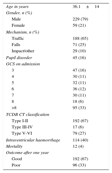
Material and methods

We retrospectively analysed 264 TBI patients to whom a MR had been performed in the first 60 days after trauma. All clinical variables related to prognosis were registered, as well as the data from the initial computed tomography. The MR imaging protocol consisted of a 3-plane localiser sequence T1-weighted and T2-weighted fast spin-echo, FLAIR and gradient-echo images (GRET2*). TAI lesions were classified according to Gentry and Firsching classifications. We calculated weighted kappa coefficients and the area under the ROC curve for each MR sequence. A multivariable analysis was performed to correlate MR findings in each sequence with the final outcome of the patients.
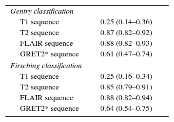
Results

TAI lesions were adequately visualised on T2, FLAIR and GRET2* sequences in more than 80% of the studies. Subcortical TAI lesions were well on FLAIR and GRET2* sequences visualised haemorrhagic TAI lesions. We saw that these MR sequences had a high inter-rater agreement for TAI diagnosis (0.8). T2 sequence presented the highest value on ROC curve in Gentry (0.68, 95%CI: 0.61–0.76, p<0.001, Nagerlkerke-R2 0.26) and Firsching classifications (0.64, 95%CI 0.57–0.72, p<0.001, Nagerlkerke-R2 0.19), followed by FLAIR and GRET2* sequences. Both classifications determined by each of these sequences were associated with poor outcome after performing a multivariable analyses adjusted for prognostic factors (p<0.02).

Conclusions

We recommend to perform conventional MR study in subacute phase including T2, FLAIR and GRET2* sequences for visualise TAI lesions. These MR findings added prognostic information in TBI patients.

Keywords:
Magnetic resonance imaging
Diffuse axonal injury
Traumatic brain injury
Severe head trauma
Resumen
Objetivo

El objetivo de este estudio es determinar qué secuencias de resonancia magnética (RM) convencional diagnostican de manera más sensible las lesiones asociadas a lesiones axonales difusas (LAD) en pacientes con TCE grave.

Material y métodos

Se analizaron de manera retrospectiva los datos de 264 pacientes con TCE grave y RM realizada dentro de las primeras 8 semanas tras el TCE. Se recogieron todas las variables clínicas potencialmente relacionadas con el pronóstico de los enfermos, así como los datos dla tomografía computarizada inicial. A todos los enfermos se les practicó un estudio de RM convencional con secuencias spin-eco potenciadas en T1 y T2, secuencia FLAIR y eco de gradiente T2 (EGRT2*). Las diferentes LAD visualizadas fueron caracterizadas según su localización y clasificadas siguiendo las escalas de Gentry y Firsching en cada secuencia. Se calculó el grado de concordancia entre la clasificación obtenida en las diferentes secuencias y la obtenida de forma definitiva por el paciente, así como el área bajo la curva ROC de cada una de ellas con respecto al pronóstico final de los pacientes.

Resultados

En las secuencias T2, FLAIR y EGRT2* se visualizan las LAD de manera adecuada en más del 80% de los casos. En FLAIR se visualizan mejor las LAD hemisféricas a nivel subcortical y el EGRT2* resalta las LAD hemorrágicas. En nuestra serie hemos visto que el grado de concordancia para diagnosticar LAD entre las secuencias T2, FLAIR y EGRT2* es alto (0,8). La secuencia T2 es la que tuvo un valor más alto en las curvas ROC tanto en la clasificación de Gentry (0,68; IC 95%: 0,61-0,76; p<0,001, Nagerlkerke-R2 0,26) como en la de Firsching (0,64; IC 95%: 0,57-0,72; p<0,001, Nagerlkerke-R2 0,19), seguida de la secuencia FLAIR y de la EGRT2*. Se observó, tras realizar un análisis multivariable, que las clasificaciones de Gentry y Firsching determinadas de forma independiente en cada secuencia se relacionaban con el pronóstico final de los enfermos al año del traumatismo (p<0,02).

Conclusiones

Para el diagnóstico adecuado de LAD en el TCE grave recomendamos la realización de una RM convencional en fase subaguda que incluya al menos las secuencias T2, FLAIR y eco de gradiente en los diferentes planos de corte. Estos hallazgos aumentan el valor pronóstico de los modelos descritos en el TCE grave.

Palabras clave:
Resonancia magnética
Lesión axonal difusa
Lesión cerebral traumática
Traumatismo craneal grave

Article

These are the options to access the full texts of the publication Neurocirugía (English edition)
Member
Member of the Sociedad Española de Neurocirugía

If it is the first time you have accessed you can obtain your credentials by contacting Elsevier Spain in suscripciones@elsevier.com or by calling our Customer Service at902 88 87 40 if you are calling from Spain or at +34 932 418 800 (from 9 to 18h., GMT + 1) if you are calling outside of Spain.

If you already have your login data, please click here .

If you have forgotten your password you can you can recover it by clicking here and selecting the option ¿I have forgotten my password¿.

Subscriber
Subscriber

If you already have your login data, please click here .

If you have forgotten your password you can you can recover it by clicking here and selecting the option “I have forgotten my password”
Subscribe
Subscribe to

Neurocirugía (English edition)

Purchase
Purchase article

Purchasing article the PDF version will be downloaded

Price 19.34 €

Purchase now
Contact
Phone for subscriptions and reporting of errors
From Monday to Friday from 9 a.m. to 6 p.m. (GMT + 1) except for the months of July and August which will be from 9 a.m. to 3 p.m.
Calls from Spain
932 415 960
Calls from outside Spain
+34 932 415 960
Email
Idiomas
Neurocirugía (English edition)
Article options
Tools