To compare the identification capability of traumatic axonal injury (TAI) by different sequences on conventional magnetic resonance (MR) studies in traumatic brain injury (TBI) patients.
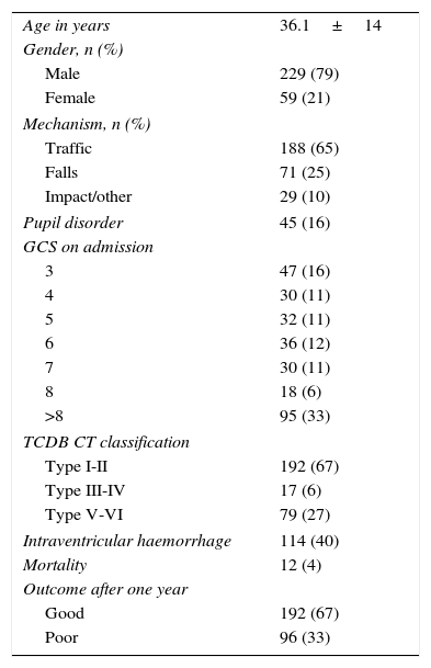
Material and methodsWe retrospectively analysed 264 TBI patients to whom a MR had been performed in the first 60 days after trauma. All clinical variables related to prognosis were registered, as well as the data from the initial computed tomography. The MR imaging protocol consisted of a 3-plane localiser sequence T1-weighted and T2-weighted fast spin-echo, FLAIR and gradient-echo images (GRET2*). TAI lesions were classified according to Gentry and Firsching classifications. We calculated weighted kappa coefficients and the area under the ROC curve for each MR sequence. A multivariable analysis was performed to correlate MR findings in each sequence with the final outcome of the patients.
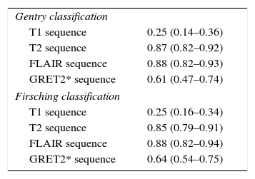
ResultsTAI lesions were adequately visualised on T2, FLAIR and GRET2* sequences in more than 80% of the studies. Subcortical TAI lesions were well on FLAIR and GRET2* sequences visualised haemorrhagic TAI lesions. We saw that these MR sequences had a high inter-rater agreement for TAI diagnosis (0.8). T2 sequence presented the highest value on ROC curve in Gentry (0.68, 95%CI: 0.61–0.76, p<0.001, Nagerlkerke-R2 0.26) and Firsching classifications (0.64, 95%CI 0.57–0.72, p<0.001, Nagerlkerke-R2 0.19), followed by FLAIR and GRET2* sequences. Both classifications determined by each of these sequences were associated with poor outcome after performing a multivariable analyses adjusted for prognostic factors (p<0.02).
ConclusionsWe recommend to perform conventional MR study in subacute phase including T2, FLAIR and GRET2* sequences for visualise TAI lesions. These MR findings added prognostic information in TBI patients.
El objetivo de este estudio es determinar qué secuencias de resonancia magnética (RM) convencional diagnostican de manera más sensible las lesiones asociadas a lesiones axonales difusas (LAD) en pacientes con TCE grave.
Material y métodosSe analizaron de manera retrospectiva los datos de 264 pacientes con TCE grave y RM realizada dentro de las primeras 8 semanas tras el TCE. Se recogieron todas las variables clínicas potencialmente relacionadas con el pronóstico de los enfermos, así como los datos dla tomografía computarizada inicial. A todos los enfermos se les practicó un estudio de RM convencional con secuencias spin-eco potenciadas en T1 y T2, secuencia FLAIR y eco de gradiente T2 (EGRT2*). Las diferentes LAD visualizadas fueron caracterizadas según su localización y clasificadas siguiendo las escalas de Gentry y Firsching en cada secuencia. Se calculó el grado de concordancia entre la clasificación obtenida en las diferentes secuencias y la obtenida de forma definitiva por el paciente, así como el área bajo la curva ROC de cada una de ellas con respecto al pronóstico final de los pacientes.
ResultadosEn las secuencias T2, FLAIR y EGRT2* se visualizan las LAD de manera adecuada en más del 80% de los casos. En FLAIR se visualizan mejor las LAD hemisféricas a nivel subcortical y el EGRT2* resalta las LAD hemorrágicas. En nuestra serie hemos visto que el grado de concordancia para diagnosticar LAD entre las secuencias T2, FLAIR y EGRT2* es alto (0,8). La secuencia T2 es la que tuvo un valor más alto en las curvas ROC tanto en la clasificación de Gentry (0,68; IC 95%: 0,61-0,76; p<0,001, Nagerlkerke-R2 0,26) como en la de Firsching (0,64; IC 95%: 0,57-0,72; p<0,001, Nagerlkerke-R2 0,19), seguida de la secuencia FLAIR y de la EGRT2*. Se observó, tras realizar un análisis multivariable, que las clasificaciones de Gentry y Firsching determinadas de forma independiente en cada secuencia se relacionaban con el pronóstico final de los enfermos al año del traumatismo (p<0,02).
ConclusionesPara el diagnóstico adecuado de LAD en el TCE grave recomendamos la realización de una RM convencional en fase subaguda que incluya al menos las secuencias T2, FLAIR y eco de gradiente en los diferentes planos de corte. Estos hallazgos aumentan el valor pronóstico de los modelos descritos en el TCE grave.
Article

If it is the first time you have accessed you can obtain your credentials by contacting Elsevier Spain in suscripciones@elsevier.com or by calling our Customer Service at902 88 87 40 if you are calling from Spain or at +34 932 418 800 (from 9 to 18h., GMT + 1) if you are calling outside of Spain.
If you already have your login data, please click here .
If you have forgotten your password you can you can recover it by clicking here and selecting the option ¿I have forgotten my password¿.


