array:23 [
  "pii" => "S2529849622000600"
  "issn" => "25298496"
  "doi" => "10.1016/j.neucie.2021.06.007"
  "estado" => "S300"
  "fechaPublicacion" => "2022-11-01"
  "aid" => "510"
  "copyright" => "Sociedad Española de Neurocirugía"
  "copyrightAnyo" => "2021"
  "documento" => "simple-article"
  "crossmark" => 1
  "subdocumento" => "crp"
  "cita" => "Neurocirugia. 2022;33:350-5"
  "abierto" => array:3 [
    "ES" => false
    "ES2" => false
    "LATM" => false
  ]
  "gratuito" => false
  "lecturas" => array:1 [
    "total" => 0
  ]
  "itemSiguiente" => array:18 [
    "pii" => "S2529849622000612"
    "issn" => "25298496"
    "doi" => "10.1016/j.neucie.2021.06.008"
    "estado" => "S300"
    "fechaPublicacion" => "2022-11-01"
    "aid" => "515"
    "copyright" => "Sociedad Española de Neurocirugía"
    "documento" => "simple-article"
    "crossmark" => 1
    "subdocumento" => "crp"
    "cita" => "Neurocirugia. 2022;33:356-60"
    "abierto" => array:3 [
      "ES" => false
      "ES2" => false
      "LATM" => false
    ]
    "gratuito" => false
    "lecturas" => array:1 [
      "total" => 0
    ]
    "en" => array:13 [
      "idiomaDefecto" => true
      "cabecera" => "<span class="elsevierStyleTextfn">Case Report</span>"
      "titulo" => "Molecular and clinical characterization of H3 K27M-mutant &#8220;non-midline&#8221; glioblastoma&#58; A case report and literature review"
      "tienePdf" => "en"
      "tieneTextoCompleto" => "en"
      "tieneResumen" => array:2 [
        0 => "en"
        1 => "es"
      ]
      "paginas" => array:1 [
        0 => array:2 [
          "paginaInicial" => "356"
          "paginaFinal" => "360"
        ]
      ]
      "titulosAlternativos" => array:1 [
        "es" => array:1 [
          "titulo" => "Estudio molecular y presentaci&#243;n cl&#237;nica del glioblastoma con mutaci&#243;n H3 K27M &#171;no de l&#237;nea media&#187;&#58; descripci&#243;n de un caso y revisi&#243;n de la literatura"
        ]
      ]
      "contieneResumen" => array:2 [
        "en" => true
        "es" => true
      ]
      "contieneTextoCompleto" => array:1 [
        "en" => true
      ]
      "contienePdf" => array:1 [
        "en" => true
      ]
      "resumenGrafico" => array:2 [
        "original" => 0
        "multimedia" => array:7 [
          "identificador" => "fig0010"
          "etiqueta" => "Fig&#46; 2"
          "tipo" => "MULTIMEDIAFIGURA"
          "mostrarFloat" => true
          "mostrarDisplay" => false
          "figura" => array:1 [
            0 => array:4 [
              "imagen" => "gr2.jpeg"
              "Alto" => 615
              "Ancho" => 1500
              "Tamanyo" => 298736
            ]
          ]
          "descripcion" => array:1 [
            "en" => "<p id="spar0020" class="elsevierStyleSimplePara elsevierViewall">HE staining showed hypercellular tumor with marked nuclear pleomorphism&#44; high mitotic activity and necrosis &#40;A&#41;&#46; Immunostaining demonstrated positive for H3 K27M &#40;B&#41;&#44; MMP-9 &#40;E&#41; antibodies&#46; And the tumor was negative for H3 K27me3 &#40;C&#41;&#44; H3&#46;3 G34R &#40;D&#41;&#46;</p>"
          ]
        ]
      ]
      "autores" => array:1 [
        0 => array:2 [
          "autoresLista" => "Shumpei Onishi, Shinji Ohba, Kazuya Kuraoka, Takashi Kurashige, Kazuhiko Sugiyama, Fumiyuki Yamasaki"
          "autores" => array:6 [
            0 => array:2 [
              "nombre" => "Shumpei"
              "apellidos" => "Onishi"
            ]
            1 => array:2 [
              "nombre" => "Shinji"
              "apellidos" => "Ohba"
            ]
            2 => array:2 [
              "nombre" => "Kazuya"
              "apellidos" => "Kuraoka"
            ]
            3 => array:2 [
              "nombre" => "Takashi"
              "apellidos" => "Kurashige"
            ]
            4 => array:2 [
              "nombre" => "Kazuhiko"
              "apellidos" => "Sugiyama"
            ]
            5 => array:2 [
              "nombre" => "Fumiyuki"
              "apellidos" => "Yamasaki"
            ]
          ]
        ]
      ]
    ]
    "idiomaDefecto" => "en"
    "EPUB" => "https://multimedia.elsevier.es/PublicationsMultimediaV1/item/epub/S2529849622000612?idApp=UINPBA00004B"
    "url" => "/25298496/0000003300000006/v3_202212060641/S2529849622000612/v3_202212060641/en/main.assets"
  ]
  "itemAnterior" => array:18 [
    "pii" => "S2529849622000594"
    "issn" => "25298496"
    "doi" => "10.1016/j.neucie.2021.04.010"
    "estado" => "S300"
    "fechaPublicacion" => "2022-11-01"
    "aid" => "504"
    "copyright" => "Sociedad Espa&#241;ola de Neurocirug&#237;a"
    "documento" => "simple-article"
    "crossmark" => 1
    "subdocumento" => "crp"
    "cita" => "Neurocirugia. 2022;33:345-9"
    "abierto" => array:3 [
      "ES" => false
      "ES2" => false
      "LATM" => false
    ]
    "gratuito" => false
    "lecturas" => array:1 [
      "total" => 0
    ]
    "en" => array:13 [
      "idiomaDefecto" => true
      "cabecera" => "<span class="elsevierStyleTextfn">Case Report</span>"
      "titulo" => "Discrepancy in the Ki67 labeling index of brain and orbital metastatic lesions from gastrointestinal neuroendocrine tumors&#58; A case report"
      "tienePdf" => "en"
      "tieneTextoCompleto" => "en"
      "tieneResumen" => array:2 [
        0 => "en"
        1 => "es"
      ]
      "paginas" => array:1 [
        0 => array:2 [
          "paginaInicial" => "345"
          "paginaFinal" => "349"
        ]
      ]
      "titulosAlternativos" => array:1 [
        "es" => array:1 [
          "titulo" => "Discrepancia en el &#237;ndice de etiquetado Ki67 de las lesiones metast&#225;sicas cerebrales y orbitales de los tumores neuroendocrinos gastrointestinales&#58; Un informe de caso"
        ]
      ]
      "contieneResumen" => array:2 [
        "en" => true
        "es" => true
      ]
      "contieneTextoCompleto" => array:1 [
        "en" => true
      ]
      "contienePdf" => array:1 [
        "en" => true
      ]
      "resumenGrafico" => array:2 [
        "original" => 0
        "multimedia" => array:7 [
          "identificador" => "fig0010"
          "etiqueta" => "Fig&#46; 2"
          "tipo" => "MULTIMEDIAFIGURA"
          "mostrarFloat" => true
          "mostrarDisplay" => false
          "figura" => array:1 [
            0 => array:4 [
              "imagen" => "gr2.jpeg"
              "Alto" => 427
              "Ancho" => 1405
              "Tamanyo" => 190583
            ]
          ]
          "descripcion" => array:1 [
            "en" => "<p id="spar0020" class="elsevierStyleSimplePara elsevierViewall">Histopathological examination of the resected brain &#40;A&#8211;E&#41; and orbital &#40;F&#8211;J&#41; tumor&#46; Hematoxylin&#8211;eosin staining&#46; Proliferating monotonous cells with round and bland nuclei in cord&#44; nest&#44; rosette&#44; or tubular structures along with fibrous stroma are observed &#40;A&#41;&#46; The tumor cells are positive for CD56 &#40;B&#41;&#44; synaptophysin &#40;C&#41;&#44; and chromogranin A &#40;D&#41;&#46; The Ki67 labeling index is 8&#37; &#40;E&#41;&#46; &#40;F&#41; Hematoxylin&#8211;eosin staining&#46; Invasive proliferation of atypical small cells with hyperchromatic nuclei and scant cytoplasm arranged in a solid nest are observed&#46; The tumor cells are positive for CD56 &#40;G&#41; and synaptophysin &#40;H&#41; and negative for chromogranin A &#40;I&#41;&#46; The Ki67 labeling index is 22&#37; &#40;J&#41;&#46;</p>"
          ]
        ]
      ]
      "autores" => array:1 [
        0 => array:2 [
          "autoresLista" => "Satoshi Matsuo, Toshiyuki Amano, Yuichiro Miyamatsu, Daisuke Hayashi, Sojiro Yamashita, Seiya Momosaki, Ken Kawabe, Akira Nakamizo"
          "autores" => array:8 [
            0 => array:2 [
              "nombre" => "Satoshi"
              "apellidos" => "Matsuo"
            ]
            1 => array:2 [
              "nombre" => "Toshiyuki"
              "apellidos" => "Amano"
            ]
            2 => array:2 [
              "nombre" => "Yuichiro"
              "apellidos" => "Miyamatsu"
            ]
            3 => array:2 [
              "nombre" => "Daisuke"
              "apellidos" => "Hayashi"
            ]
            4 => array:2 [
              "nombre" => "Sojiro"
              "apellidos" => "Yamashita"
            ]
            5 => array:2 [
              "nombre" => "Seiya"
              "apellidos" => "Momosaki"
            ]
            6 => array:2 [
              "nombre" => "Ken"
              "apellidos" => "Kawabe"
            ]
            7 => array:2 [
              "nombre" => "Akira"
              "apellidos" => "Nakamizo"
            ]
          ]
        ]
      ]
    ]
    "idiomaDefecto" => "en"
    "EPUB" => "https://multimedia.elsevier.es/PublicationsMultimediaV1/item/epub/S2529849622000594?idApp=UINPBA00004B"
    "url" => "/25298496/0000003300000006/v3_202212060641/S2529849622000594/v3_202212060641/en/main.assets"
  ]
  "en" => array:19 [
    "idiomaDefecto" => true
    "cabecera" => "<span class="elsevierStyleTextfn">Case Report</span>"
    "titulo" => "Primary central nervous system Hodgkin lymphoma&#58; A case report and review of the literature"
    "tieneTextoCompleto" => true
    "paginas" => array:1 [
      0 => array:2 [
        "paginaInicial" => "350"
        "paginaFinal" => "355"
      ]
    ]
    "autores" => array:1 [
      0 => array:4 [
        "autoresLista" => "David Aliaga, Juan Mayorga, Franscisco Verd&#250;-L&#243;pez, Jos&#233; Mar&#237;a Gallego, Laura Castell&#225;, Vicente Sabater"
        "autores" => array:6 [
          0 => array:4 [
            "nombre" => "David"
            "apellidos" => "Aliaga"
            "email" => array:1 [
              0 => "Aliagac0810@gmail.com"
            ]
            "referencia" => array:2 [
              0 => array:2 [
                "etiqueta" => "<span class="elsevierStyleSup">a</span>"
                "identificador" => "aff0005"
              ]
              1 => array:2 [
                "etiqueta" => "<span class="elsevierStyleSup">&#42;</span>"
                "identificador" => "cor0005"
              ]
            ]
          ]
          1 => array:3 [
            "nombre" => "Juan"
            "apellidos" => "Mayorga"
            "referencia" => array:1 [
              0 => array:2 [
                "etiqueta" => "<span class="elsevierStyleSup">a</span>"
                "identificador" => "aff0005"
              ]
            ]
          ]
          2 => array:3 [
            "nombre" => "Franscisco"
            "apellidos" => "Verd&#250;-L&#243;pez"
            "referencia" => array:1 [
              0 => array:2 [
                "etiqueta" => "<span class="elsevierStyleSup">a</span>"
                "identificador" => "aff0005"
              ]
            ]
          ]
          3 => array:3 [
            "nombre" => "Jos&#233; Mar&#237;a"
            "apellidos" => "Gallego"
            "referencia" => array:1 [
              0 => array:2 [
                "etiqueta" => "<span class="elsevierStyleSup">a</span>"
                "identificador" => "aff0005"
              ]
            ]
          ]
          4 => array:3 [
            "nombre" => "Laura"
            "apellidos" => "Castell&#225;"
            "referencia" => array:1 [
              0 => array:2 [
                "etiqueta" => "<span class="elsevierStyleSup">b</span>"
                "identificador" => "aff0010"
              ]
            ]
          ]
          5 => array:3 [
            "nombre" => "Vicente"
            "apellidos" => "Sabater"
            "referencia" => array:1 [
              0 => array:2 [
                "etiqueta" => "<span class="elsevierStyleSup">b</span>"
                "identificador" => "aff0010"
              ]
            ]
          ]
        ]
        "afiliaciones" => array:2 [
          0 => array:3 [
            "entidad" => "Department of Neurosurgery&#44; Consorcio Hospital General Universitario de Valencia&#44; 46014 Valencia&#44; Spain"
            "etiqueta" => "a"
            "identificador" => "aff0005"
          ]
          1 => array:3 [
            "entidad" => "Department of Diagnostic Pathology&#44; Consorcio Hospital General Universitario de Valencia&#44; 46014 Valencia&#44; Spain"
            "etiqueta" => "b"
            "identificador" => "aff0010"
          ]
        ]
        "correspondencia" => array:1 [
          0 => array:3 [
            "identificador" => "cor0005"
            "etiqueta" => "&#8270;"
            "correspondencia" => "<span class="elsevierStyleItalic">Corresponding author</span>&#46;"
          ]
        ]
      ]
    ]
    "titulosAlternativos" => array:1 [
      "es" => array:1 [
        "titulo" => "Linfoma de Hodgkin primario del sistema nervioso central&#46; Presentaci&#243;n de un caso y revisi&#243;n de la literatura"
      ]
    ]
    "resumenGrafico" => array:2 [
      "original" => 0
      "multimedia" => array:7 [
        "identificador" => "fig0015"
        "etiqueta" => "Fig&#46; 3"
        "tipo" => "MULTIMEDIAFIGURA"
        "mostrarFloat" => true
        "mostrarDisplay" => false
        "figura" => array:1 [
          0 => array:4 [
            "imagen" => "gr3.jpeg"
            "Alto" => 797
            "Ancho" => 1505
            "Tamanyo" => 308123
          ]
        ]
        "descripcion" => array:1 [
          "en" => "<p id="spar0045" class="elsevierStyleSimplePara elsevierViewall">&#40;A&#41; Immunostaining for CD45 stains non-neoplastic bystander lymphoid cells&#44; whereas the neoplastic RS cells remain unlabelled &#40;&#8593;&#41;&#46; &#40;B&#41; CD30 is typically present in a membrane pattern&#44; with accentuation in the Golgi area of the cytoplasm &#40;&#8593;&#41; &#40;perinuclear dot-like staining&#41;&#46; &#40;C&#41; CD15 is selectively expressed in neoplastic cells &#40;&#9652;&#41;&#46; &#40;D&#41; Immunostaining for PAX5 labels the nuclei of the RS cells &#40;&#9652; and those of the non-neoplastic by-stander B cells &#40;&#8593;&#41;&#46; &#40;E&#41; Ki-67 is positive in RS cells&#46; &#40;F&#41; CD163 highlights the clusters of foamy histiocytes &#40;&#42;&#41; next to a negative RS cell &#40;&#8593;&#41;&#46;</p>"
        ]
      ]
    ]
    "textoCompleto" => "<span class="elsevierStyleSections"><span id="sec0005" class="elsevierStyleSection elsevierViewall"><span class="elsevierStyleSectionTitle" id="sect0030">Introduction</span><p id="par0005" class="elsevierStylePara elsevierViewall">CNS involvement in HL is quite rare&#44; with an estimated frequency of 0&#46;02&#8211;0&#46;07&#37;&#46;<a class="elsevierStyleCrossRefs" href="#bib0110"><span class="elsevierStyleSup">1&#8211;3</span></a> Even though CNS involvement has historically been considered to occur in the context of relapsed&#47;refractory disease&#44; recent studies have shown that primary and isolated CNS involvement could be the clinical onset in a significant number of patients that could even present a better prognosis&#46;<a class="elsevierStyleCrossRef" href="#bib0125"><span class="elsevierStyleSup">4</span></a> Nevertheless&#44; primary CNS&#8211;HL is exceedingly rare&#44; with just over twenty cases reported in the literature&#46; The diagnosis of primary CNS&#8211;HL is based on well-defined morphological and immunohistochemical characteristics&#44; in addition to staging studies demonstrating a lack of disease elsewhere&#46;</p><p id="par0010" class="elsevierStylePara elsevierViewall">We present the case of a 55-year-old woman with isolated primary right frontal intracranial HL&#44; who underwent surgical resection followed by intrathecal chemotherapy and WBRT&#46; The literature on primary CNS&#8211;HL is also reviewed&#46;</p></span><span id="sec0010" class="elsevierStyleSection elsevierViewall"><span class="elsevierStyleSectionTitle" id="sect0035">Case report</span><p id="par0015" class="elsevierStylePara elsevierViewall">A 55-year-old immunocompetent woman&#44; with a medical history of juvenile dermatomyositis&#44; Sj&#246;gren syndrome and histologic diagnosis of axillary and inguinal reactive lymphadenitis&#44; three years before this episode&#44; presented with subacute confusion&#44; bradypsychia&#44; gait instability&#44; loss of appetite and fatigue&#46; Neurological assessment showed left-sided hemiparesis&#46; No evidence of peripheral lymphadenopathy or hepatosplenomegaly was noted on the physical examination&#46; Routine laboratory analysis showed no abnormal findings&#46;</p><p id="par0020" class="elsevierStylePara elsevierViewall">She underwent computed tomography &#40;CT&#41; and magnetic resonance imaging &#40;MRI&#41; scans of her brain&#44; which showed a 31<span class="elsevierStyleHsp" style=""></span>&#215;<span class="elsevierStyleHsp" style=""></span>34<span class="elsevierStyleHsp" style=""></span>mm dural-based&#44; contrast-enhancing right frontal lesion with significant surrounding vasogenic edema and mass effect&#44; and without restricted diffusion &#40;<a class="elsevierStyleCrossRef" href="#fig0005">Fig&#46; 1</a>&#41;&#46; She was started on Dexamethasone 4<span class="elsevierStyleHsp" style=""></span>mg every 8<span class="elsevierStyleHsp" style=""></span>h&#44; showing progressive clinical improvement&#46; CT scans of the chest&#44; abdomen and pelvis were performed to investigate for a primary source of malignancy&#46; Nevertheless&#44; the results of the tests were negative&#46; At this point&#44; meningioma and intracranial metastasis were the two main options for the differential diagnosis&#46;</p><elsevierMultimedia ident="fig0005"></elsevierMultimedia><p id="par0025" class="elsevierStylePara elsevierViewall">The patient underwent a right frontal craniotomy for gross total resection &#40;<a class="elsevierStyleCrossRef" href="#fig0005">Fig&#46; 1</a>&#41;&#46; Histologic sections showed a mixed proliferative tissue with lymphocytes&#44; plasma cells and histiocytes&#44; together with a large background of coagulative necrosis and fibrosis&#46; Large multinuclear cells composed of pale chromatin&#44; prominent eosinophilic nucleoli and moderate cytoplasm&#44; consistent with Reed&#8211;Sternberg &#40;RS&#41; cells&#44; were present&#46; An occasional mononuclear RS variant&#44; referred to as a &#8220;Hodgkin cell&#8221; and &#8220;lacunar cell&#8221; &#40;retracted cytoplasm&#41; were also seen&#46; Immunohistochemical studies confirmed the RS cells to be immunoreactive for CD15 and CD30&#44; although negative for CD45 and CD20&#46; The histiocytes were highlighted by a CD163 stain&#46; Scattered CD38-positive plasma cells were present&#44; primarily at the periphery of the lesion&#46; Polymerase chain reaction for Epstein&#8211;Barr virus &#40;EBV&#41; was positive in the RS cells &#40;<a class="elsevierStyleCrossRefs" href="#fig0010">Figs&#46; 2 and 3</a>&#41;&#46;</p><elsevierMultimedia ident="fig0010"></elsevierMultimedia><elsevierMultimedia ident="fig0015"></elsevierMultimedia><p id="par0030" class="elsevierStylePara elsevierViewall">After the histological examination was concluded&#44; our patient underwent a new brain and spinal MRI&#44; which showed neither brain tumoral remainder&#44; nor spinal involvement&#46; A lumbar puncture revealed normal opening pressure&#44; protein&#44; and glucose&#46; Cerebrospinal fluid cytology was negative and cell counts were within normal limits&#46; A bone marrow biopsy presented as normocellular&#44; without malignant cells or fibrosis&#46; This result was confirmed with immunohistochemical studies&#46; Positron emission tomography-computed tomography showed no systemic involvement&#46;</p><p id="par0035" class="elsevierStylePara elsevierViewall">Our patient was diagnosed with a nodular sclerosing variant of a primary CNS classic HL&#46; Based on neuro-oncology committee consensus and on available data&#44; the patient was given post surgically prophylactic intrathecal chemotherapy &#40;hydrocortisone&#44; methotrexate and cytarabine&#41; followed by WBRT &#40;30 Gray&#41; and 36 Gray boost at the surgical field&#44; showing fourteen months of disease-free survival at the time of this case report &#40;currently the patient is still in follow-up&#41;&#46;</p></span><span id="sec0015" class="elsevierStyleSection elsevierViewall"><span class="elsevierStyleSectionTitle" id="sect0040">Discussion</span><p id="par0040" class="elsevierStylePara elsevierViewall">Although uncommon&#44; secondary involvement of the CNS in HL is a known complication&#44; occurring in 0&#46;02&#8211;0&#46;07&#37; of patients&#46;<a class="elsevierStyleCrossRefs" href="#bib0110"><span class="elsevierStyleSup">1&#8211;3</span></a> However&#44; primary CNS&#8211;HL is even more rare&#44; with just about 20 cases reported in the literature&#46; The diagnosis of primary CNS&#8211;HL is based on distinct morphological and immunohistochemical features&#44; as shown in <a class="elsevierStyleCrossRefs" href="#fig0010">Figs&#46; 2 and 3</a>&#44; in addition to isolated CNS involvement&#46;</p><p id="par0045" class="elsevierStylePara elsevierViewall">Scarcity of reported data hinders&#44; to a large extent&#44; the possibility to accurately define some features&#44; such as the etiology&#44; the demographic distribution or the clinical presentation&#44; as well as&#44; the ability to reach a consensus regarding optimum treatment&#46;</p><p id="par0050" class="elsevierStylePara elsevierViewall">It has been claimed that this entity could be the result of the interaction between genetic &#40;based on the results of twin concordance studies and the identification of familial forms of HL&#41;<a class="elsevierStyleCrossRefs" href="#bib0130"><span class="elsevierStyleSup">5&#44;6</span></a> and environmental factors &#40;although HIV and EBV have generally been noted to play a role as risk factors in primary CNS lymphomas&#44; the association has not been definitively established in primary CNS&#8211;HL&#41;&#46;<a class="elsevierStyleCrossRef" href="#bib0125"><span class="elsevierStyleSup">4</span></a></p><p id="par0055" class="elsevierStylePara elsevierViewall">Patients suffering from autoimmune diseases have been recognized to be at increased risk of developing HL for several years&#46; In a recent large population-based case-controlled study in Denmark and Sweden&#44; Landgren and colleagues<a class="elsevierStyleCrossRef" href="#bib0140"><span class="elsevierStyleSup">7</span></a> concluded that significant increased risk of HL was associated with personal histories of several autoimmune conditions&#44; primarily those characterized by systemic involvement&#46; These included rheumatoid arthritis&#44; systemic lupus erythematosus&#44; celiac disease&#44; scleroderma and Sjogren&#39;s disease&#46; The latter and dermatomyositis were present in our case report&#46;</p><p id="par0060" class="elsevierStylePara elsevierViewall">Additionally&#44; it has been suggested that primary CNS&#8211;HL could be related to the number and activity of T-reg cells&#46;<a class="elsevierStyleCrossRef" href="#bib0145"><span class="elsevierStyleSup">8</span></a></p><p id="par0065" class="elsevierStylePara elsevierViewall">It has been argued that overactivation of T-reg cells&#44; or increase in their number&#44; may weaken the immune response and antitumor immunity and might favor tumor development or growth&#46;<a class="elsevierStyleCrossRefs" href="#bib0150"><span class="elsevierStyleSup">9&#44;10</span></a> The high ratio of T-reg cells in the microenvironment around RS cells over T-helper 2 cells would be associated with significantly shortened disease-free survival&#44; while a low ratio would improve disease-free survival&#46;<a class="elsevierStyleCrossRef" href="#bib0160"><span class="elsevierStyleSup">11</span></a> The role of immunosuppression and EBV infection in systemic and primary CNS&#8211;HL has also been suggested&#46; The presence of EBV in RS cells would correlate with increased T-reg cells in the tumor microenvironment and would decrease the antitumor immunity toward RS cells&#44; even though it would not impact the survival in these patients&#46;<a class="elsevierStyleCrossRef" href="#bib0165"><span class="elsevierStyleSup">12</span></a></p><p id="par0070" class="elsevierStylePara elsevierViewall">On neuroimaging&#44; primary CNS&#8211;HL tends to appear as a well-delimited intraparenchymal hipodense to hyperdense lesion on CT&#44; and usually show homogeneous contrast enhancement&#46; Nevertheless&#44; there are a few cases of ring- enhancing masses&#46;</p><p id="par0075" class="elsevierStylePara elsevierViewall">On MRI&#44; lesions usually show as well-delimited hipointense to isointense masses on T1 and T2-weighted images and almost all lesions show homogeneous enhancement with contrast material&#46; It is important to emphasize that many of these lesions show significant surrounding vasogenic edema&#44; so that added to the latter MRI characteristics&#44; it is important to consider CNS metastasis in the differential diagnosis&#46;</p><p id="par0080" class="elsevierStylePara elsevierViewall">As mentioned by Clark et al&#46;&#44;<a class="elsevierStyleCrossRef" href="#bib0170"><span class="elsevierStyleSup">13</span></a> although Hodgkin&#39;s disease occasionally spreads into the brain&#44; where it may involve the dura and leptomeninges&#44; producing a firm&#44; circumscribed dural-based tumor with radiographic and intraoperative features of a meningioma&#44; a meningioma-like presentation is rare&#46; It is important to point out that only 4 out of 23 cases of primary CNS&#8211;HL presented as dural based lesions&#46;<a class="elsevierStyleCrossRefs" href="#bib0125"><span class="elsevierStyleSup">4&#44;14&#8211;16</span></a> Thereby&#44; our case is the fifth documented primary CNS&#8211;HL with radiographic and intraoperative features of a meningioma&#46;</p><p id="par0085" class="elsevierStylePara elsevierViewall">Because of the rarity of intracranial Hodgkin&#39;s lymphoma&#44; and because it shares radiographic similarities with other entities such as meningiomas and metastasis&#44; an accurate histological and immunohistochemical analysis is always required&#46;</p><p id="par0090" class="elsevierStylePara elsevierViewall">In summarizing the demographics of this case series<a class="elsevierStyleCrossRefs" href="#bib0125"><span class="elsevierStyleSup">4&#44;8&#44;15&#44;17&#8211;19</span></a> &#40;23 cases of primary CNS&#8211;HL reported in the available literature&#41;&#44; there seems to be a slight male preponderance with 56&#46;5&#37; &#40;13&#47;23&#41; being male&#44; and a median age of 55 years &#40;range 34&#8211;84&#44; mean 58&#41;&#46; Clinical presentation shows wide variability&#44; and it primarily depends on the location of the mass&#46; The most common presenting symptoms included headache &#40;43&#37;&#59; 10&#47;23&#41;&#44; weakness &#40;30&#37;&#59; 7&#47;23&#41; and seizures &#40;17&#37;&#59; 4&#47;23&#41;&#46; About 52&#37; of primary CNS&#8211;HL cases were supratentorial in origin &#40;12&#47;23&#41;&#44; with only 34&#37; &#40;8&#47;23&#41; of documented lesions located infratentorially&#46; One of the three remaining cases presented simultaneously in both compartments&#44; while there is no available data regarding the other two cases&#46; The most common subtype of HL was nodular sclerosis&#46; Previously reported cases in this series describe various forms of treatment&#44; including surgical resection with radiotherapy &#40;57&#37;&#59; 13&#47;23&#41;&#44; as well as&#44; surgical resection with combined chemotherapy and radiotherapy &#40;39&#37;&#59; 9&#47;23&#41;&#46; The remaining patient was treated with surgical resection alone &#40;patient died five days after surgery&#41;&#46;</p><p id="par0095" class="elsevierStylePara elsevierViewall">A crucial factor that hinders the analysis of reported data&#44; relates to the limited patient follow up after the time that each case is published&#46; Among patients that received adjuvant treatment with radiotherapy alone&#44; disease-free survival at the time of case report was achieved by 85&#37; of patients &#40;11&#47;13&#41;&#44; with a mean follow-up time of 38 months &#40;range 1&#8211;120&#41;&#46; One of the two remaining patients was diagnosed with recurrence and disseminated advanced lesions after 14 months of disease-free survival&#44; while there is no available data regarding the other patient&#46; Among patients that received adjuvant treatment with chemotherapy plus radiotherapy&#44; disease-free survival at the time of case report was achieved by 78&#37; of patients &#40;7&#47;9&#41;&#44; with a mean follow-up time of 25 months &#40;range 12&#8211;84&#41;&#46; Among the latter group of patients&#44; one of them died after 13 months of follow up &#40;meningitis&#47;pneumonia&#41;&#44; despite being in complete remission and without evidence of disease at the time of the death&#46; There is no available data regarding the two remaining patients &#40;22&#37;&#41;&#46;</p><p id="par0100" class="elsevierStylePara elsevierViewall">Unlike non-Hodgkin lymphoma&#44; surgery seems to play an important role regarding prognosis&#46; Dujovny et al&#46;<a class="elsevierStyleCrossRef" href="#bib0205"><span class="elsevierStyleSup">20</span></a> recognized the value of surgical intervention for intracranial Hodgkin&#39;s disease&#46; Their review showed that the average postoperative survival time was measured in years as opposed to months for all other treatments&#46; However&#44; the longest duration of survival in the series of Dujovny et al&#46; was obtained when surgical removal was combined with radiation therapy&#46;</p><p id="par0105" class="elsevierStylePara elsevierViewall">Because of the radiosensitivity of Hodgkin&#39;s disease and other malignant lymphomas&#44; irradiation is the preferred adjunctive treatment for CNS involvement in Hodgkin&#39;s disease&#46; Whole brain irradiation of 20&#8211;30<span class="elsevierStyleHsp" style=""></span>Gy supplemented by a local boost field to bring the dose to known sites of intracranial involvement from 40 to 50<span class="elsevierStyleHsp" style=""></span>Gy has been recommended for patients presenting with intracranial disease&#46;<a class="elsevierStyleCrossRef" href="#bib0210"><span class="elsevierStyleSup">21</span></a></p><p id="par0110" class="elsevierStylePara elsevierViewall">Despite limited follow up in the majority of cases&#44; there seems to be good prognosis regarding patients diagnosed with primary CNS&#8211;HL that received treatment with surgery plus adjuvant radiotherapy alone&#44; or adjuvant radiotherapy and chemotherapy&#46;<a class="elsevierStyleCrossRef" href="#bib0180"><span class="elsevierStyleSup">15</span></a></p></span><span id="sec0020" class="elsevierStyleSection elsevierViewall"><span class="elsevierStyleSectionTitle" id="sect0045">Conclusions</span><p id="par0115" class="elsevierStylePara elsevierViewall">Primary CNS&#8211;HL is a remarkably rare entity&#44; which diagnosis is based on well-defined morphological and immunohistochemical characteristics&#44; in addition to staging studies demonstrating a lack of disease elsewhere&#46; In spite of limited casuistry&#44; CNS&#8211;HL appears to have good prognosis when treated with surgery plus adjuvant radiotherapy alone&#44; or adjuvant chemotherapy and radiotherapy&#44; yet additional cases are needed for reliable prognostic data&#46;</p></span><span id="sec0025" class="elsevierStyleSection elsevierViewall"><span class="elsevierStyleSectionTitle" id="sect0050">Conflict of interest</span><p id="par0120" class="elsevierStylePara elsevierViewall">The authors declare they have no conflict of interest&#46;</p></span><span id="sec0030" class="elsevierStyleSection elsevierViewall"><span class="elsevierStyleSectionTitle" id="sect0055">Patient consent</span><p id="par0125" class="elsevierStylePara elsevierViewall">The patient has consented to the submission of the case report for submission to the journal&#46;</p></span></span>"
    "textoCompletoSecciones" => array:1 [
      "secciones" => array:12 [
        0 => array:3 [
          "identificador" => "xres1814922"
          "titulo" => "Abstract"
          "secciones" => array:1 [
            0 => array:1 [
              "identificador" => "abst0005"
            ]
          ]
        ]
        1 => array:2 [
          "identificador" => "xpalclavsec1584567"
          "titulo" => "Keywords"
        ]
        2 => array:2 [
          "identificador" => "xpalclavsec1584566"
          "titulo" => "Abbreviations"
        ]
        3 => array:3 [
          "identificador" => "xres1814923"
          "titulo" => "Resumen"
          "secciones" => array:1 [
            0 => array:1 [
              "identificador" => "abst0010"
            ]
          ]
        ]
        4 => array:2 [
          "identificador" => "xpalclavsec1584565"
          "titulo" => "Palabras clave"
        ]
        5 => array:2 [
          "identificador" => "sec0005"
          "titulo" => "Introduction"
        ]
        6 => array:2 [
          "identificador" => "sec0010"
          "titulo" => "Case report"
        ]
        7 => array:2 [
          "identificador" => "sec0015"
          "titulo" => "Discussion"
        ]
        8 => array:2 [
          "identificador" => "sec0020"
          "titulo" => "Conclusions"
        ]
        9 => array:2 [
          "identificador" => "sec0025"
          "titulo" => "Conflict of interest"
        ]
        10 => array:2 [
          "identificador" => "sec0030"
          "titulo" => "Patient consent"
        ]
        11 => array:1 [
          "titulo" => "References"
        ]
      ]
    ]
    "pdfFichero" => "main.pdf"
    "tienePdf" => true
    "fechaRecibido" => "2021-01-19"
    "fechaAceptado" => "2021-06-01"
    "PalabrasClave" => array:2 [
      "en" => array:2 [
        0 => array:4 [
          "clase" => "keyword"
          "titulo" => "Keywords"
          "identificador" => "xpalclavsec1584567"
          "palabras" => array:6 [
            0 => "Hodgkin lymphoma"
            1 => "Primary lymphoma"
            2 => "Central nervous system"
            3 => "Brain neoplasm"
            4 => "Histology"
            5 => "Immunohistochemistry"
          ]
        ]
        1 => array:4 [
          "clase" => "abr"
          "titulo" => "Abbreviations"
          "identificador" => "xpalclavsec1584566"
          "palabras" => array:7 [
            0 => "HL"
            1 => "CNS"
            2 => "WBRT"
            3 => "CT"
            4 => "MRI"
            5 => "RS"
            6 => "EBV"
          ]
        ]
      ]
      "es" => array:1 [
        0 => array:4 [
          "clase" => "keyword"
          "titulo" => "Palabras clave"
          "identificador" => "xpalclavsec1584565"
          "palabras" => array:6 [
            0 => "Linfoma de Hodgkin"
            1 => "Linfoma primario"
            2 => "Sistema nervioso central"
            3 => "Tumor cerebral"
            4 => "Histolog&#237;a"
            5 => "Inmunohistoqu&#237;mica"
          ]
        ]
      ]
    ]
    "tieneResumen" => true
    "resumen" => array:2 [
      "en" => array:2 [
        "titulo" => "Abstract"
        "resumen" => "<span id="abst0005" class="elsevierStyleSection elsevierViewall"><p id="spar0005" class="elsevierStyleSimplePara elsevierViewall">Central nervous system &#40;CNS&#41; involvement in the context of relapsed&#47;refractory Hodgkin lymphoma &#40;HL&#41; is a quite rare&#44; but well-known complication&#46; Nevertheless&#44; primary CNS&#8211;HL is an exceedingly rare condition&#44; which diagnosis is based on well-defined morphological and immunohistochemical features&#44; in addition to isolated involvement of the CNS&#46;</p><p id="spar0010" class="elsevierStyleSimplePara elsevierViewall">In spite of limited casuistry &#40;just over twenty cases reported in the literature&#41;&#44; available data agree that primary and isolated CNS&#8211;HL&#44; when treated with a combination of surgery followed by some form of adjuvant therapy &#40;radiotherapy<span class="elsevierStyleHsp" style=""></span>&#177;<span class="elsevierStyleHsp" style=""></span>chemotherapy&#41;&#44; carries a better prognosis than those cases with CNS involvement in the context of relapsed&#47;refractory HL or those with CNS non-Hodgkin lymphoma&#46;</p><p id="spar0015" class="elsevierStyleSimplePara elsevierViewall">We herein report a case of a 55-year-old female patient who was diagnosed with primary CNS&#8211;HL&#46; The patient was treated with complete surgical resection followed by intrathecal chemotherapy and whole brain radiotherapy &#40;WBRT&#41;&#44; showing fourteen months of disease-free survival at the time of this case report&#46; A review of the available literature is also presented&#46;</p></span>"
      ]
      "es" => array:2 [
        "titulo" => "Resumen"
        "resumen" => "<span id="abst0010" class="elsevierStyleSection elsevierViewall"><p id="spar0020" class="elsevierStyleSimplePara elsevierViewall">La afectaci&#243;n del sistema nervioso central &#40;SNC&#41; en pacientes con diagn&#243;stico de linfoma de Hodgkin &#40;LH&#41; sist&#233;mico es una complicaci&#243;n muy poco frecuente&#44; aunque bien definida&#46; Sin embargo&#44; el LH primario del SNC es una entidad extremadamente rara&#44; cuyo diagn&#243;stico precisa la identificaci&#243;n de un patr&#243;n morfol&#243;gico e inmunohistoqu&#237;mico espec&#237;fico&#44; as&#237; como la afectaci&#243;n aislada del SNC&#46;</p><p id="spar0025" class="elsevierStyleSimplePara elsevierViewall">Pese a contar con una casu&#237;stica muy limitada &#40;apenas una veintena de casos publicados en la literatura&#41; la bibliograf&#237;a disponible coincide en que el LH con afectaci&#243;n primaria y aislada del SNC&#44; cuando es tratado con cirug&#237;a y tratamiento adyuvante &#40;radioterapia<span class="elsevierStyleHsp" style=""></span>&#177;<span class="elsevierStyleHsp" style=""></span>quimioterapia&#41;&#44; parece tener un mejor pron&#243;stico que aquellos casos en los que la afectaci&#243;n del SNC se produce en el contexto de un LH sist&#233;mico o en el contexto de un linfoma no Hodgkin&#46;</p><p id="spar0030" class="elsevierStyleSimplePara elsevierViewall">En este art&#237;culo se presenta el caso de una mujer de 55 a&#241;os con diagn&#243;stico histopatol&#243;gico de LH primario del SNC&#46; La paciente fue sometida a una ex&#233;resis completa de la lesi&#243;n y a tratamiento adyuvante con quimioterapia intratecal y radioterapia holocraneal&#44; con una supervivencia libre de enfermedad hasta la fecha de 14 meses&#46; Se presenta asimismo&#44; la revisi&#243;n de la literatura disponible&#46;</p></span>"
      ]
    ]
    "multimedia" => array:3 [
      0 => array:7 [
        "identificador" => "fig0005"
        "etiqueta" => "Fig&#46; 1"
        "tipo" => "MULTIMEDIAFIGURA"
        "mostrarFloat" => true
        "mostrarDisplay" => false
        "figura" => array:1 [
          0 => array:4 [
            "imagen" => "gr1.jpeg"
            "Alto" => 1678
            "Ancho" => 1505
            "Tamanyo" => 262308
          ]
        ]
        "descripcion" => array:1 [
          "en" => "<p id="spar0035" class="elsevierStyleSimplePara elsevierViewall">Pre-op axial CT scan &#40;upper image&#41;&#46; Right frontal hyperdense mass&#46; Surrounding vasogenic edema and mass effect&#46; Axial&#44; sagittal&#44; and coronal post-gadolinium contrasted T1-weighted MRI &#40;middle image&#41; demonstrating a dural-based&#44; contrast-enhancing right frontal lesion with significant surrounding vasogenic edema and mass effect&#46; Post-op MRI &#40;lower image&#41; showing gross total resection&#46;</p>"
        ]
      ]
      1 => array:7 [
        "identificador" => "fig0010"
        "etiqueta" => "Fig&#46; 2"
        "tipo" => "MULTIMEDIAFIGURA"
        "mostrarFloat" => true
        "mostrarDisplay" => false
        "figura" => array:1 [
          0 => array:4 [
            "imagen" => "gr2.jpeg"
            "Alto" => 797
            "Ancho" => 1505
            "Tamanyo" => 320609
          ]
        ]
        "descripcion" => array:1 [
          "en" => "<p id="spar0040" class="elsevierStyleSimplePara elsevierViewall">Nodular sclerosing classic HL&#46; &#40;A&#41; Panoramic view of the lymphoproliferative process with areas of central fibrosis &#40;&#8595;&#41; and adjacent brain parenchyma &#40;&#42;&#41;&#46; &#40;B&#41; Fibrous collagen bands divide the tumor into nodules&#46; &#40;C&#41; Typical lacunar cells in a mixed cellular infiltrate&#46; &#40;D&#41; Typical binucleated Reed&#8211;Sternberg cells &#40;&#9652;&#41;&#46; &#40;E&#41; Clusters of foamy histiocytes next to neoplastic lymphoid cellularity&#46; &#40;F&#41; Coagulative necrosis surrounded by fibrosis and remains of lymphoid cellularity&#46;</p>"
        ]
      ]
      2 => array:7 [
        "identificador" => "fig0015"
        "etiqueta" => "Fig&#46; 3"
        "tipo" => "MULTIMEDIAFIGURA"
        "mostrarFloat" => true
        "mostrarDisplay" => false
        "figura" => array:1 [
          0 => array:4 [
            "imagen" => "gr3.jpeg"
            "Alto" => 797
            "Ancho" => 1505
            "Tamanyo" => 308123
          ]
        ]
        "descripcion" => array:1 [
          "en" => "<p id="spar0045" class="elsevierStyleSimplePara elsevierViewall">&#40;A&#41; Immunostaining for CD45 stains non-neoplastic bystander lymphoid cells&#44; whereas the neoplastic RS cells remain unlabelled &#40;&#8593;&#41;&#46; &#40;B&#41; CD30 is typically present in a membrane pattern&#44; with accentuation in the Golgi area of the cytoplasm &#40;&#8593;&#41; &#40;perinuclear dot-like staining&#41;&#46; &#40;C&#41; CD15 is selectively expressed in neoplastic cells &#40;&#9652;&#41;&#46; &#40;D&#41; Immunostaining for PAX5 labels the nuclei of the RS cells &#40;&#9652; and those of the non-neoplastic by-stander B cells &#40;&#8593;&#41;&#46; &#40;E&#41; Ki-67 is positive in RS cells&#46; &#40;F&#41; CD163 highlights the clusters of foamy histiocytes &#40;&#42;&#41; next to a negative RS cell &#40;&#8593;&#41;&#46;</p>"
        ]
      ]
    ]
    "bibliografia" => array:2 [
      "titulo" => "References"
      "seccion" => array:1 [
        0 => array:2 [
          "identificador" => "bibs0015"
          "bibliografiaReferencia" => array:21 [
            0 => array:3 [
              "identificador" => "bib0110"
              "etiqueta" => "1"
              "referencia" => array:1 [
                0 => array:2 [
                  "contribucion" => array:1 [
                    0 => array:2 [
                      "titulo" => "Clinical characteristics and outcomes of patients with Hodgkin lymphoma with central nervous system involvement&#58; An international multicenter collaboration"
                      "autores" => array:1 [
                        0 => array:2 [
                          "etal" => true
                          "autores" => array:6 [
                            0 => "C&#46;Y&#46; Cheah"
                            1 => "P&#46;J&#46; Br&#246;ckelmann"
                            2 => "D&#46; Chihara"
                            3 => "A&#46;J&#46; Moskowitz"
                            4 => "A&#46; Engert"
                            5 => "M&#46; Jerkeman"
                          ]
                        ]
                      ]
                    ]
                  ]
                  "host" => array:1 [
                    0 => array:2 [
                      "doi" => "10.1002/ajh.24429"
                      "Revista" => array:6 [
                        "tituloSerie" => "Am J Hematol"
                        "fecha" => "2016"
                        "volumen" => "91"
                        "paginaInicial" => "894"
                        "paginaFinal" => "899"
                        "link" => array:1 [
                          0 => array:2 [
                            "url" => "https://www.ncbi.nlm.nih.gov/pubmed/27222367"
                            "web" => "Medline"
                          ]
                        ]
                      ]
                    ]
                  ]
                ]
              ]
            ]
            1 => array:3 [
              "identificador" => "bib0115"
              "etiqueta" => "2"
              "referencia" => array:1 [
                0 => array:2 [
                  "contribucion" => array:1 [
                    0 => array:2 [
                      "titulo" => "CNS Hodgkin lymphoma"
                      "autores" => array:1 [
                        0 => array:2 [
                          "etal" => true
                          "autores" => array:6 [
                            0 => "E&#46;R&#46; Gerstner"
                            1 => "L&#46;E&#46; Abrey"
                            2 => "D&#46; Schiff"
                            3 => "A&#46;J&#46; Ferreri"
                            4 => "A&#46; Lister"
                            5 => "S&#46; Montoto"
                          ]
                        ]
                      ]
                    ]
                  ]
                  "host" => array:1 [
                    0 => array:1 [
                      "Revista" => array:5 [
                        "tituloSerie" => "Blood"
                        "fecha" => "2008"
                        "volumen" => "112"
                        "paginaInicial" => "1658"
                        "paginaFinal" => "1661"
                      ]
                    ]
                  ]
                ]
              ]
            ]
            2 => array:3 [
              "identificador" => "bib0120"
              "etiqueta" => "3"
              "referencia" => array:1 [
                0 => array:2 [
                  "contribucion" => array:1 [
                    0 => array:2 [
                      "titulo" => "CNS involvement in Hodgkin&#39;s lymphoma"
                      "autores" => array:1 [
                        0 => array:2 [
                          "etal" => false
                          "autores" => array:5 [
                            0 => "D&#46; Re"
                            1 => "M&#46; Fuchs"
                            2 => "T&#46; Schober"
                            3 => "A&#46; Engert"
                            4 => "V&#46; Diehl"
                          ]
                        ]
                      ]
                    ]
                  ]
                  "host" => array:1 [
                    0 => array:2 [
                      "doi" => "10.1200/JCO.2007.12.5088"
                      "Revista" => array:5 [
                        "tituloSerie" => "J Clin Oncol"
                        "fecha" => "2007"
                        "volumen" => "25"
                        "paginaInicial" => "3182"
                        "link" => array:1 [
                          0 => array:2 [
                            "url" => "https://www.ncbi.nlm.nih.gov/pubmed/17634503"
                            "web" => "Medline"
                          ]
                        ]
                      ]
                    ]
                  ]
                ]
              ]
            ]
            3 => array:3 [
              "identificador" => "bib0125"
              "etiqueta" => "4"
              "referencia" => array:1 [
                0 => array:2 [
                  "contribucion" => array:1 [
                    0 => array:2 [
                      "titulo" => "Treatment and long-term follow-up of primary CNS classical Hodgkin&#39;s lymphoma &#8211; a case report and review of the literature"
                      "autores" => array:1 [
                        0 => array:2 [
                          "etal" => false
                          "autores" => array:3 [
                            0 => "P&#46;A&#46; Szelemej"
                            1 => "M&#46;G&#46; Bigder"
                            2 => "J&#46; Krcek"
                          ]
                        ]
                      ]
                    ]
                  ]
                  "host" => array:1 [
                    0 => array:1 [
                      "Revista" => array:5 [
                        "tituloSerie" => "Interdiscipl Neurosurg"
                        "fecha" => "2017"
                        "volumen" => "9"
                        "paginaInicial" => "30"
                        "paginaFinal" => "33"
                      ]
                    ]
                  ]
                ]
              ]
            ]
            4 => array:3 [
              "identificador" => "bib0130"
              "etiqueta" => "5"
              "referencia" => array:1 [
                0 => array:2 [
                  "contribucion" => array:1 [
                    0 => array:2 [
                      "titulo" => "Genetic susceptibility to Hodgkin&#39;s lymphoma associated with the human leukocyte antigen region"
                      "autores" => array:1 [
                        0 => array:2 [
                          "etal" => false
                          "autores" => array:5 [
                            0 => "A&#46; Diepstra"
                            1 => "M&#46; Niens"
                            2 => "G&#46;J&#46; te Meerman"
                            3 => "S&#46; Poppema"
                            4 => "A&#46; van den Berg"
                          ]
                        ]
                      ]
                    ]
                  ]
                  "host" => array:1 [
                    0 => array:1 [
                      "Revista" => array:5 [
                        "tituloSerie" => "Eur J Haematol Suppl"
                        "fecha" => "2005"
                        "volumen" => "66"
                        "paginaInicial" => "34"
                        "paginaFinal" => "41"
                      ]
                    ]
                  ]
                ]
              ]
            ]
            5 => array:3 [
              "identificador" => "bib0135"
              "etiqueta" => "6"
              "referencia" => array:1 [
                0 => array:2 [
                  "contribucion" => array:1 [
                    0 => array:2 [
                      "titulo" => "Concordance for Hodgkin&#39;s disease in identical twins suggesting genetic susceptibility to the young-adult form of the disease"
                      "autores" => array:1 [
                        0 => array:2 [
                          "etal" => true
                          "autores" => array:6 [
                            0 => "T&#46;M&#46; Mack"
                            1 => "W&#46; Cozen"
                            2 => "D&#46;K&#46; Shibata"
                            3 => "L&#46;M&#46; Weiss"
                            4 => "B&#46;N&#46; Nathwani"
                            5 => "A&#46;M&#46; Hernandez"
                          ]
                        ]
                      ]
                    ]
                  ]
                  "host" => array:1 [
                    0 => array:2 [
                      "doi" => "10.1056/NEJM199502163320701"
                      "Revista" => array:6 [
                        "tituloSerie" => "N Engl J Med"
                        "fecha" => "1995"
                        "volumen" => "332"
                        "paginaInicial" => "413"
                        "paginaFinal" => "418"
                        "link" => array:1 [
                          0 => array:2 [
                            "url" => "https://www.ncbi.nlm.nih.gov/pubmed/7824015"
                            "web" => "Medline"
                          ]
                        ]
                      ]
                    ]
                  ]
                ]
              ]
            ]
            6 => array:3 [
              "identificador" => "bib0140"
              "etiqueta" => "7"
              "referencia" => array:1 [
                0 => array:2 [
                  "contribucion" => array:1 [
                    0 => array:2 [
                      "titulo" => "Autoimmunity and susceptibility to Hodgkin lymphoma&#58; a population-based case-control study in Scandinavia"
                      "autores" => array:1 [
                        0 => array:2 [
                          "etal" => true
                          "autores" => array:6 [
                            0 => "O&#46; Landgren"
                            1 => "E&#46;A&#46; Engels"
                            2 => "R&#46;M&#46; Pfeiffer"
                            3 => "G&#46; Gridley"
                            4 => "L&#46; Mellemkjaer"
                            5 => "J&#46;H&#46; Olsen"
                          ]
                        ]
                      ]
                    ]
                  ]
                  "host" => array:1 [
                    0 => array:2 [
                      "doi" => "10.1093/jnci/djj361"
                      "Revista" => array:6 [
                        "tituloSerie" => "J Natl Cancer Inst"
                        "fecha" => "2006"
                        "volumen" => "98"
                        "paginaInicial" => "1321"
                        "paginaFinal" => "1330"
                        "link" => array:1 [
                          0 => array:2 [
                            "url" => "https://www.ncbi.nlm.nih.gov/pubmed/16985251"
                            "web" => "Medline"
                          ]
                        ]
                      ]
                    ]
                  ]
                ]
              ]
            ]
            7 => array:3 [
              "identificador" => "bib0145"
              "etiqueta" => "8"
              "referencia" => array:1 [
                0 => array:2 [
                  "contribucion" => array:1 [
                    0 => array:2 [
                      "titulo" => "Primary central nervous system Hodgkin lymphoma&#58; a case discussion and a hypothesis on the etiology"
                      "autores" => array:1 [
                        0 => array:2 [
                          "etal" => false
                          "autores" => array:3 [
                            0 => "A&#46; Alfaseh"
                            1 => "M&#46;N&#46; Rajeh"
                            2 => "G&#46; Hamed"
                          ]
                        ]
                      ]
                    ]
                  ]
                  "host" => array:1 [
                    0 => array:2 [
                      "doi" => "10.4103/ajm.AJM_104_18"
                      "Revista" => array:6 [
                        "tituloSerie" => "Avicenna J Med"
                        "fecha" => "2019"
                        "volumen" => "9"
                        "paginaInicial" => "28"
                        "paginaFinal" => "31"
                        "link" => array:1 [
                          0 => array:2 [
                            "url" => "https://www.ncbi.nlm.nih.gov/pubmed/30697523"
                            "web" => "Medline"
                          ]
                        ]
                      ]
                    ]
                  ]
                ]
              ]
            ]
            8 => array:3 [
              "identificador" => "bib0150"
              "etiqueta" => "9"
              "referencia" => array:1 [
                0 => array:2 [
                  "contribucion" => array:1 [
                    0 => array:2 [
                      "titulo" => "The role of regulatory T cells in cancer"
                      "autores" => array:1 [
                        0 => array:2 [
                          "etal" => false
                          "autores" => array:1 [
                            0 => "T&#46;Y&#46; Ha"
                          ]
                        ]
                      ]
                    ]
                  ]
                  "host" => array:1 [
                    0 => array:2 [
                      "doi" => "10.4110/in.2009.9.6.209"
                      "Revista" => array:6 [
                        "tituloSerie" => "Immune Netw"
                        "fecha" => "2009"
                        "volumen" => "9"
                        "paginaInicial" => "209"
                        "paginaFinal" => "235"
                        "link" => array:1 [
                          0 => array:2 [
                            "url" => "https://www.ncbi.nlm.nih.gov/pubmed/20157609"
                            "web" => "Medline"
                          ]
                        ]
                      ]
                    ]
                  ]
                ]
              ]
            ]
            9 => array:3 [
              "identificador" => "bib0155"
              "etiqueta" => "10"
              "referencia" => array:1 [
                0 => array:2 [
                  "contribucion" => array:1 [
                    0 => array:2 [
                      "titulo" => "Not an immunoprivilaged site anymore but a virtual secondary lymphoid organ"
                      "autores" => array:1 [
                        0 => array:2 [
                          "etal" => false
                          "autores" => array:3 [
                            0 => "N&#46; Negi"
                            1 => "B&#46;K&#46; Das"
                            2 => "CNS&#58;"
                          ]
                        ]
                      ]
                    ]
                  ]
                  "host" => array:1 [
                    0 => array:1 [
                      "Revista" => array:5 [
                        "tituloSerie" => "Int Rev Immunol"
                        "fecha" => "2018"
                        "volumen" => "37"
                        "paginaInicial" => "57"
                        "paginaFinal" => "68"
                      ]
                    ]
                  ]
                ]
              ]
            ]
            10 => array:3 [
              "identificador" => "bib0160"
              "etiqueta" => "11"
              "referencia" => array:1 [
                0 => array:2 [
                  "contribucion" => array:1 [
                    0 => array:2 [
                      "titulo" => "Revisiting the prognostic value of regulatory T cells in patients with cancer"
                      "autores" => array:1 [
                        0 => array:2 [
                          "etal" => false
                          "autores" => array:6 [
                            0 => "C&#46; Badoual"
                            1 => "S&#46; Hans"
                            2 => "W&#46;H&#46; Fridman"
                            3 => "D&#46; Brasnu"
                            4 => "S&#46; Erdman"
                            5 => "E&#46; Tartour"
                          ]
                        ]
                      ]
                    ]
                  ]
                  "host" => array:1 [
                    0 => array:2 [
                      "doi" => "10.1200/JCO.2009.23.0680"
                      "Revista" => array:6 [
                        "tituloSerie" => "J Clin Oncol"
                        "fecha" => "2009"
                        "volumen" => "27"
                        "paginaInicial" => "e5"
                        "paginaFinal" => "e6"
                        "link" => array:1 [
                          0 => array:2 [
                            "url" => "https://www.ncbi.nlm.nih.gov/pubmed/19470910"
                            "web" => "Medline"
                          ]
                        ]
                      ]
                    ]
                  ]
                ]
              ]
            ]
            11 => array:3 [
              "identificador" => "bib0165"
              "etiqueta" => "12"
              "referencia" => array:1 [
                0 => array:2 [
                  "contribucion" => array:1 [
                    0 => array:2 [
                      "titulo" => "Increased expression of CD4<span class="elsevierStyleHsp" style=""></span>&#43;<span class="elsevierStyleHsp" style=""></span>CD25<span class="elsevierStyleHsp" style=""></span>&#43;<span class="elsevierStyleHsp" style=""></span>FOXP3&#43; regulatory T cells correlates with Epstein&#8211;Barr virus and has no impact on survival in patients with classical Hodgkin lymphoma in Brazil"
                      "autores" => array:1 [
                        0 => array:2 [
                          "etal" => true
                          "autores" => array:6 [
                            0 => "M&#46;C&#46; Assis"
                            1 => "A&#46;H&#46; Campos"
                            2 => "J&#46;S&#46; Oliveira"
                            3 => "F&#46;A&#46; Soares"
                            4 => "J&#46;M&#46; Silva"
                            5 => "P&#46;B&#46; Silva"
                          ]
                        ]
                      ]
                    ]
                  ]
                  "host" => array:1 [
                    0 => array:2 [
                      "doi" => "10.1007/s12032-012-0299-4"
                      "Revista" => array:6 [
                        "tituloSerie" => "Med Oncol"
                        "fecha" => "2012"
                        "volumen" => "29"
                        "paginaInicial" => "3614"
                        "paginaFinal" => "3619"
                        "link" => array:1 [
                          0 => array:2 [
                            "url" => "https://www.ncbi.nlm.nih.gov/pubmed/22791223"
                            "web" => "Medline"
                          ]
                        ]
                      ]
                    ]
                  ]
                ]
              ]
            ]
            12 => array:3 [
              "identificador" => "bib0170"
              "etiqueta" => "13"
              "referencia" => array:1 [
                0 => array:2 [
                  "contribucion" => array:1 [
                    0 => array:2 [
                      "titulo" => "Primary intracranial Hodgkin&#39;s lymphoma without dural attachment"
                      "autores" => array:1 [
                        0 => array:2 [
                          "etal" => false
                          "autores" => array:4 [
                            0 => "W&#46;C&#46; Clark"
                            1 => "T&#46; Callihan"
                            2 => "L&#46; Schwartzberg"
                            3 => "J&#46; Fontanesi"
                          ]
                        ]
                      ]
                    ]
                  ]
                  "host" => array:1 [
                    0 => array:2 [
                      "doi" => "10.3171/jns.1992.76.4.0692"
                      "Revista" => array:6 [
                        "tituloSerie" => "J Neurosurg"
                        "fecha" => "1992"
                        "volumen" => "76"
                        "paginaInicial" => "692"
                        "paginaFinal" => "695"
                        "link" => array:1 [
                          0 => array:2 [
                            "url" => "https://www.ncbi.nlm.nih.gov/pubmed/1545264"
                            "web" => "Medline"
                          ]
                        ]
                      ]
                    ]
                  ]
                ]
              ]
            ]
            13 => array:3 [
              "identificador" => "bib0175"
              "etiqueta" => "14"
              "referencia" => array:1 [
                0 => array:2 [
                  "contribucion" => array:1 [
                    0 => array:2 [
                      "titulo" => "Primary intracerebral Hodgkin&#39;s disease mimicking meningioma&#58; case report"
                      "autores" => array:1 [
                        0 => array:2 [
                          "etal" => false
                          "autores" => array:6 [
                            0 => "M&#46;D&#46; Johnson"
                            1 => "M&#46;C&#46; Kinney"
                            2 => "B&#46;W&#46; Scheithauer"
                            3 => "R&#46;J&#46; Briley"
                            4 => "K&#46; Hamilton"
                            5 => "W&#46;F&#46; McPherson"
                          ]
                        ]
                      ]
                    ]
                  ]
                  "host" => array:1 [
                    0 => array:2 [
                      "doi" => "10.1097/00006123-200008000-00038"
                      "Revista" => array:6 [
                        "tituloSerie" => "Neurosurgery"
                        "fecha" => "2000"
                        "volumen" => "47"
                        "paginaInicial" => "454"
                        "paginaFinal" => "456"
                        "link" => array:1 [
                          0 => array:2 [
                            "url" => "https://www.ncbi.nlm.nih.gov/pubmed/10942021"
                            "web" => "Medline"
                          ]
                        ]
                      ]
                    ]
                  ]
                ]
              ]
            ]
            14 => array:3 [
              "identificador" => "bib0180"
              "etiqueta" => "15"
              "referencia" => array:1 [
                0 => array:2 [
                  "contribucion" => array:1 [
                    0 => array:2 [
                      "titulo" => "Primary Hodgkin lymphoma of the central nervous system&#58; two case reports and review of the literature"
                      "autores" => array:1 [
                        0 => array:2 [
                          "etal" => false
                          "autores" => array:4 [
                            0 => "J&#46;L&#46; Kresak"
                            1 => "J&#46; Nguyen"
                            2 => "K&#46; Wong"
                            3 => "R&#46; Davis"
                          ]
                        ]
                      ]
                    ]
                  ]
                  "host" => array:1 [
                    0 => array:2 [
                      "doi" => "10.1111/neup.12035"
                      "Revista" => array:6 [
                        "tituloSerie" => "Neuropathology"
                        "fecha" => "2013"
                        "volumen" => "33"
                        "paginaInicial" => "658"
                        "paginaFinal" => "662"
                        "link" => array:1 [
                          0 => array:2 [
                            "url" => "https://www.ncbi.nlm.nih.gov/pubmed/23530967"
                            "web" => "Medline"
                          ]
                        ]
                      ]
                    ]
                  ]
                ]
              ]
            ]
            15 => array:3 [
              "identificador" => "bib0185"
              "etiqueta" => "16"
              "referencia" => array:1 [
                0 => array:2 [
                  "contribucion" => array:1 [
                    0 => array:2 [
                      "titulo" => "Primary Hodgkin&#39;s disease of the falx cerebri"
                      "autores" => array:1 [
                        0 => array:2 [
                          "etal" => false
                          "autores" => array:4 [
                            0 => "K&#46; Nagashima"
                            1 => "S&#46; Mori"
                            2 => "Yoshimasu"
                            3 => "K&#46; Takahasi"
                          ]
                        ]
                      ]
                    ]
                  ]
                  "host" => array:1 [
                    0 => array:2 [
                      "doi" => "10.1007/BF00690459"
                      "Revista" => array:7 [
                        "tituloSerie" => "Acta Neuropathol"
                        "fecha" => "1980"
                        "volumen" => "51"
                        "paginaInicial" => "161"
                        "paginaFinal" => "163"
                        "link" => array:1 [
                          0 => array:2 [
                            "url" => "https://www.ncbi.nlm.nih.gov/pubmed/7435148"
                            "web" => "Medline"
                          ]
                        ]
                        "itemHostRev" => array:3 [
                          "pii" => "S0092867421000027"
                          "estado" => "S300"
                          "issn" => "00928674"
                        ]
                      ]
                    ]
                  ]
                ]
              ]
            ]
            16 => array:3 [
              "identificador" => "bib0190"
              "etiqueta" => "17"
              "referencia" => array:1 [
                0 => array:2 [
                  "contribucion" => array:1 [
                    0 => array:2 [
                      "titulo" => "Primary manifestation of Hodgkin&#39;s disease in the central nervous system"
                      "autores" => array:1 [
                        0 => array:2 [
                          "etal" => true
                          "autores" => array:6 [
                            0 => "M&#46; Deckert-Schl&#252;ter"
                            1 => "J&#46; Marek"
                            2 => "M&#46; Setl&#237;k"
                            3 => "J&#46; Markov&#225;"
                            4 => "E&#46; Pakos"
                            5 => "R&#46; Fischer"
                          ]
                        ]
                      ]
                    ]
                  ]
                  "host" => array:1 [
                    0 => array:2 [
                      "doi" => "10.1007/s004280050195"
                      "Revista" => array:7 [
                        "tituloSerie" => "Virchows Arch"
                        "fecha" => "1998"
                        "volumen" => "432"
                        "paginaInicial" => "477"
                        "paginaFinal" => "481"
                        "link" => array:1 [
                          0 => array:2 [
                            "url" => "https://www.ncbi.nlm.nih.gov/pubmed/9645450"
                            "web" => "Medline"
                          ]
                        ]
                        "itemHostRev" => array:3 [
                          "pii" => "S0168827818300217"
                          "estado" => "S300"
                          "issn" => "01688278"
                        ]
                      ]
                    ]
                  ]
                ]
              ]
            ]
            17 => array:3 [
              "identificador" => "bib0195"
              "etiqueta" => "18"
              "referencia" => array:1 [
                0 => array:2 [
                  "contribucion" => array:1 [
                    0 => array:2 [
                      "titulo" => "Primary EBV-positive Hodgkin&#39;s lymphoma of the CNS under azathioprine treatment&#58; case report and review of the literature"
                      "autores" => array:1 [
                        0 => array:2 [
                          "etal" => false
                          "autores" => array:6 [
                            0 => "C&#46; Henkenberens"
                            1 => "A&#46; Franzke"
                            2 => "P&#46; Raab"
                            3 => "I&#46; Oschlies"
                            4 => "W&#46; Klapper"
                            5 => "H&#46; Christiansen"
                          ]
                        ]
                      ]
                    ]
                  ]
                  "host" => array:1 [
                    0 => array:2 [
                      "doi" => "10.1007/s00066-014-0670-9"
                      "Revista" => array:6 [
                        "tituloSerie" => "Strahlenther Onkol"
                        "fecha" => "2014"
                        "volumen" => "190"
                        "paginaInicial" => "847"
                        "paginaFinal" => "852"
                        "link" => array:1 [
                          0 => array:2 [
                            "url" => "https://www.ncbi.nlm.nih.gov/pubmed/24823896"
                            "web" => "Medline"
                          ]
                        ]
                      ]
                    ]
                  ]
                ]
              ]
            ]
            18 => array:3 [
              "identificador" => "bib0200"
              "etiqueta" => "19"
              "referencia" => array:1 [
                0 => array:2 [
                  "contribucion" => array:1 [
                    0 => array:2 [
                      "titulo" => "Primary central nervous system Hodgkin lymphoma-like posttransplant lymphoproliferative disorder"
                      "autores" => array:1 [
                        0 => array:2 [
                          "etal" => false
                          "autores" => array:6 [
                            0 => "Y&#46;S&#46; Hori"
                            1 => "K&#46; Nagakita"
                            2 => "Y&#46; Ebisudani"
                            3 => "M&#46; Aoi"
                            4 => "T&#46; Fukuhara"
                            5 => "Y&#46; Shinno"
                          ]
                        ]
                      ]
                    ]
                  ]
                  "host" => array:1 [
                    0 => array:1 [
                      "Revista" => array:5 [
                        "tituloSerie" => "World Neurosur"
                        "fecha" => "2018"
                        "volumen" => "114"
                        "paginaInicial" => "230"
                        "paginaFinal" => "234"
                      ]
                    ]
                  ]
                ]
              ]
            ]
            19 => array:3 [
              "identificador" => "bib0205"
              "etiqueta" => "20"
              "referencia" => array:1 [
                0 => array:2 [
                  "contribucion" => array:1 [
                    0 => array:2 [
                      "titulo" => "Intracranial manifestations of Hodgkin&#39;s disease"
                      "autores" => array:1 [
                        0 => array:2 [
                          "etal" => false
                          "autores" => array:3 [
                            0 => "M&#46; Dujovny"
                            1 => "D&#46; McBride"
                            2 => "R&#46; Segal"
                          ]
                        ]
                      ]
                    ]
                  ]
                  "host" => array:1 [
                    0 => array:1 [
                      "Revista" => array:6 [
                        "tituloSerie" => "Surg Neurol"
                        "fecha" => "1980"
                        "volumen" => "13"
                        "paginaInicial" => "258"
                        "paginaFinal" => "265"
                        "link" => array:1 [
                          0 => array:2 [
                            "url" => "https://www.ncbi.nlm.nih.gov/pubmed/7376061"
                            "web" => "Medline"
                          ]
                        ]
                      ]
                    ]
                  ]
                ]
              ]
            ]
            20 => array:3 [
              "identificador" => "bib0210"
              "etiqueta" => "21"
              "referencia" => array:1 [
                0 => array:2 [
                  "contribucion" => array:1 [
                    0 => array:2 [
                      "titulo" => "Intracranial Hodgkin&#39;s disease&#58; a report of 12 cases and review of the literature"
                      "autores" => array:1 [
                        0 => array:2 [
                          "etal" => false
                          "autores" => array:2 [
                            0 => "M&#46;D&#46; Sapozink"
                            1 => "H&#46;S&#46; Kaplan"
                          ]
                        ]
                      ]
                    ]
                  ]
                  "host" => array:1 [
                    0 => array:2 [
                      "doi" => "10.1002/1097-0142(19831001)52:7<1301::aid-cncr2820520728>3.0.co;2-5"
                      "Revista" => array:6 [
                        "tituloSerie" => "Cancer"
                        "fecha" => "1983"
                        "volumen" => "52"
                        "paginaInicial" => "1301"
                        "paginaFinal" => "1307"
                        "link" => array:1 [
                          0 => array:2 [
                            "url" => "https://www.ncbi.nlm.nih.gov/pubmed/6883291"
                            "web" => "Medline"
                          ]
                        ]
                      ]
                    ]
                  ]
                ]
              ]
            ]
          ]
        ]
      ]
    ]
  ]
  "idiomaDefecto" => "en"
  "url" => "/25298496/0000003300000006/v3_202212060641/S2529849622000600/v3_202212060641/en/main.assets"
  "Apartado" => array:4 [
    "identificador" => "92441"
    "tipo" => "SECCION"
    "en" => array:2 [
      "titulo" => "Case Reports"
      "idiomaDefecto" => true
    ]
    "idiomaDefecto" => "en"
  ]
  "PDF" => "https://static.elsevier.es/multimedia/25298496/0000003300000006/v3_202212060641/S2529849622000600/v3_202212060641/en/main.pdf?idApp=UINPBA00004B&text.app=https://revistaneurocirugia.com/"
  "EPUB" => "https://multimedia.elsevier.es/PublicationsMultimediaV1/item/epub/S2529849622000600?idApp=UINPBA00004B"
]
Share
Journal Information
Vol. 33. Issue 6.
Pages 350-355 (November - December 2022)
Share
Share
Download PDF
More article options
Vol. 33. Issue 6.
Pages 350-355 (November - December 2022)
Case Report
Primary central nervous system Hodgkin lymphoma: A case report and review of the literature
Linfoma de Hodgkin primario del sistema nervioso central. Presentación de un caso y revisión de la literatura
David Aliagaa,
Corresponding author
Aliagac0810@gmail.com

Corresponding author.
, Juan Mayorgaa, Franscisco Verdú-Lópeza, José María Gallegoa, Laura Castelláb, Vicente Sabaterb
a Department of Neurosurgery, Consorcio Hospital General Universitario de Valencia, 46014 Valencia, Spain
b Department of Diagnostic Pathology, Consorcio Hospital General Universitario de Valencia, 46014 Valencia, Spain
Article information
Abstract
Full Text
Bibliography
Download PDF
Statistics
Figures (3)
Show moreShow less
Abstract

Central nervous system (CNS) involvement in the context of relapsed/refractory Hodgkin lymphoma (HL) is a quite rare, but well-known complication. Nevertheless, primary CNS–HL is an exceedingly rare condition, which diagnosis is based on well-defined morphological and immunohistochemical features, in addition to isolated involvement of the CNS.

In spite of limited casuistry (just over twenty cases reported in the literature), available data agree that primary and isolated CNS–HL, when treated with a combination of surgery followed by some form of adjuvant therapy (radiotherapy±chemotherapy), carries a better prognosis than those cases with CNS involvement in the context of relapsed/refractory HL or those with CNS non-Hodgkin lymphoma.

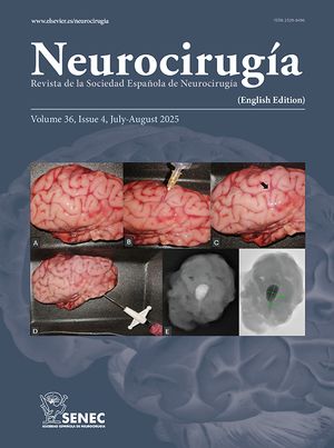
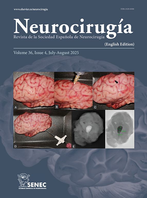
We herein report a case of a 55-year-old female patient who was diagnosed with primary CNS–HL. The patient was treated with complete surgical resection followed by intrathecal chemotherapy and whole brain radiotherapy (WBRT), showing fourteen months of disease-free survival at the time of this case report. A review of the available literature is also presented.

Keywords:
Hodgkin lymphoma
Primary lymphoma
Central nervous system
Brain neoplasm
Histology
Immunohistochemistry
Abbreviations:
HL
CNS
WBRT
CT
MRI
RS
EBV
Resumen

La afectación del sistema nervioso central (SNC) en pacientes con diagnóstico de linfoma de Hodgkin (LH) sistémico es una complicación muy poco frecuente, aunque bien definida. Sin embargo, el LH primario del SNC es una entidad extremadamente rara, cuyo diagnóstico precisa la identificación de un patrón morfológico e inmunohistoquímico específico, así como la afectación aislada del SNC.

Pese a contar con una casuística muy limitada (apenas una veintena de casos publicados en la literatura) la bibliografía disponible coincide en que el LH con afectación primaria y aislada del SNC, cuando es tratado con cirugía y tratamiento adyuvante (radioterapia±quimioterapia), parece tener un mejor pronóstico que aquellos casos en los que la afectación del SNC se produce en el contexto de un LH sistémico o en el contexto de un linfoma no Hodgkin.

En este artículo se presenta el caso de una mujer de 55 años con diagnóstico histopatológico de LH primario del SNC. La paciente fue sometida a una exéresis completa de la lesión y a tratamiento adyuvante con quimioterapia intratecal y radioterapia holocraneal, con una supervivencia libre de enfermedad hasta la fecha de 14 meses. Se presenta asimismo, la revisión de la literatura disponible.

Palabras clave:
Linfoma de Hodgkin
Linfoma primario
Sistema nervioso central
Tumor cerebral
Histología
Inmunohistoquímica

Article

These are the options to access the full texts of the publication Neurocirugía (English edition)
Member
Member of the Sociedad Española de Neurocirugía

If it is the first time you have accessed you can obtain your credentials by contacting Elsevier Spain in suscripciones@elsevier.com or by calling our Customer Service at902 88 87 40 if you are calling from Spain or at +34 932 418 800 (from 9 to 18h., GMT + 1) if you are calling outside of Spain.

If you already have your login data, please click here .

If you have forgotten your password you can you can recover it by clicking here and selecting the option ¿I have forgotten my password¿.

Subscriber
Subscriber

If you already have your login data, please click here .

If you have forgotten your password you can you can recover it by clicking here and selecting the option “I have forgotten my password”
Subscribe
Subscribe to

Neurocirugía (English edition)

Purchase
Purchase article

Purchasing article the PDF version will be downloaded

Price 19.34 €

Purchase now
Contact
Phone for subscriptions and reporting of errors
From Monday to Friday from 9 a.m. to 6 p.m. (GMT + 1) except for the months of July and August which will be from 9 a.m. to 3 p.m.
Calls from Spain
932 415 960
Calls from outside Spain
+34 932 415 960
Email
Idiomas
Neurocirugía (English edition)