array:24 [
  "pii" => "S252984961830042X"
  "issn" => "25298496"
  "doi" => "10.1016/j.neucie.2018.06.001"
  "estado" => "S300"
  "fechaPublicacion" => "2019-01-01"
  "aid" => "342"
  "copyright" => "Sociedad Española de Neurocirugía"
  "copyrightAnyo" => "2018"
  "documento" => "article"
  "crossmark" => 1
  "subdocumento" => "fla"
  "cita" => "Neurocirugia. 2019;30:11-8"
  "abierto" => array:3 [
    "ES" => false
    "ES2" => false
    "LATM" => false
  ]
  "gratuito" => false
  "lecturas" => array:1 [
    "total" => 0
  ]
  "Traduccion" => array:1 [
    "en" => array:19 [
      "pii" => "S1130147318300678"
      "issn" => "11301473"
      "doi" => "10.1016/j.neucir.2018.06.004"
      "estado" => "S300"
      "fechaPublicacion" => "2019-01-01"
      "aid" => "342"
      "copyright" => "Sociedad Española de Neurocirugía"
      "documento" => "article"
      "crossmark" => 1
      "subdocumento" => "fla"
      "cita" => "Neurocirugia. 2019;30:11-8"
      "abierto" => array:3 [
        "ES" => false
        "ES2" => false
        "LATM" => false
      ]
      "gratuito" => false
      "lecturas" => array:2 [
        "total" => 40
        "formatos" => array:2 [
          "HTML" => 28
          "PDF" => 12
        ]
      ]
      "en" => array:13 [
        "idiomaDefecto" => true
        "cabecera" => "<span class="elsevierStyleTextfn">Clinical Research</span>"
        "titulo" => "Perfusion MRI grading diffuse gliomas&#58; Impact of permeability parameters on molecular biomarkers and survival"
        "tienePdf" => "en"
        "tieneTextoCompleto" => "en"
        "tieneResumen" => array:2 [
          0 => "en"
          1 => "es"
        ]
        "paginas" => array:1 [
          0 => array:2 [
            "paginaInicial" => "11"
            "paginaFinal" => "18"
          ]
        ]
        "titulosAlternativos" => array:1 [
          "es" => array:1 [
            "titulo" => "Utilidad de la RM perfusi&#243;n en la gradaci&#243;n de los gliomas difusos&#58; relaci&#243;n entre par&#225;metros de permeabilidad&#44; marcadores moleculares y supervivencia"
          ]
        ]
        "contieneResumen" => array:2 [
          "en" => true
          "es" => true
        ]
        "contieneTextoCompleto" => array:1 [
          "en" => true
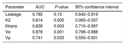
        ]
        "contienePdf" => array:1 [
          "en" => true
        ]
        "resumenGrafico" => array:2 [
          "original" => 0
          "multimedia" => array:7 [
            "identificador" => "fig0020"
            "etiqueta" => "Fig&#46; 4"
            "tipo" => "MULTIMEDIAFIGURA"
            "mostrarFloat" => true
            "mostrarDisplay" => false
            "figura" => array:1 [
              0 => array:4 [
                "imagen" => "gr4.jpeg"
                "Alto" => 1196
                "Ancho" => 1621
                "Tamanyo" => 69629
              ]
            ]
            "descripcion" => array:1 [
              "en" => "<p id="spar0060" class="elsevierStyleSimplePara elsevierViewall">Kaplan&#8211;Meier survival curves of 49 patients with diffuse gliomas&#46; Progression-free survival curves are plotted according to classification based on the cut off value of 1&#46;2 for leakage parameter&#46; Leakage values had also a significant influence on progression-free survival&#44; with a median survival of 15 months for patients with leakage values &#60;1&#46;2 and 6 months for patients with leakage values &#62;1&#46;2&#46;</p>"
            ]
          ]
        ]
        "autores" => array:1 [
          0 => array:2 [
            "autoresLista" => "Amaya Hilario, Aurelio Hernandez-Lain, Juan Manuel Sepulveda, Alfonso Lagares, Angel Perez-Nu&#241;ez, Ana Ramos"
            "autores" => array:6 [
              0 => array:2 [
                "nombre" => "Amaya"
                "apellidos" => "Hilario"
              ]
              1 => array:2 [
                "nombre" => "Aurelio"
                "apellidos" => "Hernandez-Lain"
              ]
              2 => array:2 [
                "nombre" => "Juan Manuel"
                "apellidos" => "Sepulveda"
              ]
              3 => array:2 [
                "nombre" => "Alfonso"
                "apellidos" => "Lagares"
              ]
              4 => array:2 [
                "nombre" => "Angel"
                "apellidos" => "Perez-Nu&#241;ez"
              ]
              5 => array:2 [
                "nombre" => "Ana"
                "apellidos" => "Ramos"
              ]
            ]
          ]
        ]
      ]
      "idiomaDefecto" => "en"
      "Traduccion" => array:1 [
        "en" => array:9 [
          "pii" => "S252984961830042X"
          "doi" => "10.1016/j.neucie.2018.06.001"
          "estado" => "S300"
          "subdocumento" => ""
          "abierto" => array:3 [
            "ES" => false
            "ES2" => false
            "LATM" => false
          ]
          "gratuito" => false
          "lecturas" => array:1 [
            "total" => 0
          ]
          "idiomaDefecto" => "en"
          "EPUB" => "https://multimedia.elsevier.es/PublicationsMultimediaV1/item/epub/S252984961830042X?idApp=UINPBA00004B"
        ]
      ]
      "EPUB" => "https://multimedia.elsevier.es/PublicationsMultimediaV1/item/epub/S1130147318300678?idApp=UINPBA00004B"
      "url" => "/11301473/0000003000000001/v1_201901030610/S1130147318300678/v1_201901030610/en/main.assets"
    ]
  ]
  "itemSiguiente" => array:19 [
    "pii" => "S2529849618300418"
    "issn" => "25298496"
    "doi" => "10.1016/j.neucie.2018.09.003"
    "estado" => "S300"
    "fechaPublicacion" => "2019-01-01"
    "aid" => "350"
    "copyright" => "Sociedad Espa&#241;ola de Neurocirug&#237;a"
    "documento" => "article"
    "crossmark" => 1
    "subdocumento" => "fla"
    "cita" => "Neurocirugia. 2019;30:19-27"
    "abierto" => array:3 [
      "ES" => false
      "ES2" => false
      "LATM" => false
    ]
    "gratuito" => false
    "lecturas" => array:1 [
      "total" => 0
    ]
    "en" => array:13 [
      "idiomaDefecto" => true
      "cabecera" => "<span class="elsevierStyleTextfn">Clinical Research</span>"
      "titulo" => "Assessment of the impact of glioma diagnostic reclassification following the new 2016 WHO classification on a series of cases"
      "tienePdf" => "en"
      "tieneTextoCompleto" => "en"
      "tieneResumen" => array:2 [
        0 => "en"
        1 => "es"
      ]
      "paginas" => array:1 [
        0 => array:2 [
          "paginaInicial" => "19"
          "paginaFinal" => "27"
        ]
      ]
      "titulosAlternativos" => array:1 [
        "es" => array:1 [
          "titulo" => "Evaluaci&#243;n del impacto del cambio diagn&#243;stico de los gliomas aplicando la nueva clasificaci&#243;n de la OMS de 2016 sobre una serie de casos"
        ]
      ]
      "contieneResumen" => array:2 [
        "en" => true
        "es" => true
      ]
      "contieneTextoCompleto" => array:1 [
        "en" => true
      ]
      "contienePdf" => array:1 [
        "en" => true
      ]
      "resumenGrafico" => array:2 [
        "original" => 0
        "multimedia" => array:8 [
          "identificador" => "fig0005"
          "etiqueta" => "Fig&#46; 1"
          "tipo" => "MULTIMEDIAFIGURA"
          "mostrarFloat" => true
          "mostrarDisplay" => false
          "fuente" => "<span class="elsevierStyleItalic">Source</span>&#58; Louis et al&#46;<a class="elsevierStyleCrossRef" href="#bib0235"><span class="elsevierStyleSup">13</span></a>"
          "figura" => array:1 [
            0 => array:4 [
              "imagen" => "gr1.jpeg"
              "Alto" => 1813
              "Ancho" => 2917
              "Tamanyo" => 325250
            ]
          ]
          "descripcion" => array:1 [
            "en" => "<p id="spar0045" class="elsevierStyleSimplePara elsevierViewall">Diagnostic algorithm of the new 2016 WHO classification of gliomas&#46;</p>"
          ]
        ]
      ]
      "autores" => array:1 [
        0 => array:2 [
          "autoresLista" => "Marc Valera-Mel&#233;, Olga Mateo Sierra, Emma Sola Vendrell, Juan Ad&#225;n Guzm&#225;n de Villoria L&#46;, Lorena Carvajal D&#237;az, &#211;scar Lucas Gil de Sagredo del Corral, Roberto Garc&#237;a Leal"
          "autores" => array:7 [
            0 => array:2 [
              "nombre" => "Marc"
              "apellidos" => "Valera-Mel&#233;"
            ]
            1 => array:2 [
              "nombre" => "Olga"
              "apellidos" => "Mateo Sierra"
            ]
            2 => array:2 [
              "nombre" => "Emma"
              "apellidos" => "Sola Vendrell"
            ]
            3 => array:2 [
              "nombre" => "Juan Ad&#225;n"
              "apellidos" => "Guzm&#225;n de Villoria L&#46;"
            ]
            4 => array:2 [
              "nombre" => "Lorena"
              "apellidos" => "Carvajal D&#237;az"
            ]
            5 => array:2 [
              "nombre" => "&#211;scar Lucas"
              "apellidos" => "Gil de Sagredo del Corral"
            ]
            6 => array:2 [
              "nombre" => "Roberto"
              "apellidos" => "Garc&#237;a Leal"
            ]
          ]
        ]
      ]
    ]
    "idiomaDefecto" => "en"
    "Traduccion" => array:1 [
      "es" => array:9 [
        "pii" => "S1130147318300903"
        "doi" => "10.1016/j.neucir.2018.09.002"
        "estado" => "S300"
        "subdocumento" => ""
        "abierto" => array:3 [
          "ES" => false
          "ES2" => false
          "LATM" => false
        ]
        "gratuito" => false
        "lecturas" => array:1 [
          "total" => 0
        ]
        "idiomaDefecto" => "es"
        "EPUB" => "https://multimedia.elsevier.es/PublicationsMultimediaV1/item/epub/S1130147318300903?idApp=UINPBA00004B"
      ]
    ]
    "EPUB" => "https://multimedia.elsevier.es/PublicationsMultimediaV1/item/epub/S2529849618300418?idApp=UINPBA00004B"
    "url" => "/25298496/0000003000000001/v1_201901030633/S2529849618300418/v1_201901030633/en/main.assets"
  ]
  "itemAnterior" => array:19 [
    "pii" => "S252984961830039X"
    "issn" => "25298496"
    "doi" => "10.1016/j.neucie.2018.10.001"
    "estado" => "S300"
    "fechaPublicacion" => "2019-01-01"
    "aid" => "346"
    "copyright" => "Sociedad Espa&#241;ola de Neurocirug&#237;a"
    "documento" => "article"
    "crossmark" => 1
    "subdocumento" => "fla"
    "cita" => "Neurocirugia. 2019;30:1-10"
    "abierto" => array:3 [
      "ES" => false
      "ES2" => false
      "LATM" => false
    ]
    "gratuito" => false
    "lecturas" => array:1 [
      "total" => 0
    ]
    "en" => array:13 [
      "idiomaDefecto" => true
      "cabecera" => "<span class="elsevierStyleTextfn">Clinical Research</span>"
      "titulo" => "Cystic meningiomas&#58; Radiological and pathological correlation with surgical implications"
      "tienePdf" => "en"
      "tieneTextoCompleto" => "en"
      "tieneResumen" => array:2 [
        0 => "en"
        1 => "es"
      ]
      "paginas" => array:1 [
        0 => array:2 [
          "paginaInicial" => "1"
          "paginaFinal" => "10"
        ]
      ]
      "titulosAlternativos" => array:1 [
        "es" => array:1 [
          "titulo" => "Meningiomas qu&#237;sticos&#58; correlaci&#243;n radiol&#243;gica y patol&#243;gica con implicaciones quir&#250;rgicas"
        ]
      ]
      "contieneResumen" => array:2 [
        "en" => true
        "es" => true
      ]
      "contieneTextoCompleto" => array:1 [
        "en" => true
      ]
      "contienePdf" => array:1 [
        "en" => true
      ]
      "resumenGrafico" => array:2 [
        "original" => 0
        "multimedia" => array:7 [
          "identificador" => "fig0005"
          "etiqueta" => "Fig&#46; 1"
          "tipo" => "MULTIMEDIAFIGURA"
          "mostrarFloat" => true
          "mostrarDisplay" => false
          "figura" => array:1 [
            0 => array:4 [
              "imagen" => "gr1.jpeg"
              "Alto" => 1105
              "Ancho" => 1250
              "Tamanyo" => 155351
            ]
          ]
          "descripcion" => array:1 [
            "en" => "<p id="spar0050" class="elsevierStyleSimplePara elsevierViewall">Morphological classification of CMs based on the scheme by Nauta et al&#46; The following are differentiated among tumoural cysts in the strictest sense&#58; &#40;A&#41; those with a central location &#40;type 1&#41; from &#40;B&#41; those with an eccentric location &#40;type 2&#41;&#44; where the wall may in extreme cases be fibrotic with nodules of cancerous cells &#40;arrows&#41;&#46; The following are distinguished with regard to peri-tumoural cysts&#58; &#40;C&#41; those that form within the cerebral parenchyma&#44; giving rise to a parenchymatous pseudocapsule &#40;arrows&#41; of gliotic tissue without encompassing the leptomeninges &#40;type 3&#41;&#44; from &#40;D&#41; those formed through the trapping of CSF &#40;arrow&#41; in the subarachnoid space &#40;type 4&#41;&#46; Type 3 may contain proteinaceous material with shed cancer cells &#40;asterisk in C&#41;&#46; Q&#58; cyst&#59; the solid component of the tumour is shown in orange &#40;dark grey in black and white version&#41;&#46;</p>"
          ]
        ]
      ]
      "autores" => array:1 [
        0 => array:2 [
          "autoresLista" => "Rodrigo Carrasco Moro, Herbert D&#46; Jim&#233;nez Zapata, H&#233;ctor Pian Arias, Juan S&#46; Mart&#237;nez San Mill&#225;n, Mar&#237;a A&#46; Mart&#237;nez Rodrigo, Jos&#233; M&#46; Pascual Garvi"
          "autores" => array:6 [
            0 => array:2 [
              "nombre" => "Rodrigo"
              "apellidos" => "Carrasco Moro"
            ]
            1 => array:2 [
              "nombre" => "Herbert D&#46;"
              "apellidos" => "Jim&#233;nez Zapata"
            ]
            2 => array:2 [
              "nombre" => "H&#233;ctor"
              "apellidos" => "Pian Arias"
            ]
            3 => array:2 [
              "nombre" => "Juan S&#46;"
              "apellidos" => "Mart&#237;nez San Mill&#225;n"
            ]
            4 => array:2 [
              "nombre" => "Mar&#237;a A&#46;"
              "apellidos" => "Mart&#237;nez Rodrigo"
            ]
            5 => array:2 [
              "nombre" => "Jos&#233; M&#46;"
              "apellidos" => "Pascual Garvi"
            ]
          ]
        ]
      ]
    ]
    "idiomaDefecto" => "en"
    "Traduccion" => array:1 [
      "es" => array:9 [
        "pii" => "S1130147318300861"
        "doi" => "10.1016/j.neucir.2018.08.002"
        "estado" => "S300"
        "subdocumento" => ""
        "abierto" => array:3 [
          "ES" => false
          "ES2" => false
          "LATM" => false
        ]
        "gratuito" => false
        "lecturas" => array:1 [
          "total" => 0
        ]
        "idiomaDefecto" => "es"
        "EPUB" => "https://multimedia.elsevier.es/PublicationsMultimediaV1/item/epub/S1130147318300861?idApp=UINPBA00004B"
      ]
    ]
    "EPUB" => "https://multimedia.elsevier.es/PublicationsMultimediaV1/item/epub/S252984961830039X?idApp=UINPBA00004B"
    "url" => "/25298496/0000003000000001/v1_201901030633/S252984961830039X/v1_201901030633/en/main.assets"
  ]
  "en" => array:19 [
    "idiomaDefecto" => true
    "cabecera" => "<span class="elsevierStyleTextfn">Clinical Research</span>"
    "titulo" => "Perfusion MRI grading diffuse gliomas&#58; Impact of permeability parameters on molecular biomarkers and survival"
    "tieneTextoCompleto" => true
    "paginas" => array:1 [
      0 => array:2 [
        "paginaInicial" => "11"
        "paginaFinal" => "18"
      ]
    ]
    "autores" => array:1 [
      0 => array:4 [
        "autoresLista" => "Amaya Hilario, Aurelio Hernandez-Lain, Juan Manuel Sepulveda, Alfonso Lagares, Angel Perez-Nu&#241;ez, Ana Ramos"
        "autores" => array:6 [
          0 => array:4 [
            "nombre" => "Amaya"
            "apellidos" => "Hilario"
            "email" => array:1 [
              0 => "amayahilario&#64;yahoo&#46;es"
            ]
            "referencia" => array:2 [
              0 => array:2 [
                "etiqueta" => "<span class="elsevierStyleSup">a</span>"
                "identificador" => "aff0005"
              ]
              1 => array:2 [
                "etiqueta" => "<span class="elsevierStyleSup">&#42;</span>"
                "identificador" => "cor0005"
              ]
            ]
          ]
          1 => array:3 [
            "nombre" => "Aurelio"
            "apellidos" => "Hernandez-Lain"
            "referencia" => array:1 [
              0 => array:2 [
                "etiqueta" => "<span class="elsevierStyleSup">b</span>"
                "identificador" => "aff0010"
              ]
            ]
          ]
          2 => array:3 [
            "nombre" => "Juan Manuel"
            "apellidos" => "Sepulveda"
            "referencia" => array:1 [
              0 => array:2 [
                "etiqueta" => "<span class="elsevierStyleSup">c</span>"
                "identificador" => "aff0015"
              ]
            ]
          ]
          3 => array:3 [
            "nombre" => "Alfonso"
            "apellidos" => "Lagares"
            "referencia" => array:1 [
              0 => array:2 [
                "etiqueta" => "<span class="elsevierStyleSup">d</span>"
                "identificador" => "aff0020"
              ]
            ]
          ]
          4 => array:3 [
            "nombre" => "Angel"
            "apellidos" => "Perez-Nu&#241;ez"
            "referencia" => array:1 [
              0 => array:2 [
                "etiqueta" => "<span class="elsevierStyleSup">d</span>"
                "identificador" => "aff0020"
              ]
            ]
          ]
          5 => array:3 [
            "nombre" => "Ana"
            "apellidos" => "Ramos"
            "referencia" => array:1 [
              0 => array:2 [
                "etiqueta" => "<span class="elsevierStyleSup">a</span>"
                "identificador" => "aff0005"
              ]
            ]
          ]
        ]
        "afiliaciones" => array:4 [
          0 => array:3 [
            "entidad" => "Department of Radiology&#44; Neuroradiology Section&#44; Universitary Hospital 12 de Octubre&#44; Madrid&#44; Spain"
            "etiqueta" => "a"
            "identificador" => "aff0005"
          ]
          1 => array:3 [
            "entidad" => "Department of Neuropathology&#44; Universitary Hospital 12 de Octubre&#44; Madrid&#44; Spain"
            "etiqueta" => "b"
            "identificador" => "aff0010"
          ]
          2 => array:3 [
            "entidad" => "Department of Medical Oncology&#44; Universitary Hospital 12 de Octubre&#44; Madrid&#44; Spain"
            "etiqueta" => "c"
            "identificador" => "aff0015"
          ]
          3 => array:3 [
            "entidad" => "Department of Neurosurgery&#44; Universitary Hospital 12 de Octubre&#44; Madrid&#44; Spain"
            "etiqueta" => "d"
            "identificador" => "aff0020"
          ]
        ]
        "correspondencia" => array:1 [
          0 => array:3 [
            "identificador" => "cor0005"
            "etiqueta" => "&#8270;"
            "correspondencia" => "<span class="elsevierStyleItalic">Corresponding author</span>&#46;"
          ]
        ]
      ]
    ]
    "titulosAlternativos" => array:1 [
      "es" => array:1 [
        "titulo" => "Utilidad de la RM perfusi&#243;n en la gradaci&#243;n de los gliomas difusos&#58; relaci&#243;n entre par&#225;metros de permeabilidad&#44; marcadores moleculares y supervivencia"
      ]
    ]
    "resumenGrafico" => array:2 [
      "original" => 0
      "multimedia" => array:7 [
        "identificador" => "fig0005"
        "etiqueta" => "Fig&#46; 1"
        "tipo" => "MULTIMEDIAFIGURA"
        "mostrarFloat" => true
        "mostrarDisplay" => false
        "figura" => array:1 [
          0 => array:4 [
            "imagen" => "gr1.jpeg"
            "Alto" => 1452
            "Ancho" => 935
            "Tamanyo" => 264818
          ]
        ]
        "descripcion" => array:1 [
          "en" => "<p id="spar0045" class="elsevierStyleSimplePara elsevierViewall">Immunostaining for the mutated form of isocitrate dehydrogenase &#40;A&#41; and for the loss of ATRX protein expression &#40;B&#41;&#46;</p>"
        ]
      ]
    ]
    "textoCompleto" => "<span class="elsevierStyleSections"><span id="sec0005" class="elsevierStyleSection elsevierViewall"><span class="elsevierStyleSectionTitle" id="sect0070">Introduction</span><p id="par0005" class="elsevierStylePara elsevierViewall">Diffuse gliomas are the most common primary cerebral tumor of adulthood&#46;<a class="elsevierStyleCrossRef" href="#bib0125"><span class="elsevierStyleSup">1</span></a> In the management of gliomas&#44; the World Health Organization &#40;WHO&#41; tumor grade is an important prognostic factor along with clinical and radiologic findings&#46;<a class="elsevierStyleCrossRef" href="#bib0130"><span class="elsevierStyleSup">2</span></a> Molecular and genetic biomarkers of gliomas are also emerging prognostic factors predicting the biologic behavior of tumors&#46;<a class="elsevierStyleCrossRef" href="#bib0135"><span class="elsevierStyleSup">3</span></a></p><p id="par0010" class="elsevierStylePara elsevierViewall">Dynamic susceptibility-weighted &#40;DSC&#41; imaging remains the most commonly used perfusion technique in glioma grading&#44; and cerebral blood volume &#40;CBV&#41; has been recently identified as an important prognostic marker for survival independent of tumor grade<a class="elsevierStyleCrossRef" href="#bib0140"><span class="elsevierStyleSup">4</span></a>&#59; however&#44; those perfusion measurements may be influenced by extravascular contrast leakage from tumor vessels&#46;<a class="elsevierStyleCrossRef" href="#bib0145"><span class="elsevierStyleSup">5</span></a> Dynamic contrast-enhanced &#40;DCE&#41; perfusion Magnetic resonance imaging &#40;MRI&#41; is an alternative technique for glioma grading providing hemodynamic parameters such as volume transfer coefficient &#40;Ktrans&#41;&#44; fractional volume of the extravascular extracellular space &#40;Ve&#41; and fractional blood plasma volume &#40;Vp&#41; that characterize the microvascular environment&#46;<a class="elsevierStyleCrossRefs" href="#bib0125"><span class="elsevierStyleSup">1&#44;2&#44;5</span></a> Like CBV&#44; Ktrans has demonstrated to be able to differentiate high- and low-grade tumors and predicts prognosis in diffuse gliomas&#46;<a class="elsevierStyleCrossRef" href="#bib0150"><span class="elsevierStyleSup">6</span></a></p><p id="par0015" class="elsevierStylePara elsevierViewall">Previously in the literature&#44; there are few studies published comparing DCE and DSC perfusion imaging in diffuse gliomas&#46;<a class="elsevierStyleCrossRef" href="#bib0145"><span class="elsevierStyleSup">5</span></a> Although Law et al&#46;<a class="elsevierStyleCrossRef" href="#bib0155"><span class="elsevierStyleSup">7</span></a> have previously reported a weak but positive correlation between Ktrans and CBV&#59; leakage parameters extracted from DSC and their relationship to the DCE-measures of vascular permeability remains unclear&#46;<a class="elsevierStyleCrossRef" href="#bib0160"><span class="elsevierStyleSup">8</span></a> Moreover&#44; to our knowledge&#44; permeability parameters have not yet been analyzed in function of the molecular status in diffuse gliomas&#46;</p><p id="par0020" class="elsevierStylePara elsevierViewall">The objectives of this study were&#58; &#40;1&#41; compare DCE and DSC permeability parameters&#44; &#40;2&#41; evaluate diagnostic accuracy of permeability parameters when discriminating high- and low-grade gliomas&#44; &#40;3&#41; analyze relationship of permeability with overall survival &#40;OS&#41; and progression-free survival &#40;PFS&#41;&#44; and &#40;4&#41; assess permeability differences in high-grade tumors classified on the basis of isocitrate dehydrogenase &#40;IDH&#41;&#44; O6-methylguanine-dna-methyltransferase promoter methylation &#40;MGMT&#41; and alpha-thalassemia&#47;mental retardation syndrome X-linked &#40;ATRX&#41; status&#46;</p></span><span id="sec0010" class="elsevierStyleSection elsevierViewall"><span class="elsevierStyleSectionTitle" id="sect0075">Material and methods</span><span id="sec0015" class="elsevierStyleSection elsevierViewall"><span class="elsevierStyleSectionTitle" id="sect0080">Patient selection and histopathological analysis</span><p id="par0025" class="elsevierStylePara elsevierViewall">From February 2014 to July 2016&#44; we conducted a retrospective analysis of forty-nine patients with histologically proved diffuse gliomas&#46; Following surgical resection or biopsy&#44; two experienced neuropathologists provided histopathologic diagnosis by using the 2007 WHO classification&#46;<a class="elsevierStyleCrossRef" href="#bib0165"><span class="elsevierStyleSup">9</span></a> Histologic specimens were classified into grade II-IV gliomas based on the presence of astrocytic or oligodendroglial morphology&#44; cell density&#44; atypia&#44; mitotic activity&#44; vascular proliferation and necrosis&#46; From a prognostic and therapeutic point of view&#44; a variable percentage of high-grade gliomas were also subclassified according to IDH &#40;87&#37; analyzed&#44; <span class="elsevierStyleItalic">n</span><span class="elsevierStyleHsp" style=""></span>&#61;<span class="elsevierStyleHsp" style=""></span>36&#41;&#44; MGMT &#40;48&#46;7&#37; analyzed&#44; <span class="elsevierStyleItalic">n</span><span class="elsevierStyleHsp" style=""></span>&#61;<span class="elsevierStyleHsp" style=""></span>20&#41; and ATRX status &#40;39&#37; analyzed&#44; <span class="elsevierStyleItalic">n</span><span class="elsevierStyleHsp" style=""></span>&#61;<span class="elsevierStyleHsp" style=""></span>16&#41;&#46;</p></span><span id="sec0020" class="elsevierStyleSection elsevierViewall"><span class="elsevierStyleSectionTitle" id="sect0085">Immunohistochemistry</span><p id="par0030" class="elsevierStylePara elsevierViewall">To detect the IDH1 R132H mutation&#44; a mouse monoclonal antibody &#40;dilution 1&#58;100&#44; DIA H09&#44; Dianova&#44; Germany&#41; was used&#46; ATRX immunohistochemistry &#40;HPA001906 rabbit polyclonal&#44; dilution 1&#58;100&#44; Sigma Aldrich&#44; USA&#41; was performed using an automated immunostainer&#46; IDH1 and IDH2 mutations were detected by direct Sanger sequencing in an ABI PRISM 310 DNA Analyzer &#40;Applied Biosystems&#44; Foster City&#44; CA&#44; USA&#41; or by pyrosequencing assays using the Pyromark Q24 ID instrument &#40;Qiagen&#41;&#46; <a class="elsevierStyleCrossRef" href="#fig0005">Fig&#46; 1</a> represents positive staining results for IDH and ATRX mutation&#46;</p><elsevierMultimedia ident="fig0005"></elsevierMultimedia></span><span id="sec0025" class="elsevierStyleSection elsevierViewall"><span class="elsevierStyleSectionTitle" id="sect0090">MGMT methylation-specific PCR &#40;MSP&#41;</span><p id="par0035" class="elsevierStylePara elsevierViewall">For MGMT promoter methylation analysis&#44; DNA was bisulfite-treated using the EZ DNA methylation Kit &#40;Zymo Research&#44; Orange&#44; CA&#44; USA&#41; according to the manufacturer&#39;s instructions&#46;</p></span><span id="sec0030" class="elsevierStyleSection elsevierViewall"><span class="elsevierStyleSectionTitle" id="sect0095">Magnetic resonance imaging data acquisition</span><p id="par0040" class="elsevierStylePara elsevierViewall">All patients were examined with the same imaging acquisition protocol at a 1&#46;5<span class="elsevierStyleHsp" style=""></span>T scanner &#40;Signa Excite&#59; GE Healthcare&#44; Milwaukee&#44; Wisconsin&#41; by using an 8-channel encoding head coil&#46; Preoperative MRI protocol included pre-contrast T1-weighted 3D fast spoiled gradient-recalled acquisition in the steady state &#40;FSPGR&#41; &#40;TR&#44; 7<span class="elsevierStyleHsp" style=""></span>ms&#59; TE&#44; 2&#46;2<span class="elsevierStyleHsp" style=""></span>ms&#59; slice thickness&#44; 1&#46;6<span class="elsevierStyleHsp" style=""></span>mm&#59; flip angle&#44; 12&#176;&#59; matrix 320<span class="elsevierStyleHsp" style=""></span>&#215;<span class="elsevierStyleHsp" style=""></span>320&#41;&#44; axial T2-weighted fast spin-echo &#40;TR<span class="elsevierStyleHsp" style=""></span>&#61;<span class="elsevierStyleHsp" style=""></span>4100&#44; TE<span class="elsevierStyleHsp" style=""></span>&#61;<span class="elsevierStyleHsp" style=""></span>85&#41;&#44; axial FLAIR &#40;TR<span class="elsevierStyleHsp" style=""></span>&#61;<span class="elsevierStyleHsp" style=""></span>8000&#44; TE<span class="elsevierStyleHsp" style=""></span>&#61;<span class="elsevierStyleHsp" style=""></span>120&#44; TI<span class="elsevierStyleHsp" style=""></span>&#61;<span class="elsevierStyleHsp" style=""></span>2000&#41;&#44; and diffusion-weighted imaging &#40;DWI&#41; &#40;single-shot spin-echo echo planar imaging with 4-mm contiguous sections and <span class="elsevierStyleItalic">b</span> values of 0 and 1000<span class="elsevierStyleHsp" style=""></span>s&#47;mm<span class="elsevierStyleSup">2</span>&#41;&#46; Data from T2WI&#44; FLAIR were obtained using 4-mm-thick sections with a 0&#46;4-mm skip&#44; a 320<span class="elsevierStyleHsp" style=""></span>&#215;<span class="elsevierStyleHsp" style=""></span>256 matrix and a FOV of 24<span class="elsevierStyleHsp" style=""></span>&#215;<span class="elsevierStyleHsp" style=""></span>24<span class="elsevierStyleHsp" style=""></span>cm&#46;</p><p id="par0045" class="elsevierStylePara elsevierViewall">DCE-MRI was performed with a dynamic gradient-echo T1-weighted sequence using the following parameters&#58; TR&#47;TE&#44; 3&#46;1&#47;1&#46;4<span class="elsevierStyleHsp" style=""></span>ms&#59; flip angle&#44; 15&#176;&#59; matrix&#44; 160<span class="elsevierStyleHsp" style=""></span>&#215;<span class="elsevierStyleHsp" style=""></span>160&#59; section thickness&#44; 2&#46;4<span class="elsevierStyleHsp" style=""></span>mm&#46; After pre-contrast axial sequence&#44; 30-dynamic contrast-enhanced T1-weighted images were obtained with an injection of 5<span class="elsevierStyleHsp" style=""></span>mL of gadobutrol &#40;Gadovist&#44; 1<span class="elsevierStyleHsp" style=""></span>mmol&#47;mL&#59; Berlex Laboratories&#44; Wayne&#44; NJ&#41; at a rate of 3<span class="elsevierStyleHsp" style=""></span>mL&#47;s &#40;Spectris Solaris EP&#44; MR Injection System&#41;&#46; After DCE perfusion imaging&#44; dynamic contrast-enhanced T2&#42;-weighted perfusion MRI was performed by using a gradient-echo EPI acquisition during the first pass of a bolus of gadobutrol at a dose of 0&#46;1<span class="elsevierStyleHsp" style=""></span>mmol&#47;kg&#46; We used the 5<span class="elsevierStyleHsp" style=""></span>mL of gadobutrol used for DCE perfusion MRI as a preload for DSC imaging&#46;<a class="elsevierStyleCrossRef" href="#bib0170"><span class="elsevierStyleSup">10</span></a> A series of 40 multisection acquisitions was acquired at 0&#46;2-s interval&#46; The first 8 acquisitions were performed before the contrast agent was injected to establish a pre-contrast baseline&#46; For the 8<span class="elsevierStyleSup">th</span> acquisition&#44; gadobutrol was injected at a rate of 5<span class="elsevierStyleHsp" style=""></span>mL&#47;s followed by a 20-mL bolus of saline at 5<span class="elsevierStyleHsp" style=""></span>mL&#47;s&#46; After DSC perfusion imaging&#44; we acquired the post-contrast T1-weighted 3DFSPGR images&#46;</p></span><span id="sec0035" class="elsevierStyleSection elsevierViewall"><span class="elsevierStyleSectionTitle" id="sect0100">Image post-processing and perfusion MRI analysis</span><p id="par0050" class="elsevierStylePara elsevierViewall">Post-processing of the 3D T1-weighted with contrast enhancement&#44; DCE and DSC perfusion MRI data was performed by using Olea Sphere v 2&#46;3 software &#40;Olea Medical&#44; La Ciotat&#44; France&#41;&#46; Segmentation of the preoperative enhancing tumor &#40;excluding macroscopic necrosis and cyst&#41; was performed on each slice of the volumetric post-contrast T1-weighetd images according to a semi-automated region-growing segmentation method&#46; Pharmacokinetic analysis of DCE-MRI was performed based on the extended Tofts model&#46;<a class="elsevierStyleCrossRef" href="#bib0175"><span class="elsevierStyleSup">11</span></a> Parametric maps of Ktrans&#44; Ve&#44; Vp&#44; rate constant between EES and blood plasma &#40;Kep&#41;&#44; CBV and leakage-corrected CBV were generated for each patient&#46; Those maps were calculated following automatic co-registration of perfusion parameters with post-contrast T1-weighted images&#44; which included the previously segmented enhancing tumor region of interest &#40;ROIs&#41;&#46; Then&#44; we estimated the &#8220;leakage&#8221; from the subtraction between CBV and leakage-corrected CBV at preoperative MRI based on the information obtained from all ROIs of each slice&#46;</p></span><span id="sec0040" class="elsevierStyleSection elsevierViewall"><span class="elsevierStyleSectionTitle" id="sect0105">Statistical analysis</span><p id="par0055" class="elsevierStylePara elsevierViewall">All statistical analysis was performed using the software Statistical Package for the Social Sciences &#40;SPSS v&#46;12&#44; Chicago&#44; IL&#41;&#46; Using the Pearson correlation we performed comparisons of DSC &#40;mean leakage&#41; and DCE-derived permeability parameters &#40;mean Ktrans&#44; Ve&#44; Vp and Kep&#41;&#46; A Mann&#8211;Whitney <span class="elsevierStyleItalic">U</span> test was used to compare the different permeability parameters between high- and low-grade tumors&#46; Receiver operating characteristic &#40;ROC&#41; curves analysis was used to determine the permeability parameters that best discriminate between high- and low-grade gliomas&#46; The area under curve &#40;AUC&#41; was obtained to indicate the degree of the relationship between permeability parameters and grade of gliomas&#46; Optimal threshold value for the parameter leakage was identified using ROC analysis for discriminating high- and low-grade tumors with the Youden&#39;s Index &#40;YI&#41; method&#44; where the YI of a cutpoint <span class="elsevierStyleItalic">c</span> is defined as YI&#40;<span class="elsevierStyleItalic">c</span>&#41; &#188; max &#40;Sensitivity&#40;<span class="elsevierStyleItalic">c</span>&#41;<span class="elsevierStyleHsp" style=""></span>&#43;<span class="elsevierStyleHsp" style=""></span>Specificity&#40;<span class="elsevierStyleItalic">c</span>&#41;<span class="elsevierStyleHsp" style=""></span>&#8722;<span class="elsevierStyleHsp" style=""></span>1 &#40;25&#41;&#46; Kaplan&#8211;Meier analysis and Cox proportional hazards models were constructed to test the relation of permeability parameters with survival calculating hazard ratios&#46; Mann&#8211;Whitney <span class="elsevierStyleItalic">U</span> test was used to evaluate the relationship of DSC and DCE-derived perfusion parameters with molecular biomarkers &#40;IDH&#44; MGMT and ATRX&#41;&#46; A value of <span class="elsevierStyleItalic">P</span><span class="elsevierStyleHsp" style=""></span>&#60;<span class="elsevierStyleHsp" style=""></span>0&#46;05 was regarded as statistically significant&#46;</p></span></span><span id="sec0045" class="elsevierStyleSection elsevierViewall"><span class="elsevierStyleSectionTitle" id="sect0110">Results</span><span id="sec0050" class="elsevierStyleSection elsevierViewall"><span class="elsevierStyleSectionTitle" id="sect0115">Study population</span><p id="par0060" class="elsevierStylePara elsevierViewall">A total of forty-nine patients with diffuse gliomas were enrolled in the study&#46; This retrospective analysis included 28 male and 21 female patients &#40;age ranged 16&#8211;78 years&#41;&#46; Gliomas were classified into forty-one high-grade and 8 low-grade tumors&#46; Moreover&#44; in the group of high-grade gliomas&#44; 27&#46;78&#37; &#40;<span class="elsevierStyleItalic">n</span><span class="elsevierStyleHsp" style=""></span>&#61;<span class="elsevierStyleHsp" style=""></span>10&#41; were positive for IDH mutation&#44; 65&#37; &#40;<span class="elsevierStyleItalic">n</span><span class="elsevierStyleHsp" style=""></span>&#61;<span class="elsevierStyleHsp" style=""></span>13&#41; had methylated MGMT&#44; and 31&#46;25&#37; &#40;<span class="elsevierStyleItalic">n</span><span class="elsevierStyleHsp" style=""></span>&#61;<span class="elsevierStyleHsp" style=""></span>5&#41; had ATX loss&#46; Mean follow-up was 15 months&#46;</p></span><span id="sec0055" class="elsevierStyleSection elsevierViewall"><span class="elsevierStyleSectionTitle" id="sect0120">Correlation of DSC and DCE permeability parameters</span><p id="par0065" class="elsevierStylePara elsevierViewall">Leakage showed a significant positive correlation with Ktrans &#40;<span class="elsevierStyleItalic">R</span><span class="elsevierStyleHsp" style=""></span>&#61;<span class="elsevierStyleHsp" style=""></span>0&#46;310&#44; <span class="elsevierStyleItalic">P</span><span class="elsevierStyleHsp" style=""></span>&#61;<span class="elsevierStyleHsp" style=""></span>0&#46;030&#41; and Vp &#40;<span class="elsevierStyleItalic">R</span><span class="elsevierStyleHsp" style=""></span>&#61;<span class="elsevierStyleHsp" style=""></span>0&#46;289&#44; <span class="elsevierStyleItalic">P</span><span class="elsevierStyleHsp" style=""></span>&#61;<span class="elsevierStyleHsp" style=""></span>0&#46;044&#41;&#46; Vp also presented a significant correlation with Ve &#40;<span class="elsevierStyleItalic">R</span><span class="elsevierStyleHsp" style=""></span>&#61;<span class="elsevierStyleHsp" style=""></span>0&#46;388&#44; <span class="elsevierStyleItalic">P</span><span class="elsevierStyleHsp" style=""></span>&#61;<span class="elsevierStyleHsp" style=""></span>0&#46;006&#41;&#46; The correlation was specifically significant between Ktrans and Ve &#40;<span class="elsevierStyleItalic">R</span><span class="elsevierStyleHsp" style=""></span>&#61;<span class="elsevierStyleHsp" style=""></span>0&#46;727&#44; <span class="elsevierStyleItalic">P</span><span class="elsevierStyleHsp" style=""></span>&#60;<span class="elsevierStyleHsp" style=""></span>0&#46;001&#41;&#44; and between Ktrans and Kep &#40;<span class="elsevierStyleItalic">R</span><span class="elsevierStyleHsp" style=""></span>&#61;<span class="elsevierStyleHsp" style=""></span>0&#46;670&#44; <span class="elsevierStyleItalic">P</span><span class="elsevierStyleHsp" style=""></span>&#60;<span class="elsevierStyleHsp" style=""></span>0&#46;001&#41;&#46; Correlations between DSC and DCE permeability parameters are displayed in <a class="elsevierStyleCrossRef" href="#tbl0005">Table 1</a></p><elsevierMultimedia ident="tbl0005"></elsevierMultimedia></span><span id="sec0060" class="elsevierStyleSection elsevierViewall"><span class="elsevierStyleSectionTitle" id="sect0125">Accuracy of DSC and DCE-derived permeability parameters according to tumor grade</span><p id="par0070" class="elsevierStylePara elsevierViewall">We have demonstrated significant differences in the values of leakage &#40;<span class="elsevierStyleItalic">P</span><span class="elsevierStyleHsp" style=""></span>&#61;<span class="elsevierStyleHsp" style=""></span>0&#46;01&#41;&#44; Ktrans &#40;<span class="elsevierStyleItalic">P</span><span class="elsevierStyleHsp" style=""></span>&#61;<span class="elsevierStyleHsp" style=""></span>0&#46;002&#41;&#44; Vp &#40;<span class="elsevierStyleItalic">P</span><span class="elsevierStyleHsp" style=""></span>&#61;<span class="elsevierStyleHsp" style=""></span>0&#46;032&#41; and Ve &#40;<span class="elsevierStyleItalic">P</span><span class="elsevierStyleHsp" style=""></span>&#60;<span class="elsevierStyleHsp" style=""></span>0&#46;001&#41; between high-grade and low-grade diffuse gliomas&#46; High-grade tumors showed higher values of leakage &#40;median 1&#46;17&#59; interquartile range &#40;IQR&#41; 0&#46;47&#8211;1&#46;88&#41;&#44; Ktrans &#40;median 0&#46;24&#59; IQR 0&#46;11&#8211;0&#46;42&#41;&#44; Ve &#40;median 0&#46;18&#59; IQR 0&#46;09&#8211;0&#46;35&#41; and Vp &#40;median 0&#46;08&#59; IQR 0&#46;05&#8211;0&#46;11&#41;&#46; On the other hand&#44; low-grade tumors exhibited lower values of leakage &#40;median 0&#46;34&#59; IQR &#8722;0&#46;02&#8211;0&#46;88&#41;&#44; Ktrans &#40;median 0&#46;060&#59; IQR 0&#46;02&#8211;0&#46;13&#41;&#44; Ve &#40;median 0&#46;04&#59; IQR 0&#46;01&#8211;0&#46;07&#41; and Vp &#40;median 0&#46;035&#59; IQR 0&#46;03&#8211;0&#46;08&#41;&#46; We have not demonstrated significant differences in Kep values between high- and low-grade tumors&#46;</p><p id="par0075" class="elsevierStylePara elsevierViewall">ROC curve analysis was also performed to assess DSC and DCE as a tool to differentiate high- and low-grade gliomas&#46; The highest AUC was demonstrated by the DCE permeability parameters Ktrans &#40;AUC<span class="elsevierStyleHsp" style=""></span>&#61;<span class="elsevierStyleHsp" style=""></span>0&#46;838&#44; CI95&#37; 0&#46;710&#8211;0&#46;967&#44; <span class="elsevierStyleItalic">P</span><span class="elsevierStyleHsp" style=""></span>&#61;<span class="elsevierStyleHsp" style=""></span>0&#46;003&#41; and Ve &#40;AUC<span class="elsevierStyleHsp" style=""></span>&#61;<span class="elsevierStyleHsp" style=""></span>0&#46;878&#44; CI95&#37; 0&#46;768&#8211;0&#46;988&#44; <span class="elsevierStyleItalic">P</span><span class="elsevierStyleHsp" style=""></span>&#61;<span class="elsevierStyleHsp" style=""></span>0&#46;001&#41; &#40;<a class="elsevierStyleCrossRef" href="#fig0010">Fig&#46; 2</a>&#41;&#46; In our series&#44; the cut off value of 0&#46;075 for the perfusion parameter Ve generated the best combination of sensitivity &#40;80&#37;&#41;&#44; specificity &#40;75&#37;&#41;&#44; positive predictive value &#40;94&#46;29&#37;&#41; and negative predictive value &#40;42&#46;86&#37;&#41; when differentiating high and low-grade tumors&#46; <a class="elsevierStyleCrossRef" href="#tbl0010">Table 2</a> summarizes AUC values permeability parameters discriminating high- and low-grade tumors&#46;</p><elsevierMultimedia ident="fig0010"></elsevierMultimedia><elsevierMultimedia ident="tbl0010"></elsevierMultimedia></span><span id="sec0065" class="elsevierStyleSection elsevierViewall"><span class="elsevierStyleSectionTitle" id="sect0130">Impact of permeability parameters on survival</span><p id="par0080" class="elsevierStylePara elsevierViewall">DSC-derived leakage was the unique permeability parameter that showed a significant influence on overall survival &#40;<span class="elsevierStyleItalic">P</span><span class="elsevierStyleHsp" style=""></span>&#61;<span class="elsevierStyleHsp" style=""></span>0&#46;006&#59; hazard ratio &#40;HR&#41; 1&#46;560&#44; 95&#37; CI 1&#46;137&#8211;2&#46;164&#41;&#41; and progression-free survival &#40;<span class="elsevierStyleItalic">P</span><span class="elsevierStyleHsp" style=""></span>&#61;<span class="elsevierStyleHsp" style=""></span>0&#46;012&#59; HR 1&#46;432&#44; 95&#37; CI 1&#46;082&#8211;1&#46;895&#41;&#46; In our series&#44; including both high- and low-grade tumors&#44; median OS was 20 months &#40;CI95&#37; 7&#46;846&#8211;32&#46;154&#41; and median PFS was 9 months &#40;CI95&#37; 5&#46;32&#8211;12&#46;679&#41;&#46; For Kaplan Meier analysis&#44; the optimal cut off value of 1&#46;2 for the parameter leakage was identified using ROC curves with Youden index method&#46;</p><p id="par0085" class="elsevierStylePara elsevierViewall">Taking into account tumor enhancement&#44; all non-enhancing gliomas had leakage values &#60;1&#46;2&#46; When tumor enhancement was present&#44; 42&#46;58&#37; had leakage values &#62;1&#46;2 and the remainder 56&#46;42&#37; had leakage values &#62;1&#46;2&#46; We have not demonstrated a relationship between tumor enhancement and permeability parameters&#46;</p><p id="par0090" class="elsevierStylePara elsevierViewall">All low-grade tumors had leakage values lower than 1&#46;2&#46; In high-grade tumors&#44; 51&#46;2&#37; had leakage values &#60;1&#46;2 and the remainder 48&#46;8&#37; had leakage values &#62;1&#46;2&#46;</p><p id="par0095" class="elsevierStylePara elsevierViewall">Kaplan&#8211;Meier estimates of survival indicated that patients with a leakage mean of less than 1&#46;2 had a significant better OS &#40;Log-rank 8&#46;789&#59; <span class="elsevierStyleItalic">P</span><span class="elsevierStyleHsp" style=""></span>&#61;<span class="elsevierStyleHsp" style=""></span>0&#46;003&#41; and PFS &#40;Log-rank 6&#46;612&#59; <span class="elsevierStyleItalic">P</span><span class="elsevierStyleHsp" style=""></span>&#61;<span class="elsevierStyleHsp" style=""></span>0&#46;010&#41;&#46; The group of gliomas with leakage values &#62;1&#46;2 had a median overall and progression-free survival of 12 months &#40;95&#37; CI&#44; 3&#46;918&#8211;20&#46;082&#41; and 6 months &#40;95&#37; CI&#44; 4&#46;028&#8211;7&#46;792&#41;&#44; respectively&#46; Patients with leakage values &#60;1&#46;2 had a median progression-free survival of 15 months &#40;95&#37; CI&#44; 3&#46;470&#8211;26&#46;530&#41;&#46; At the end of the follow-up&#44; patients with leakage values lower than 1&#46;2 had not reached median overall survival&#46;</p><p id="par0100" class="elsevierStylePara elsevierViewall"><a class="elsevierStyleCrossRefs" href="#fig0015">Figs&#46; 3 and 4</a> represent respectively the significant differences in OS and PFS in diffuse gliomas classified on the basis of the threshold value of 1&#46;2 for the permeability parameter leakage&#46;</p><elsevierMultimedia ident="fig0015"></elsevierMultimedia><elsevierMultimedia ident="fig0020"></elsevierMultimedia><p id="par0105" class="elsevierStylePara elsevierViewall">The remainder permeability parameters showed no significant correlation to survival&#46;</p></span><span id="sec0070" class="elsevierStyleSection elsevierViewall"><span class="elsevierStyleSectionTitle" id="sect0135">Permeability differences in high-grade tumors classified according to molecular status</span><p id="par0110" class="elsevierStylePara elsevierViewall">In addition&#44; we assessed differences in DSC and DCE-derived permeability parameters in high-grade tumors classified on the basis of IDH&#44; ATRX and MGMT biomarkers&#46;</p><p id="par0115" class="elsevierStylePara elsevierViewall">Among IDH mutant &#40;<span class="elsevierStyleItalic">n</span><span class="elsevierStyleHsp" style=""></span>&#61;<span class="elsevierStyleHsp" style=""></span>10&#41; and non-mutant &#40;<span class="elsevierStyleItalic">n</span><span class="elsevierStyleHsp" style=""></span>&#61;<span class="elsevierStyleHsp" style=""></span>26&#41; high-grade gliomas&#44; there were significant differences in leakage &#40;<span class="elsevierStyleItalic">P</span><span class="elsevierStyleHsp" style=""></span>&#61;<span class="elsevierStyleHsp" style=""></span>0&#46;004&#41; and Ktrans values &#40;<span class="elsevierStyleItalic">P</span><span class="elsevierStyleHsp" style=""></span>&#61;<span class="elsevierStyleHsp" style=""></span>0&#46;028&#41;&#46; Median leakage values were respectively 0&#46;32 and 1&#46;46 for IDH mutant and non-mutant gliomas&#46; Median Ktrans values were 0&#46;13 for IDH mutant and 0&#46;26 for IDH non-mutant high-grade gliomas&#46; IDH mutant high-grade tumors showed lower leakage and Ktrans values&#46;</p><p id="par0120" class="elsevierStylePara elsevierViewall">Among ATRX mutant &#40;<span class="elsevierStyleItalic">n</span><span class="elsevierStyleHsp" style=""></span>&#61;<span class="elsevierStyleHsp" style=""></span>5&#41; and non-mutant &#40;<span class="elsevierStyleItalic">n</span><span class="elsevierStyleHsp" style=""></span>&#61;<span class="elsevierStyleHsp" style=""></span>11&#41; high-grade tumors&#44; there were significant differences in leakage &#40;<span class="elsevierStyleItalic">P</span><span class="elsevierStyleHsp" style=""></span>&#61;<span class="elsevierStyleHsp" style=""></span>0&#46;005&#41; and Vp values &#40;<span class="elsevierStyleItalic">P</span><span class="elsevierStyleHsp" style=""></span>&#61;<span class="elsevierStyleHsp" style=""></span>0&#46;027&#41;&#46; Median leakage values were 0&#46;12 for ATRX mutant and 0&#46;69 for ATRX non-mutant high-grade gliomas&#46; Regarding fractional blood plasma volume&#44; median Vp values were respectively 0&#46;05 and 0&#46;1 for ATRX mutant and non-mutant gliomas&#46; High-grade ATRX mutant tumors presented lower leakage and Vp values&#46;</p><p id="par0125" class="elsevierStylePara elsevierViewall">We have not demonstrated significant differences in perfusion parameters in MGMT methylated &#40;<span class="elsevierStyleItalic">n</span><span class="elsevierStyleHsp" style=""></span>&#61;<span class="elsevierStyleHsp" style=""></span>13&#41; and non-methylated tumors &#40;<span class="elsevierStyleItalic">n</span><span class="elsevierStyleHsp" style=""></span>&#61;<span class="elsevierStyleHsp" style=""></span>7&#41;&#46;</p><p id="par0130" class="elsevierStylePara elsevierViewall">In our series&#44; leakage was the best parameter discriminating high-grade gliomas classified based on IDH and ATRX status&#46;</p></span></span><span id="sec0075" class="elsevierStyleSection elsevierViewall"><span class="elsevierStyleSectionTitle" id="sect0140">Discussion</span><p id="par0135" class="elsevierStylePara elsevierViewall">Conventional contrast-enhanced MRI is not always precise in differentiating high- from low-grade gliomas&#46; Contrast enhancement indicates breakdown of the blood&#8211;brain-barrier&#44; and although is frequently associated with high-grade tumors&#44; it is not always accurate in predicting tumor grade&#46;<a class="elsevierStyleCrossRefs" href="#bib0145"><span class="elsevierStyleSup">5&#44;7</span></a> For this reason&#44; DSC and DCE perfusion MRI have become prognostic biomarkers providing measurements of neoangiogenesis in the preoperative grading of diffuse gliomas&#46;<a class="elsevierStyleCrossRefs" href="#bib0140"><span class="elsevierStyleSup">4&#44;12</span></a> In this study&#44; we have demonstrated a significant correlation between DSC-derived leakage and DCE-derived Ktrans and Vp&#46; Within DCE perfusion MRI&#44; the correlation was specifically significant between Ktrans and Ve &#40;<span class="elsevierStyleItalic">P</span><span class="elsevierStyleHsp" style=""></span>&#60;<span class="elsevierStyleHsp" style=""></span>0&#46;001&#41;&#44; and between Ktrans and Kep &#40;<span class="elsevierStyleItalic">P</span><span class="elsevierStyleHsp" style=""></span>&#60;<span class="elsevierStyleHsp" style=""></span>0&#46;001&#41;&#46; We are in agreement with the results previously published by Jia and cols&#44;<a class="elsevierStyleCrossRef" href="#bib0185"><span class="elsevierStyleSup">13</span></a> demonstrating a relationship between Ktrans and Ve&#44; which can be attributed to the fact that Ve values may depend markedly on the permeability of tumor vessels&#46; Unlike Alcaide-Leon and cols&#44;<a class="elsevierStyleCrossRef" href="#bib0190"><span class="elsevierStyleSup">14</span></a> we have not demonstrated a positive correlation between Vp and Ktrans&#44; which may be due to the analysis of both high- and low grade tumors in a relatively small sample series&#46;</p><p id="par0140" class="elsevierStylePara elsevierViewall">Neovascular proliferation is a hallmark in the evaluation of biological aggressiveness and malignancy of gliomas&#46;<a class="elsevierStyleCrossRef" href="#bib0185"><span class="elsevierStyleSup">13</span></a> When contrast agents move from plasma to extravascular extracellular space &#40;EES&#41; through the disrupted endothelial membrane of tumor vessels&#44; the distribution of contrast and magnetic relaxation will change in the intravascular and extravascular spaces&#46;<a class="elsevierStyleCrossRef" href="#bib0195"><span class="elsevierStyleSup">15</span></a> Therefore&#44; the preoperative non-invasive estimation of vascularity is critical in the management of diffuse gliomas&#46;<a class="elsevierStyleCrossRef" href="#bib0185"><span class="elsevierStyleSup">13</span></a> In our study&#44; we have demonstrated significant differences in leakage&#44; Ktrans&#44; Ve and Vp values between high- and low-grade tumors&#46; Higher-grade tumors showed greater values of both DSC and DCE perfusion parameters&#46; Our results&#44; which confirm the usefulness of both DSC and DCE perfusion in the preoperative grading of gliomas&#44; are in agreement with most of the series published to date&#46;<a class="elsevierStyleCrossRefs" href="#bib0145"><span class="elsevierStyleSup">5&#44;13&#44;16</span></a> These results may be explained by the fact that during the development of gliomas&#44; the microvasculature becomes aberrant&#46;<a class="elsevierStyleCrossRef" href="#bib0205"><span class="elsevierStyleSup">17</span></a> Thus&#44; there are more immature and hyperpermeable vessels&#44; and therefore with higher values in permeability parameters&#44; in high-grade than in low-grade gliomas&#46;</p><p id="par0145" class="elsevierStylePara elsevierViewall">As well as Choi et al&#46;<a class="elsevierStyleCrossRef" href="#bib0205"><span class="elsevierStyleSup">17</span></a> published in 2013&#44; Ve appeared to be comparable with Ktrans in differentiating high-grade from low-grade gliomas&#46; In our series&#44; in addition&#44; Ve was the perfusion parameter that best discriminated high- and low-grade tumors &#40;<span class="elsevierStyleItalic">P</span><span class="elsevierStyleHsp" style=""></span>&#61;<span class="elsevierStyleHsp" style=""></span>0&#46;001&#41;&#46;</p><p id="par0150" class="elsevierStylePara elsevierViewall">The utility of DSC perfusion MRI as a prognostic marker in diffuse gliomas is well recognized&#44; relating relative CBV with overall survival&#46;<a class="elsevierStyleCrossRef" href="#bib0180"><span class="elsevierStyleSup">12</span></a> Previous studies published by Jain<a class="elsevierStyleCrossRef" href="#bib0210"><span class="elsevierStyleSup">18</span></a> and Hirai et al&#46;<a class="elsevierStyleCrossRef" href="#bib0215"><span class="elsevierStyleSup">19</span></a> found that maximum CBV was a predictor of overall survival in glioblastoma&#46; In a previous study of our research team&#44; we have also demonstrated a significant association between CBV and survival in high- and low-grade tumors&#46;<a class="elsevierStyleCrossRef" href="#bib0180"><span class="elsevierStyleSup">12</span></a> However&#44; the use of DCE as a potential biomarker for prognosis has not been so well studied&#46; While Ktrans has been found to increase with glioma grade&#44; its prognostic value remains unclear&#46;<a class="elsevierStyleCrossRef" href="#bib0130"><span class="elsevierStyleSup">2</span></a> In this series&#44; we have analyzed not only the relationship of DCE-derived perfusion parameters with survival&#44; but also the relationship of the parameter leakage to survival&#44; not studied to date&#46;</p><p id="par0155" class="elsevierStylePara elsevierViewall">In our series&#44; DSC-derived leakage was the unique permeability parameter that showed a significant influence on OS &#40;<span class="elsevierStyleItalic">P</span><span class="elsevierStyleHsp" style=""></span>&#61;<span class="elsevierStyleHsp" style=""></span>0&#46;006&#41; and PFS &#40;<span class="elsevierStyleItalic">P</span><span class="elsevierStyleHsp" style=""></span>&#61;<span class="elsevierStyleHsp" style=""></span>0&#46;012&#41;&#46; In addition&#44; patients who had leakage values below 1&#46;2 had a significant better OS and PFS&#46; This result could be explained in part by the fact that low-grade gliomas are tumors with lower leakage values&#44; which in turn show a more favorable prognosis than high-grade tumors&#46;</p><p id="par0160" class="elsevierStylePara elsevierViewall">The major goal of this study is the assessment of permeability differences in high-grade tumors classified on the basis of molecular biomarkers&#46; To our knowledge&#44; this is the first time that the relationship between ATRX and permeability parameters is analyzed&#46; In gliomas&#44; the most frequent missense mutations in IDH genes are present at the 132 residue in IDH1 &#40;85&#37;&#41; and at 172 in IDH2 &#40;3&#37;&#41;&#46; They have been mainly found in secondary glioblastomas &#40;73&#8211;85&#37;&#41;&#44; and along with grades II and III astrocytic and oligodendroglial tumors &#40;72&#8211;100&#37;&#41;&#46;<a class="elsevierStyleCrossRef" href="#bib0220"><span class="elsevierStyleSup">20</span></a></p><p id="par0165" class="elsevierStylePara elsevierViewall">We have demonstrated significant differences in leakage and Ktrans values among IDH-mutated and non-mutated high-grade gliomas&#46; IDH mutant tumors showed lower leakage and Ktrans values&#46; The presence of IDH mutations has been shown to correlate with better survival in patients with glioblastoma&#46;<a class="elsevierStyleCrossRef" href="#bib0220"><span class="elsevierStyleSup">20</span></a> Since lower leakage values are also associated with a longer survival&#44; it would be expected that high-grade IDH-mutated would show lower values in the permeability parameters&#46;</p><p id="par0170" class="elsevierStylePara elsevierViewall">Alterations in the telomere maintenance-related gene ATRX have also been the subject of many investigations into glioma classification and prognosis&#46;<a class="elsevierStyleCrossRef" href="#bib0225"><span class="elsevierStyleSup">21</span></a> ATRX mutations&#44; which are characterized by loss of ATRX protein expression on immunochemistry&#44; occur in nearly 75&#37; of grade II&#8211;III astrocytomas and secondary glioblastomas&#46;<a class="elsevierStyleCrossRef" href="#bib0230"><span class="elsevierStyleSup">22</span></a> Previously&#44; it has been demonstrated that modifications in ATRX gene is not only associated with histologic subgroups&#44; but also related with survival stratifying glioblastomas into prognostically relevant subgroups&#46;<a class="elsevierStyleCrossRef" href="#bib0235"><span class="elsevierStyleSup">23</span></a> Chaurasia et al&#46;<a class="elsevierStyleCrossRef" href="#bib0235"><span class="elsevierStyleSup">23</span></a> have recently exhibited that aberrant expression of ATRX and IDH1 proteins was associated with a significantly increased survival rates&#46;</p><p id="par0175" class="elsevierStylePara elsevierViewall">In our series&#44; we have exhibited that high-grade tumors with ATRX loss presented significantly lower leakage and Vp values&#46; In the same way as with IDH mutation&#44; and probably because most tumors with ATRX loss have also IDH mutation&#44; high-grade gliomas with loss of ATRX expression showed lower leakage values at DSC perfusion MRI&#46; In our series&#44; leakage was the best parameter discriminating high-grade gliomas classified based on IDH and ATRX status&#46;</p><p id="par0180" class="elsevierStylePara elsevierViewall">Disorders of MGMT promoter methylation are related to transcriptional silencing of the MGMT gene&#44; thus permitting alkylating agents to be more effective in patients with MGMT promoter hypermethylation&#46;<a class="elsevierStyleCrossRef" href="#bib0220"><span class="elsevierStyleSup">20</span></a> In our study&#44; we have not found significant differences in permeability parameters in MGMT methylated and non-methylated tumors&#46; Our results are in disagreement with those published in 2014 by Ahn et al&#46;<a class="elsevierStyleCrossRef" href="#bib0240"><span class="elsevierStyleSup">24</span></a> who demonstrated in a group of glioblastomas that Ktrans values were significantly higher in the MGMT methylated group&#46; MGMT methylation is not related to tumor permeability&#44; but predisposes GBM treated with alkylating agents to a better prognosis&#46;</p><p id="par0185" class="elsevierStylePara elsevierViewall">This study has some limitations&#44; such as the retrospective nature&#44; the amount of enrolled patients and the composition of our sample&#44; with a relatively low number of grade II tumors and absence of molecular markers of all high-grade tumors&#46; Through promising&#44; these data need to be confirmed in a larger cohort of patients in an attempt to find permeability differences in MGMT methylated and non-methylated tumors&#46;</p></span><span id="sec0080" class="elsevierStyleSection elsevierViewall"><span class="elsevierStyleSectionTitle" id="sect0145">Conclusion</span><p id="par0190" class="elsevierStylePara elsevierViewall">The results of our study demonstrate a significant correlation between the permeability parameters leakage &#40;DSC&#41;&#44; Ktrans and Vp &#40;DCE&#41;&#46; Both DSC and DCE perfusion MRI-derived parameters can serve as a noninvasive method for approximating tumor grade&#46; Ve and Ktrans were the best parameters distinguishing high- and low-grade gliomas&#46; Leakage was the only permeability parameter significantly related to OS and PFS&#59; with prolonged survival for patients with leakage values lower than 1&#46;2&#46; Leakage was also the best parameter discriminating high-grade gliomas classified on the basis of IDH and ATRX status&#46;</p></span><span id="sec0085" class="elsevierStyleSection elsevierViewall"><span class="elsevierStyleSectionTitle" id="sect0150">Conflict of interest</span><p id="par0195" class="elsevierStylePara elsevierViewall">None declared&#46;</p></span></span>"
    "textoCompletoSecciones" => array:1 [
      "secciones" => array:12 [
        0 => array:3 [
          "identificador" => "xres1133120"
          "titulo" => "Abstract"
          "secciones" => array:4 [
            0 => array:2 [
              "identificador" => "abst0005"
              "titulo" => "Background and purpose"
            ]
            1 => array:2 [
              "identificador" => "abst0010"
              "titulo" => "Materials and methods"
            ]
            2 => array:2 [
              "identificador" => "abst0015"
              "titulo" => "Results"
            ]
            3 => array:2 [
              "identificador" => "abst0020"
              "titulo" => "Conclusions"
            ]
          ]
        ]
        1 => array:2 [
          "identificador" => "xpalclavsec1065527"
          "titulo" => "Keywords"
        ]
        2 => array:2 [
          "identificador" => "xpalclavsec1065526"
          "titulo" => "Abbreviations"
        ]
        3 => array:3 [
          "identificador" => "xres1133119"
          "titulo" => "Resumen"
          "secciones" => array:4 [
            0 => array:2 [
              "identificador" => "abst0025"
              "titulo" => "Introducci&#243;n y objetivos"
            ]
            1 => array:2 [
              "identificador" => "abst0030"
              "titulo" => "Material y m&#233;todos"
            ]
            2 => array:2 [
              "identificador" => "abst0035"
              "titulo" => "Resultados"
            ]
            3 => array:2 [
              "identificador" => "abst0040"
              "titulo" => "Conclusi&#243;n"
            ]
          ]
        ]
        4 => array:2 [
          "identificador" => "xpalclavsec1065528"
          "titulo" => "Palabras clave"
        ]
        5 => array:2 [
          "identificador" => "sec0005"
          "titulo" => "Introduction"
        ]
        6 => array:3 [
          "identificador" => "sec0010"
          "titulo" => "Material and methods"
          "secciones" => array:6 [
            0 => array:2 [
              "identificador" => "sec0015"
              "titulo" => "Patient selection and histopathological analysis"
            ]
            1 => array:2 [
              "identificador" => "sec0020"
              "titulo" => "Immunohistochemistry"
            ]
            2 => array:2 [
              "identificador" => "sec0025"
              "titulo" => "MGMT methylation-specific PCR &#40;MSP&#41;"
            ]
            3 => array:2 [
              "identificador" => "sec0030"
              "titulo" => "Magnetic resonance imaging data acquisition"
            ]
            4 => array:2 [
              "identificador" => "sec0035"
              "titulo" => "Image post-processing and perfusion MRI analysis"
            ]
            5 => array:2 [
              "identificador" => "sec0040"
              "titulo" => "Statistical analysis"
            ]
          ]
        ]
        7 => array:3 [
          "identificador" => "sec0045"
          "titulo" => "Results"
          "secciones" => array:5 [
            0 => array:2 [
              "identificador" => "sec0050"
              "titulo" => "Study population"
            ]
            1 => array:2 [
              "identificador" => "sec0055"
              "titulo" => "Correlation of DSC and DCE permeability parameters"
            ]
            2 => array:2 [
              "identificador" => "sec0060"
              "titulo" => "Accuracy of DSC and DCE-derived permeability parameters according to tumor grade"
            ]
            3 => array:2 [
              "identificador" => "sec0065"
              "titulo" => "Impact of permeability parameters on survival"
            ]
            4 => array:2 [
              "identificador" => "sec0070"
              "titulo" => "Permeability differences in high-grade tumors classified according to molecular status"
            ]
          ]
        ]
        8 => array:2 [
          "identificador" => "sec0075"
          "titulo" => "Discussion"
        ]
        9 => array:2 [
          "identificador" => "sec0080"
          "titulo" => "Conclusion"
        ]
        10 => array:2 [
          "identificador" => "sec0085"
          "titulo" => "Conflict of interest"
        ]
        11 => array:1 [
          "titulo" => "References"
        ]
      ]
    ]
    "pdfFichero" => "main.pdf"
    "tienePdf" => true
    "fechaRecibido" => "2017-12-20"
    "fechaAceptado" => "2018-06-01"
    "PalabrasClave" => array:2 [
      "en" => array:2 [
        0 => array:4 [
          "clase" => "keyword"
          "titulo" => "Keywords"
          "identificador" => "xpalclavsec1065527"
          "palabras" => array:5 [
            0 => "Brain"
            1 => "Glioma"
            2 => "Neoplasms"
            3 => "Perfusion"
            4 => "Permeability"
          ]
        ]
        1 => array:4 [
          "clase" => "abr"
          "titulo" => "Abbreviations"
          "identificador" => "xpalclavsec1065526"
          "palabras" => array:21 [
            0 => "ATRX"
            1 => "AUC"
            2 => "CBV"
            3 => "DCE"
            4 => "DSC"
            5 => "DWI"
            6 => "EES"
            7 => "HR"
            8 => "IDH"
            9 => "IQR"
            10 => "Kep"
            11 => "Ktrans"
            12 => "MGMT"
            13 => "MRI"
            14 => "OS"
            15 => "PFS"
            16 => "ROC"
            17 => "ROI"
            18 => "Vp"
            19 => "Ve"
            20 => "WHO"
          ]
        ]
      ]
      "es" => array:1 [
        0 => array:4 [
          "clase" => "keyword"
          "titulo" => "Palabras clave"
          "identificador" => "xpalclavsec1065528"
          "palabras" => array:5 [
            0 => "Cerebro"
            1 => "Gliomas"
            2 => "Neoplasias"
            3 => "Perfusi&#243;n"
            4 => "Permeabilidad"
          ]
        ]
      ]
    ]
    "tieneResumen" => true
    "resumen" => array:2 [
      "en" => array:3 [
        "titulo" => "Abstract"
        "resumen" => "<span id="abst0005" class="elsevierStyleSection elsevierViewall"><span class="elsevierStyleSectionTitle" id="sect0010">Background and purpose</span><p id="spar0005" class="elsevierStyleSimplePara elsevierViewall">Our objectives were&#58; &#40;1&#41; compare dynamic susceptibility-weighted &#40;DSC&#41; and dynamic contrast-enhanced &#40;DCE&#41; permeability parameters&#44; &#40;2&#41; evaluate diagnostic accuracy of DSC and DCE discriminating high- and low-grade tumors&#44; &#40;3&#41; analyze relationship of permeability parameters with overall &#40;OS&#41; and progression-free survival &#40;PFS&#41; and &#40;4&#41; assess differences in high-grade tumors classified according to molecular biomarkers&#46;</p></span> <span id="abst0010" class="elsevierStyleSection elsevierViewall"><span class="elsevierStyleSectionTitle" id="sect0015">Materials and methods</span><p id="spar0010" class="elsevierStyleSimplePara elsevierViewall">49 patients with histologically proved diffuse gliomas underwent DSC and DCE imaging&#46; Parametric maps of cerebral blood volume &#40;CBV&#41;&#44; CBV-leakage corrected&#44; volume transfer coefficient &#40;Ktrans&#41;&#44; fractional volume of the extravascular extracellular space &#40;EES&#41; &#40;Ve&#41;&#44; fractional blood plasma volume &#40;Vp&#41; and rate constant between EES and blood plasma &#40;Kep&#41; were calculated&#46; High-grade gliomas were also classified according to isocitrate dehydrogenase &#40;IDH&#41;&#44; alpha-thalassemia&#47;mental retardation syndrome X-linked &#40;ATRX&#41; and O6-methylguanine-dna-methyltransferase promoter methylation &#40;MGMT&#41; status&#46;</p></span> <span id="abst0015" class="elsevierStyleSection elsevierViewall"><span class="elsevierStyleSectionTitle" id="sect0020">Results</span><p id="spar0015" class="elsevierStyleSimplePara elsevierViewall">There is correlation between parameters leakage&#44; Ktrans and Vp&#46; ROC curve analysis showed significance in both Ktrans and Ve for glioma grading&#46; Threshold value of 0&#46;075 for Ve generated the best combination of sensitivity &#40;80&#37;&#41; and specificity &#40;75&#37;&#41; in tumor gradation&#46; Leakage was the only permeability parameter related to OS &#40;<span class="elsevierStyleItalic">P</span><span class="elsevierStyleHsp" style=""></span>&#61;<span class="elsevierStyleHsp" style=""></span>0&#46;006&#41; and PFS &#40;0&#46;012&#41;&#59; with prolonged survival for leakage values lower than 1&#46;2&#46; IDH-mutated high-grade tumors showed lower leakage and Ktrans values&#46; High-grade tumors with loss of ATRX presented lower leakage and Vp values&#46;</p></span> <span id="abst0020" class="elsevierStyleSection elsevierViewall"><span class="elsevierStyleSectionTitle" id="sect0025">Conclusions</span><p id="spar0020" class="elsevierStyleSimplePara elsevierViewall">Both DSC and DCE permeability parameters serve as non-invasive method for glioma grading&#46; Leakage was the unique permeability parameter related to survival and the best discriminating high-grade gliomas classified according to IDH and ATRX status&#46;</p></span>"
        "secciones" => array:4 [
          0 => array:2 [
            "identificador" => "abst0005"
            "titulo" => "Background and purpose"
          ]
          1 => array:2 [
            "identificador" => "abst0010"
            "titulo" => "Materials and methods"
          ]
          2 => array:2 [
            "identificador" => "abst0015"
            "titulo" => "Results"
          ]
          3 => array:2 [
            "identificador" => "abst0020"
            "titulo" => "Conclusions"
          ]
        ]
      ]
      "es" => array:3 [
        "titulo" => "Resumen"
        "resumen" => "<span id="abst0025" class="elsevierStyleSection elsevierViewall"><span class="elsevierStyleSectionTitle" id="sect0035">Introducci&#243;n y objetivos</span><p id="spar0025" class="elsevierStyleSimplePara elsevierViewall">1&#41; Evaluar la utilidad de los par&#225;metros de permeabilidad para clasificar gliomas de alto y bajo grado&#59; 2&#41; Analizar diferencias de permeabilidad de glioblastomas clasificados seg&#250;n marcadores moleculares&#59; 3&#41; Analizar la relaci&#243;n de la permeabilidad con la supervivencia global &#40;SG&#41; y libre de progresi&#243;n &#40;SLP&#41;&#44; y 4&#41; Comparar par&#225;metros de perfusi&#243;n T1 y T2&#42;&#46;</p></span> <span id="abst0030" class="elsevierStyleSection elsevierViewall"><span class="elsevierStyleSectionTitle" id="sect0040">Material y m&#233;todos</span><p id="spar0030" class="elsevierStyleSimplePara elsevierViewall">Cuarenta y nueve pacientes con gliomas difusos confirmados histol&#243;gicamente fueron estudiados con perfusi&#243;n T1 y T2&#42;&#46; Calculamos los mapas param&#233;tricos del volumen sangu&#237;neo cerebral &#40;CBV&#41;&#44; CBV-leakage corregido&#44; constante de permeabilidad &#40;Ktrans&#41;&#44; fracci&#243;n de volumen vascular &#40;Vp&#41;&#44; fracci&#243;n de volumen del espacio intersticial &#40;Ve&#41; y coeficiente de extracci&#243;n &#40;Kep&#41;&#46; El an&#225;lisis histol&#243;gico se bas&#243; en criterios OMS 2007 y los glioblastomas se clasificaron seg&#250;n mutaci&#243;n de genes isocitrato deshidrogenasa &#40;IDH&#41;&#44; Alpha Thalassemia&#47;Mental Retardation Syndrome X-Linked &#40;ATRX&#41; y metilguanidina-ADN metiltransferasa &#40;MGMT&#41;&#46;</p></span> <span id="abst0035" class="elsevierStyleSection elsevierViewall"><span class="elsevierStyleSectionTitle" id="sect0045">Resultados</span><p id="spar0035" class="elsevierStyleSimplePara elsevierViewall">Incluimos 28 varones y 21 mujeres &#40;16-78 a&#241;os&#41;&#46; Los gliomas se clasificaron en 41 tumores de alto grado y 8 de bajo grado&#46; Leakage se correlacion&#243; con SG y SLP&#59; y mostr&#243; correlaci&#243;n lineal con Ktrans y Vp&#46; Los gliomas de alto y de bajo grado mostraron diferencias en los valores de leakage&#44; Ktrans&#44; Vp y Ve&#46; Leakage fue el par&#225;metro que mejor discrimin&#243; los glioblastomas clasificados seg&#250;n mutaci&#243;n IDH y ATRX&#46;</p></span> <span id="abst0040" class="elsevierStyleSection elsevierViewall"><span class="elsevierStyleSectionTitle" id="sect0050">Conclusi&#243;n</span><p id="spar0040" class="elsevierStyleSimplePara elsevierViewall">La perfusi&#243;n T1 y T2&#42; sirven para clasificar a los gliomas difusos en funci&#243;n del grado tumoral&#46; Leakage se relaciona con la SG y SLP y es el par&#225;metro que mejor discrimina glioblastomas clasificados en funci&#243;n de IDH y ATRX&#46;</p></span>"
        "secciones" => array:4 [
          0 => array:2 [
            "identificador" => "abst0025"
            "titulo" => "Introducci&#243;n y objetivos"
          ]
          1 => array:2 [
            "identificador" => "abst0030"
            "titulo" => "Material y m&#233;todos"
          ]
          2 => array:2 [
            "identificador" => "abst0035"
            "titulo" => "Resultados"
          ]
          3 => array:2 [
            "identificador" => "abst0040"
            "titulo" => "Conclusi&#243;n"
          ]
        ]
      ]
    ]
    "multimedia" => array:6 [
      0 => array:7 [
        "identificador" => "fig0005"
        "etiqueta" => "Fig&#46; 1"
        "tipo" => "MULTIMEDIAFIGURA"
        "mostrarFloat" => true
        "mostrarDisplay" => false
        "figura" => array:1 [
          0 => array:4 [
            "imagen" => "gr1.jpeg"
            "Alto" => 1452
            "Ancho" => 935
            "Tamanyo" => 264818
          ]
        ]
        "descripcion" => array:1 [
          "en" => "<p id="spar0045" class="elsevierStyleSimplePara elsevierViewall">Immunostaining for the mutated form of isocitrate dehydrogenase &#40;A&#41; and for the loss of ATRX protein expression &#40;B&#41;&#46;</p>"
        ]
      ]
      1 => array:7 [
        "identificador" => "fig0010"
        "etiqueta" => "Fig&#46; 2"
        "tipo" => "MULTIMEDIAFIGURA"
        "mostrarFloat" => true
        "mostrarDisplay" => false
        "figura" => array:1 [
          0 => array:4 [
            "imagen" => "gr2.jpeg"
            "Alto" => 1114
            "Ancho" => 1599
            "Tamanyo" => 87686
          ]
        ]
        "descripcion" => array:1 [
          "en" => "<p id="spar0050" class="elsevierStyleSimplePara elsevierViewall">Receiver operating characteristic curves depicting the true positive rate &#40;specificity&#41; and the false positive rate &#40;sensitivity&#41; of DSC and DCE-MRI perfusion permeability parameters in classifying low- and high-grade gliomas&#46;</p>"
        ]
      ]
      2 => array:7 [
        "identificador" => "fig0015"
        "etiqueta" => "Fig&#46; 3"
        "tipo" => "MULTIMEDIAFIGURA"
        "mostrarFloat" => true
        "mostrarDisplay" => false
        "figura" => array:1 [
          0 => array:4 [
            "imagen" => "gr3.jpeg"
            "Alto" => 1179
            "Ancho" => 1616
            "Tamanyo" => 67064
          ]
        ]
        "descripcion" => array:1 [
          "en" => "<p id="spar0055" class="elsevierStyleSimplePara elsevierViewall">Overall survival calculated by using Kaplan&#8211;Meier curves for all patients stratified by the leakage threshold value of 1&#46;2&#46; Leakage values had a significant influence on overall survival&#46; In our series&#44; the median survival for diffuse gliomas with leakage values higher than 1&#46;2 was 12 months&#46; At the end of the follow-up&#44; patients with leakage values lower than 1&#46;2 had no reached median overall survival&#46;</p>"
        ]
      ]
      3 => array:7 [
        "identificador" => "fig0020"
        "etiqueta" => "Fig&#46; 4"
        "tipo" => "MULTIMEDIAFIGURA"
        "mostrarFloat" => true
        "mostrarDisplay" => false
        "figura" => array:1 [
          0 => array:4 [
            "imagen" => "gr4.jpeg"
            "Alto" => 1196
            "Ancho" => 1621
            "Tamanyo" => 69629
          ]
        ]
        "descripcion" => array:1 [
          "en" => "<p id="spar0060" class="elsevierStyleSimplePara elsevierViewall">Kaplan&#8211;Meier survival curves of 49 patients with diffuse gliomas&#46; Progression-free survival curves are plotted according to classification based on the cut off value of 1&#46;2 for leakage parameter&#46; Leakage values had also a significant influence on progression-free survival&#44; with a median survival of 15 months for patients with leakage values &#60;1&#46;2 and 6 months for patients with leakage values &#62;1&#46;2&#46;</p>"
        ]
      ]
      4 => array:8 [
        "identificador" => "tbl0005"
        "etiqueta" => "Table 1"
        "tipo" => "MULTIMEDIATABLA"
        "mostrarFloat" => true
        "mostrarDisplay" => false
        "detalles" => array:1 [
          0 => array:3 [
            "identificador" => "at1"
            "detalle" => "Table "
            "rol" => "short"
          ]
        ]
        "tabla" => array:1 [
          "tablatextoimagen" => array:1 [
            0 => array:2 [
              "tabla" => array:1 [
                0 => """
                  <table border="0" frame="\n
                  \t\t\t\t\tvoid\n
                  \t\t\t\t" class=""><thead title="thead"><tr title="table-row"><th class="td" title="table-head  " align="" valign="top" scope="col">&nbsp;\t\t\t\t\t\t\n
                  \t\t\t\t</th><th class="td" title="table-head  " align="center" valign="top" scope="col">Leakage and Ktrans&nbsp;\t\t\t\t\t\t\n
                  \t\t\t\t</th><th class="td" title="table-head  " align="center" valign="top" scope="col">Leakage and Vp&nbsp;\t\t\t\t\t\t\n
                  \t\t\t\t</th><th class="td" title="table-head  " align="center" valign="top" scope="col">Ktrans and Ve&nbsp;\t\t\t\t\t\t\n
                  \t\t\t\t</th><th class="td" title="table-head  " align="center" valign="top" scope="col">Ktrans and Kep&nbsp;\t\t\t\t\t\t\n
                  \t\t\t\t</th><th class="td" title="table-head  " align="center" valign="top" scope="col">Ve and Vp&nbsp;\t\t\t\t\t\t\n
                  \t\t\t\t</th></tr></thead><tbody title="tbody"><tr title="table-row"><td class="td-with-role" title="table-entry ; entry_with_role_rowhead " align="left" valign="top">Pearson correlation coefficient&nbsp;\t\t\t\t\t\t\n
                  \t\t\t\t</td><td class="td" title="table-entry  " align="left" valign="top">0&#46;310 &#40;<span class="elsevierStyleItalic">P</span><span class="elsevierStyleHsp" style=""></span>&#61;<span class="elsevierStyleHsp" style=""></span>0&#46;030&#41;&nbsp;\t\t\t\t\t\t\n
                  \t\t\t\t</td><td class="td" title="table-entry  " align="left" valign="top">0&#46;289 &#40;<span class="elsevierStyleItalic">P</span><span class="elsevierStyleHsp" style=""></span>&#61;<span class="elsevierStyleHsp" style=""></span>0&#46;044&#41;&nbsp;\t\t\t\t\t\t\n
                  \t\t\t\t</td><td class="td" title="table-entry  " align="left" valign="top">0&#46;727 &#40;<span class="elsevierStyleItalic">P</span><span class="elsevierStyleHsp" style=""></span>&#60;<span class="elsevierStyleHsp" style=""></span>0&#46;001&#41;&nbsp;\t\t\t\t\t\t\n
                  \t\t\t\t</td><td class="td" title="table-entry  " align="left" valign="top">0&#46;670 &#40;<span class="elsevierStyleItalic">P</span><span class="elsevierStyleHsp" style=""></span>&#60;<span class="elsevierStyleHsp" style=""></span>0&#46;001&#41;&nbsp;\t\t\t\t\t\t\n
                  \t\t\t\t</td><td class="td" title="table-entry  " align="left" valign="top">0&#46;388 &#40;<span class="elsevierStyleItalic">P</span><span class="elsevierStyleHsp" style=""></span>&#61;<span class="elsevierStyleHsp" style=""></span>0&#46;006&#41;&nbsp;\t\t\t\t\t\t\n
                  \t\t\t\t</td></tr></tbody></table>
                  """
              ]
              "imagenFichero" => array:1 [
                0 => "xTab1933270.png"
              ]
            ]
          ]
        ]
        "descripcion" => array:1 [
          "en" => "<p id="spar0065" class="elsevierStyleSimplePara elsevierViewall">Results of the correlation study performed between DSC and DCE perfusion parameters&#46;</p>"
        ]
      ]
      5 => array:8 [
        "identificador" => "tbl0010"
        "etiqueta" => "Table 2"
        "tipo" => "MULTIMEDIATABLA"
        "mostrarFloat" => true
        "mostrarDisplay" => false
        "detalles" => array:1 [
          0 => array:3 [
            "identificador" => "at2"
            "detalle" => "Table "
            "rol" => "short"
          ]
        ]
        "tabla" => array:1 [
          "tablatextoimagen" => array:1 [
            0 => array:2 [
              "tabla" => array:1 [
                0 => """
                  <table border="0" frame="\n
                  \t\t\t\t\tvoid\n
                  \t\t\t\t" class=""><thead title="thead"><tr title="table-row"><th class="td" title="table-head  " align="left" valign="top" scope="col" style="border-bottom: 2px solid black">Parameter&nbsp;\t\t\t\t\t\t\n
                  \t\t\t\t</th><th class="td" title="table-head  " align="center" valign="top" scope="col" style="border-bottom: 2px solid black">AUC&nbsp;\t\t\t\t\t\t\n
                  \t\t\t\t</th><th class="td" title="table-head  " align="center" valign="top" scope="col" style="border-bottom: 2px solid black"><span class="elsevierStyleItalic">P</span> value&nbsp;\t\t\t\t\t\t\n
                  \t\t\t\t</th><th class="td" title="table-head  " align="center" valign="top" scope="col" style="border-bottom: 2px solid black">95&#37; confidence interval&nbsp;\t\t\t\t\t\t\n
                  \t\t\t\t</th></tr></thead><tbody title="tbody"><tr title="table-row"><td class="td-with-role" title="table-entry ; entry_with_role_rowhead " align="left" valign="top">Leakage&nbsp;\t\t\t\t\t\t\n
                  \t\t\t\t</td><td class="td" title="table-entry  " align="char" valign="top">0&#46;780&nbsp;\t\t\t\t\t\t\n
                  \t\t\t\t</td><td class="td" title="table-entry  " align="char" valign="top">0&#46;13&nbsp;\t\t\t\t\t\t\n
                  \t\t\t\t</td><td class="td" title="table-entry  " align="char" valign="top">0&#46;642&#8211;0&#46;919&nbsp;\t\t\t\t\t\t\n
                  \t\t\t\t</td></tr><tr title="table-row"><td class="td-with-role" title="table-entry ; entry_with_role_rowhead " align="left" valign="top">K2&nbsp;\t\t\t\t\t\t\n
                  \t\t\t\t</td><td class="td" title="table-entry  " align="char" valign="top">0&#46;814&nbsp;\t\t\t\t\t\t\n
                  \t\t\t\t</td><td class="td" title="table-entry  " align="char" valign="top">0&#46;005&nbsp;\t\t\t\t\t\t\n
                  \t\t\t\t</td><td class="td" title="table-entry  " align="char" valign="top">0&#46;065&#8211;0&#46;307&nbsp;\t\t\t\t\t\t\n
                  \t\t\t\t</td></tr><tr title="table-row"><td class="td-with-role" title="table-entry ; entry_with_role_rowhead " align="left" valign="top">Ktrans&nbsp;\t\t\t\t\t\t\n
                  \t\t\t\t</td><td class="td" title="table-entry  " align="char" valign="top">0&#46;838&nbsp;\t\t\t\t\t\t\n
                  \t\t\t\t</td><td class="td" title="table-entry  " align="char" valign="top">0&#46;003&nbsp;\t\t\t\t\t\t\n
                  \t\t\t\t</td><td class="td" title="table-entry  " align="char" valign="top">0&#46;710&#8211;0&#46;967&nbsp;\t\t\t\t\t\t\n
                  \t\t\t\t</td></tr><tr title="table-row"><td class="td-with-role" title="table-entry ; entry_with_role_rowhead " align="left" valign="top">Ve&nbsp;\t\t\t\t\t\t\n
                  \t\t\t\t</td><td class="td" title="table-entry  " align="char" valign="top">0&#46;878&nbsp;\t\t\t\t\t\t\n
                  \t\t\t\t</td><td class="td" title="table-entry  " align="char" valign="top">0&#46;001&nbsp;\t\t\t\t\t\t\n
                  \t\t\t\t</td><td class="td" title="table-entry  " align="char" valign="top">0&#46;768&#8211;0&#46;988&nbsp;\t\t\t\t\t\t\n
                  \t\t\t\t</td></tr><tr title="table-row"><td class="td-with-role" title="table-entry ; entry_with_role_rowhead " align="left" valign="top">Vp&nbsp;\t\t\t\t\t\t\n
                  \t\t\t\t</td><td class="td" title="table-entry  " align="char" valign="top">0&#46;741&nbsp;\t\t\t\t\t\t\n
                  \t\t\t\t</td><td class="td" title="table-entry  " align="char" valign="top">0&#46;033&nbsp;\t\t\t\t\t\t\n
                  \t\t\t\t</td><td class="td" title="table-entry  " align="char" valign="top">0&#46;550&#8211;0&#46;931&nbsp;\t\t\t\t\t\t\n
                  \t\t\t\t</td></tr></tbody></table>
                  """
              ]
              "imagenFichero" => array:1 [
                0 => "xTab1933271.png"
              ]
            ]
          ]
        ]
        "descripcion" => array:1 [
          "en" => "<p id="spar0070" class="elsevierStyleSimplePara elsevierViewall">ROC curve analysis of permeability parameters for differentiation between high- and low-grade gliomas&#46;</p>"
        ]
      ]
    ]
    "bibliografia" => array:2 [
      "titulo" => "References"
      "seccion" => array:1 [
        0 => array:2 [
          "identificador" => "bibs0015"
          "bibliografiaReferencia" => array:24 [
            0 => array:3 [
              "identificador" => "bib0125"
              "etiqueta" => "1"
              "referencia" => array:1 [
                0 => array:2 [
                  "contribucion" => array:1 [
                    0 => array:2 [
                      "titulo" => "Mitotic activity in glioblastoma correlates with estimated extravascular space derived from dynamic contrast-enhanced MR imaging"
                      "autores" => array:1 [
                        0 => array:2 [
                          "etal" => true
                          "autores" => array:6 [
                            0 => "S&#46;J&#46; Mills"
                            1 => "D&#46; du Plessis"
                            2 => "Pal P&#46;"
                            3 => "Thompson G&#46;"
                            4 => "Buonacorrsi G&#46;"
                            5 => "Soh C&#46;"
                          ]
                        ]
                      ]
                    ]
                  ]
                  "host" => array:1 [
                    0 => array:1 [
                      "Revista" => array:5 [
                        "tituloSerie" => "Am J Neuroradiol"
                        "fecha" => "2016"
                        "volumen" => "37"
                        "paginaInicial" => "811"
                        "paginaFinal" => "817"
                      ]
                    ]
                  ]
                ]
              ]
            ]
            1 => array:3 [
              "identificador" => "bib0130"
              "etiqueta" => "2"
              "referencia" => array:1 [
                0 => array:2 [
                  "contribucion" => array:1 [
                    0 => array:2 [
                      "titulo" => "Preoperative prognostic value of dynamic contrast-enhanced MRI-derived contrast transfer coefficient and plasma volume in patients with cerebral gliomas"
                      "autores" => array:1 [
                        0 => array:2 [
                          "etal" => false
                          "autores" => array:6 [
                            0 => "T&#46;B&#46; Nguyen"
                            1 => "G&#46;O&#46; Cron"
                            2 => "J&#46;F&#46;&#60; Mercier"
                            3 => "C&#46; Foottit"
                            4 => "C&#46;H&#46; Torres"
                            5 => "S&#46;ET-AL&#62; Chakraborty"
                          ]
                        ]
                      ]
                    ]
                  ]
                  "host" => array:1 [
                    0 => array:2 [
                      "doi" => "10.3174/ajnr.A4006"
                      "Revista" => array:6 [
                        "tituloSerie" => "Am J Neuroradiol"
                        "fecha" => "2015"
                        "volumen" => "36"
                        "paginaInicial" => "63"
                        "paginaFinal" => "69"
                        "link" => array:1 [
                          0 => array:2 [
                            "url" => "https://www.ncbi.nlm.nih.gov/pubmed/24948500"
                            "web" => "Medline"
                          ]
                        ]
                      ]
                    ]
                  ]
                ]
              ]
            ]
            2 => array:3 [
              "identificador" => "bib0135"
              "etiqueta" => "3"
              "referencia" => array:1 [
                0 => array:2 [
                  "contribucion" => array:1 [
                    0 => array:2 [
                      "titulo" => "The added prognostic value of preoperative dynamic contrast-enhanced MRI histogram analysis in patients with glioblastoma&#58; analysis of overall and progression-free survival"
                      "autores" => array:1 [
                        0 => array:2 [
                          "etal" => true
                          "autores" => array:6 [
                            0 => "Y&#46;S&#46; Choi"
                            1 => "D&#46;W&#46; Kim"
                            2 => "S&#46;K&#46; Lee"
                            3 => "J&#46;H&#46; Chang"
                            4 => "S&#46;G&#46; Kang"
                            5 => "E&#46;H&#46; Kim"
                          ]
                        ]
                      ]
                    ]
                  ]
                  "host" => array:1 [
                    0 => array:2 [
                      "doi" => "10.3174/ajnr.A4449"
                      "Revista" => array:6 [
                        "tituloSerie" => "Am J Neuroradiol"
                        "fecha" => "2015"
                        "volumen" => "36"
                        "paginaInicial" => "2235"
                        "paginaFinal" => "2241"
                        "link" => array:1 [
                          0 => array:2 [
                            "url" => "https://www.ncbi.nlm.nih.gov/pubmed/26338911"
                            "web" => "Medline"
                          ]
                        ]
                      ]
                    ]
                  ]
                ]
              ]
            ]
            3 => array:3 [
              "identificador" => "bib0140"
              "etiqueta" => "4"
              "referencia" => array:1 [
                0 => array:2 [
                  "contribucion" => array:1 [
                    0 => array:2 [
                      "titulo" => "The added value of apparent diffusion coefficient to cerebral blood volumen in the preoperative grading of diffuse gliomas"
                      "autores" => array:1 [
                        0 => array:2 [
                          "etal" => true
                          "autores" => array:6 [
                            0 => "A&#46; Hilario"
                            1 => "A&#46; Ramos"
                            2 => "A&#46; Perez-Nu&#241;ez"
                            3 => "E&#46; Salvador"
                            4 => "J&#46;M&#46; Millan"
                            5 => "A&#46; Lagares"
                          ]
                        ]
                      ]
                    ]
                  ]
                  "host" => array:1 [
                    0 => array:2 [
                      "doi" => "10.3174/ajnr.A2846"
                      "Revista" => array:6 [
                        "tituloSerie" => "Am J Neuroradiol"
                        "fecha" => "2012"
                        "volumen" => "33"
                        "paginaInicial" => "701"
                        "paginaFinal" => "707"
                        "link" => array:1 [
                          0 => array:2 [
                            "url" => "https://www.ncbi.nlm.nih.gov/pubmed/22207304"
                            "web" => "Medline"
                          ]
                        ]
                      ]
                    ]
                  ]
                ]
              ]
            ]
            4 => array:3 [
              "identificador" => "bib0145"
              "etiqueta" => "5"
              "referencia" => array:1 [
                0 => array:2 [
                  "contribucion" => array:1 [
                    0 => array:2 [
                      "titulo" => "Comparison of the diagnostic accuracy of DSC- and dynamic contrast-enhanced MRI in the preoperative grading of astrocytomas"
                      "autores" => array:1 [
                        0 => array:2 [
                          "etal" => true
                          "autores" => array:6 [
                            0 => "T&#46;B&#46; Nguyen"
                            1 => "G&#46;O&#46; Cron"
                            2 => "K&#46; Perdrizet"
                            3 => "K&#46; Bezzina"
                            4 => "C&#46;H&#46; Torres"
                            5 => "S&#46; Chakraborty"
                          ]
                        ]
                      ]
                    ]
                  ]
                  "host" => array:1 [
                    0 => array:1 [
                      "Revista" => array:5 [
                        "tituloSerie" => "Am J Neuroradiol"
                        "fecha" => "2015"
                        "volumen" => "36"
                        "paginaInicial" => "2017"
                        "paginaFinal" => "2022"
                      ]
                    ]
                  ]
                ]
              ]
            ]
            5 => array:3 [
              "identificador" => "bib0150"
              "etiqueta" => "6"
              "referencia" => array:1 [
                0 => array:2 [
                  "contribucion" => array:1 [
                    0 => array:2 [
                      "titulo" => "Do cerebral blood volume and contrast transfer coefficient predict prognosis in human glioma&#63;"
                      "autores" => array:1 [
                        0 => array:2 [
                          "etal" => false
                          "autores" => array:6 [
                            0 => "S&#46;J&#46; Mills"
                            1 => "T&#46;A&#46; Patankar"
                            2 => "H&#46;A&#46; Haroon"
                            3 => "D&#46; Bal&#233;riaux"
                            4 => "R&#46; Swindell"
                            5 => "A&#46; Jackson"
                          ]
                        ]
                      ]
                    ]
                  ]
                  "host" => array:1 [
                    0 => array:1 [
                      "Revista" => array:6 [
                        "tituloSerie" => "Am J Neuroradiol"
                        "fecha" => "2006"
                        "volumen" => "27"
                        "paginaInicial" => "853"
                        "paginaFinal" => "858"
                        "link" => array:1 [
                          0 => array:2 [
                            "url" => "https://www.ncbi.nlm.nih.gov/pubmed/16611778"
                            "web" => "Medline"
                          ]
                        ]
                      ]
                    ]
                  ]
                ]
              ]
            ]
            6 => array:3 [
              "identificador" => "bib0155"
              "etiqueta" => "7"
              "referencia" => array:1 [
                0 => array:2 [
                  "contribucion" => array:1 [
                    0 => array:2 [
                      "titulo" => "Comparison of cerebral blood volume and vascular permeability from dynamic susceptibility contrast-enhanced perfusion MR imaging with glioma grade"
                      "autores" => array:1 [
                        0 => array:2 [
                          "etal" => true
                          "autores" => array:6 [
                            0 => "M&#46; Law"
                            1 => "S&#46; Yang"
                            2 => "J&#46;S&#46; Babb"
                            3 => "E&#46;A&#46; Knopp"
                            4 => "J&#46;G&#46; Golfinos"
                            5 => "D&#46; Zagzag"
                          ]
                        ]
                      ]
                    ]
                  ]
                  "host" => array:1 [
                    0 => array:1 [
                      "Revista" => array:6 [
                        "tituloSerie" => "Am J Neuroradiol"
                        "fecha" => "2004"
                        "volumen" => "25"
                        "paginaInicial" => "746"
                        "paginaFinal" => "755"
                        "link" => array:1 [
                          0 => array:2 [
                            "url" => "https://www.ncbi.nlm.nih.gov/pubmed/15140713"
                            "web" => "Medline"
                          ]
                        ]
                      ]
                    ]
                  ]
                ]
              ]
            ]
            7 => array:3 [
              "identificador" => "bib0160"
              "etiqueta" => "8"
              "referencia" => array:1 [
                0 => array:2 [
                  "contribucion" => array:1 [
                    0 => array:2 [
                      "titulo" => "On the use of DSC-MRI for measuring vascular permeability"
                      "autores" => array:1 [
                        0 => array:2 [
                          "etal" => false
                          "autores" => array:4 [
                            0 => "J&#46;T&#46; Skinner"
                            1 => "P&#46;L&#46; Moots"
                            2 => "G&#46;D&#46; Ayers"
                            3 => "C&#46;C&#46; Quarles"
                          ]
                        ]
                      ]
                    ]
                  ]
                  "host" => array:1 [
                    0 => array:2 [
                      "doi" => "10.3174/ajnr.A4478"
                      "Revista" => array:6 [
                        "tituloSerie" => "Am J Neuroradiol"
                        "fecha" => "2016"
                        "volumen" => "37"
                        "paginaInicial" => "80"
                        "paginaFinal" => "87"
                        "link" => array:1 [
                          0 => array:2 [
                            "url" => "https://www.ncbi.nlm.nih.gov/pubmed/26427833"
                            "web" => "Medline"
                          ]
                        ]
                      ]
                    ]
                  ]
                ]
              ]
            ]
            8 => array:3 [
              "identificador" => "bib0165"
              "etiqueta" => "9"
              "referencia" => array:1 [
                0 => array:2 [
                  "contribucion" => array:1 [
                    0 => array:2 [
                      "titulo" => "The 2007 WHO classification of tumors of the central nervous system"
                      "autores" => array:1 [
                        0 => array:2 [
                          "etal" => true
                          "autores" => array:6 [
                            0 => "D&#46;N&#46; Louis"
                            1 => "H&#46; Ohgaki"
                            2 => "O&#46;D&#46; Wiestler"
                            3 => "W&#46;K&#46; Cavenee"
                            4 => "P&#46;C&#46; Burger"
                            5 => "A&#46; Jouvet"
                          ]
                        ]
                      ]
                    ]
                  ]
                  "host" => array:1 [
                    0 => array:2 [
                      "doi" => "10.1007/s00401-007-0243-4"
                      "Revista" => array:7 [
                        "tituloSerie" => "Acta Neuropathol"
                        "fecha" => "2007"
                        "volumen" => "114"
                        "paginaInicial" => "97"
                        "paginaFinal" => "109"
                        "link" => array:1 [
                          0 => array:2 [
                            "url" => "https://www.ncbi.nlm.nih.gov/pubmed/17618441"
                            "web" => "Medline"
                          ]
                        ]
                        "itemHostRev" => array:3 [
                          "pii" => "S0264410X09012754"
                          "estado" => "S300"
                          "issn" => "0264410X"
                        ]
                      ]
                    ]
                  ]
                ]
              ]
            ]
            9 => array:3 [
              "identificador" => "bib0170"
              "etiqueta" => "10"
              "referencia" => array:1 [
                0 => array:2 [
                  "contribucion" => array:1 [
                    0 => array:2 [
                      "titulo" => "Magnetic resonance perfusion and permeability imaging in brain tumors"
                      "autores" => array:1 [
                        0 => array:2 [
                          "etal" => false
                          "autores" => array:2 [
                            0 => "S&#46; Lacerda"
                            1 => "M&#46; Law"
                          ]
                        ]
                      ]
                    ]
                  ]
                  "host" => array:1 [
                    0 => array:1 [
                      "Revista" => array:5 [
                        "tituloSerie" => "Neuroimag Clin N Am"
                        "fecha" => "2009"
                        "volumen" => "19"
                        "paginaInicial" => "527"
                        "paginaFinal" => "557"
                      ]
                    ]
                  ]
                ]
              ]
            ]
            10 => array:3 [
              "identificador" => "bib0175"
              "etiqueta" => "11"
              "referencia" => array:1 [
                0 => array:2 [
                  "contribucion" => array:1 [
                    0 => array:2 [
                      "titulo" => "Estimating kinetic parameters from dynamic contrast-enhanced T&#40;1&#41;-weighetd MRI of a diffusible tracer&#58; standardized quantities and symbols"
                      "autores" => array:1 [
                        0 => array:2 [
                          "etal" => true
                          "autores" => array:6 [
                            0 => "P&#46;S&#46; Tofts"
                            1 => "G&#46; Brix"
                            2 => "D&#46;L&#46; Buckley"
                            3 => "J&#46;L&#46; Evelhoch"
                            4 => "E&#46; Henderson"
                            5 => "M&#46;V&#46; Knopp"
                          ]
                        ]
                      ]
                    ]
                  ]
                  "host" => array:1 [
                    0 => array:1 [
                      "Revista" => array:5 [
                        "tituloSerie" => "J Magn Reson Imag"
                        "fecha" => "1999"
                        "volumen" => "10"
                        "paginaInicial" => "223"
                        "paginaFinal" => "232"
                      ]
                    ]
                  ]
                ]
              ]
            ]
            11 => array:3 [
              "identificador" => "bib0180"
              "etiqueta" => "12"
              "referencia" => array:1 [
                0 => array:2 [
                  "contribucion" => array:1 [
                    0 => array:2 [
                      "titulo" => "A prognostic model based on preoperative MRI predicts overall survival in patients with diffuse gliomas"
                      "autores" => array:1 [
                        0 => array:2 [
                          "etal" => true
                          "autores" => array:6 [
                            0 => "A&#46; Hilario"
                            1 => "J&#46;M&#46; Sepulveda"
                            2 => "A&#46; Perez-Nu&#241;ez"
                            3 => "E&#46; Salvador"
                            4 => "J&#46;M&#46; Millan"
                            5 => "A&#46; Hernandez-Lain"
                          ]
                        ]
                      ]
                    ]
                  ]
                  "host" => array:1 [
                    0 => array:2 [
                      "doi" => "10.3174/ajnr.A3837"
                      "Revista" => array:6 [
                        "tituloSerie" => "Am J Neuroradiol"
                        "fecha" => "2014"
                        "volumen" => "35"
                        "paginaInicial" => "1096"
                        "paginaFinal" => "1102"
                        "link" => array:1 [
                          0 => array:2 [
                            "url" => "https://www.ncbi.nlm.nih.gov/pubmed/24457819"
                            "web" => "Medline"
                          ]
                        ]
                      ]
                    ]
                  ]
                ]
              ]
            ]
            12 => array:3 [
              "identificador" => "bib0185"
              "etiqueta" => "13"
              "referencia" => array:1 [
                0 => array:2 [
                  "contribucion" => array:1 [
                    0 => array:2 [
                      "titulo" => "Quantitative analysis of neovascular permeability in glioma by dynamic contrat-enhanced MR imaging"
                      "autores" => array:1 [
                        0 => array:2 [
                          "etal" => false
                          "autores" => array:5 [
                            0 => "Z&#46; Jia"
                            1 => "D&#46; Geng"
                            2 => "T&#46; Xie"
                            3 => "J&#46; Zhang"
                            4 => "Y&#46; Liu"
                          ]
                        ]
                      ]
                    ]
                  ]
                  "host" => array:1 [
                    0 => array:2 [
                      "doi" => "10.1016/j.jocn.2011.08.030"
                      "Revista" => array:6 [
                        "tituloSerie" => "J Clin Neurosci"
                        "fecha" => "2012"
                        "volumen" => "19"
                        "paginaInicial" => "820"
                        "paginaFinal" => "823"
                        "link" => array:1 [
                          0 => array:2 [
                            "url" => "https://www.ncbi.nlm.nih.gov/pubmed/22381582"
                            "web" => "Medline"
                          ]
                        ]
                      ]
                    ]
                  ]
                ]
              ]
            ]
            13 => array:3 [
              "identificador" => "bib0190"
              "etiqueta" => "14"
              "referencia" => array:1 [
                0 => array:2 [
                  "contribucion" => array:1 [
                    0 => array:2 [
                      "titulo" => "Pixel-by-pixel comparison of volume transfer constant and estimates of cerebral blood volume from dynamic contrast-enhanced and dynamic susceptibility contrast-enhanced MR imaging in high-grade gliomas"
                      "autores" => array:1 [
                        0 => array:2 [
                          "etal" => false
                          "autores" => array:6 [
                            0 => "P&#46; Alcaide-Leon"
                            1 => "D&#46; Pareto"
                            2 => "E&#46; Martinez-Saez"
                            3 => "C&#46; Auger"
                            4 => "A&#46; Bharatha"
                            5 => "A&#46; Rovira"
                          ]
                        ]
                      ]
                    ]
                  ]
                  "host" => array:1 [
                    0 => array:2 [
                      "doi" => "10.3174/ajnr.A4231"
                      "Revista" => array:6 [
                        "tituloSerie" => "Am J Neuroradiol"
                        "fecha" => "2015"
                        "volumen" => "36"
                        "paginaInicial" => "871"
                        "paginaFinal" => "876"
                        "link" => array:1 [
                          0 => array:2 [
                            "url" => "https://www.ncbi.nlm.nih.gov/pubmed/25634715"
                            "web" => "Medline"
                          ]
                        ]
                      ]
                    ]
                  ]
                ]
              ]
            ]
            14 => array:3 [
              "identificador" => "bib0195"
              "etiqueta" => "15"
              "referencia" => array:1 [
                0 => array:2 [
                  "contribucion" => array:1 [
                    0 => array:2 [
                      "titulo" => "Theoretical basis of hemodynamic MR imaging techniques to measure cerebral blood volume&#44; cerebral blood flow&#44; and permeability"
                      "autores" => array:1 [
                        0 => array:2 [
                          "etal" => false
                          "autores" => array:1 [
                            0 => "G&#46; Zaharchuck"
                          ]
                        ]
                      ]
                    ]
                  ]
                  "host" => array:1 [
                    0 => array:2 [
                      "doi" => "10.3174/ajnr.A0831"
                      "Revista" => array:6 [
                        "tituloSerie" => "Am J Neuroradiol"
                        "fecha" => "2007"
                        "volumen" => "28"
                        "paginaInicial" => "1850"
                        "paginaFinal" => "1858"
                        "link" => array:1 [
                          0 => array:2 [
                            "url" => "https://www.ncbi.nlm.nih.gov/pubmed/17998415"
                            "web" => "Medline"
                          ]
                        ]
                      ]
                    ]
                  ]
                ]
              ]
            ]
            15 => array:3 [
              "identificador" => "bib0200"
              "etiqueta" => "16"
              "referencia" => array:1 [
                0 => array:2 [
                  "contribucion" => array:1 [
                    0 => array:2 [
                      "titulo" => "Glioma grading capability&#58; comparisons among parameters from dynamic contrast-enhanced MRI and ADC value on DWI"
                      "autores" => array:1 [
                        0 => array:2 [
                          "etal" => false
                          "autores" => array:6 [
                            0 => "H&#46;S&#46; Choi"
                            1 => "A&#46;H&#46; Kim"
                            2 => "S&#46;S&#46; Ahn"
                            3 => "N&#46;Y&#46; Shin"
                            4 => "J&#46; Kim"
                            5 => "S&#46;K&#46; Lee"
                          ]
                        ]
                      ]
                    ]
                  ]
                  "host" => array:1 [
                    0 => array:2 [
                      "doi" => "10.3348/kjr.2013.14.3.487"
                      "Revista" => array:6 [
                        "tituloSerie" => "Korean J Radiol"
                        "fecha" => "2013"
                        "volumen" => "14"
                        "paginaInicial" => "487"
                        "paginaFinal" => "492"
                        "link" => array:1 [
                          0 => array:2 [
                            "url" => "https://www.ncbi.nlm.nih.gov/pubmed/23690718"
                            "web" => "Medline"
                          ]
                        ]
                      ]
                    ]
                  ]
                ]
              ]
            ]
            16 => array:3 [
              "identificador" => "bib0205"
              "etiqueta" => "17"
              "referencia" => array:1 [
                0 => array:2 [
                  "contribucion" => array:1 [
                    0 => array:2 [
                      "titulo" => "Microvascular adaptive changes in experimental endogenous brain gliomas"
                      "autores" => array:1 [
                        0 => array:2 [
                          "etal" => false
                          "autores" => array:3 [
                            0 => "S&#46; Bulnes"
                            1 => "J&#46; Bilbao"
                            2 => "J&#46;V&#46; LaFuente"
                          ]
                        ]
                      ]
                    ]
                  ]
                  "host" => array:1 [
                    0 => array:2 [
                      "doi" => "10.14670/HH-24.693"
                      "Revista" => array:7 [
                        "tituloSerie" => "Histol Histopathol"
                        "fecha" => "2009"
                        "volumen" => "24"
                        "paginaInicial" => "693"
                        "paginaFinal" => "706"
                        "link" => array:1 [
                          0 => array:2 [
                            "url" => "https://www.ncbi.nlm.nih.gov/pubmed/19337968"
                            "web" => "Medline"
                          ]
                        ]
                        "itemHostRev" => array:3 [
                          "pii" => "S0264410X15003990"
                          "estado" => "S300"
                          "issn" => "0264410X"
                        ]
                      ]
                    ]
                  ]
                ]
              ]
            ]
            17 => array:3 [
              "identificador" => "bib0210"
              "etiqueta" => "18"
              "referencia" => array:1 [
                0 => array:2 [
                  "contribucion" => array:1 [
                    0 => array:2 [
                      "titulo" => "Genomic mapping and survival prediction in glioblastoma&#58; molecular subclassification strengthened by hemodynamic imaging biomarkers"
                      "autores" => array:1 [
                        0 => array:2 [
                          "etal" => true
                          "autores" => array:6 [
                            0 => "R&#46; Jain"
                            1 => "L&#46;M&#46; Poisson"
                            2 => "J&#46; Narang"
                            3 => "D&#46; Gutman"
                            4 => "L&#46; Scarpace"
                            5 => "S&#46;N&#46; Hwang"
                          ]
                        ]
                      ]
                    ]
                  ]
                  "host" => array:1 [
                    0 => array:2 [
                      "doi" => "10.1148/radiol.12120846"
                      "Revista" => array:6 [
                        "tituloSerie" => "Radiology"
                        "fecha" => "2013"
                        "volumen" => "267"
                        "paginaInicial" => "212"
                        "paginaFinal" => "220"
                        "link" => array:1 [
                          0 => array:2 [
                            "url" => "https://www.ncbi.nlm.nih.gov/pubmed/23238158"
                            "web" => "Medline"
                          ]
                        ]
                      ]
                    ]
                  ]
                ]
              ]
            ]
            18 => array:3 [
              "identificador" => "bib0215"
              "etiqueta" => "19"
              "referencia" => array:1 [
                0 => array:2 [
                  "contribucion" => array:1 [
                    0 => array:2 [
                      "titulo" => "Prognostic value of perfusion MR imaging of high-grade astrocytomas&#58; long-term follow-up study"
                      "autores" => array:1 [
                        0 => array:2 [
                          "etal" => true
                          "autores" => array:6 [
                            0 => "T&#46; Hirai"
                            1 => "R&#46; Murakami"
                            2 => "H&#46; Nakamura"
                            3 => "M&#46; Kitajima"
                            4 => "H&#46; Fukuoka"
                            5 => "A&#46; Sasao"
                          ]
                        ]
                      ]
                    ]
                  ]
                  "host" => array:1 [
                    0 => array:2 [
                      "doi" => "10.3174/ajnr.A1121"
                      "Revista" => array:6 [
                        "tituloSerie" => "Am J Neuroradiol"
                        "fecha" => "2008"
                        "volumen" => "29"
                        "paginaInicial" => "1505"
                        "paginaFinal" => "1510"
                        "link" => array:1 [
                          0 => array:2 [
                            "url" => "https://www.ncbi.nlm.nih.gov/pubmed/18556364"
                            "web" => "Medline"
                          ]
                        ]
                      ]
                    ]
                  ]
                ]
              ]
            ]
            19 => array:3 [
              "identificador" => "bib0220"
              "etiqueta" => "20"
              "referencia" => array:1 [
                0 => array:2 [
                  "contribucion" => array:1 [
                    0 => array:2 [
                      "titulo" => "Diagnostic and therapeutic biomarkers in glioblastoma&#58; current status and future perspectives"
                      "autores" => array:1 [
                        0 => array:2 [
                          "etal" => false
                          "autores" => array:4 [
                            0 => "W&#46; Szopa"
                            1 => "T&#46;A&#46; Burley"
                            2 => "G&#46; Kramer-Marek"
                            3 => "W&#46; Kaspera"
                          ]
                        ]
                      ]
                    ]
                  ]
                  "host" => array:1 [
                    0 => array:2 [
                      "doi" => "10.1155/2017/8013575"
                      "Revista" => array:3 [
                        "tituloSerie" => "BioMed Res Int"
                        "fecha" => "2017"
                        "volumen" => "20"
                      ]
                    ]
                  ]
                ]
              ]
            ]
            20 => array:3 [
              "identificador" => "bib0225"
              "etiqueta" => "21"
              "referencia" => array:1 [
                0 => array:2 [
                  "contribucion" => array:1 [
                    0 => array:2 [
                      "titulo" => "Adult infiltrating gliomas with WHO 2016 integrated diagnosis&#58; additional prognostic roles of ATRX and TERT"
                      "autores" => array:1 [
                        0 => array:2 [
                          "etal" => true
                          "autores" => array:6 [
                            0 => "M&#46; Pekmezci"
                            1 => "T&#46; Rice"
                            2 => "A&#46;M&#46; Molinaro"
                            3 => "K&#46;M&#46; Walsh"
                            4 => "P&#46;A&#46; Decker"
                            5 => "H&#46; Hansen"
                          ]
                        ]
                      ]
                    ]
                  ]
                  "host" => array:1 [
                    0 => array:2 [
                      "doi" => "10.1007/s00401-017-1690-1"
                      "Revista" => array:2 [
                        "tituloSerie" => "Acta Neuropathol"
                        "fecha" => "2017"
                      ]
                    ]
                  ]
                ]
              ]
            ]
            21 => array:3 [
              "identificador" => "bib0230"
              "etiqueta" => "22"
              "referencia" => array:1 [
                0 => array:2 [
                  "contribucion" => array:1 [
                    0 => array:2 [
                      "titulo" => "Glioma groups based on 1p&#47;19q IDH&#44; and TERT promoter mutations in tumors"
                      "autores" => array:1 [
                        0 => array:2 [
                          "etal" => false
                          "autores" => array:3 [
                            0 => "J&#46;E&#46; Eckel-Passow"
                            1 => "D&#46;H&#46; Lachance"
                            2 => "A&#46;M&#46; Molinaro"
                          ]
                        ]
                      ]
                    ]
                  ]
                  "host" => array:1 [
                    0 => array:2 [
                      "doi" => "10.1056/NEJMoa1407279"
                      "Revista" => array:6 [
                        "tituloSerie" => "N Engl J Med"
                        "fecha" => "2015"
                        "volumen" => "372"
                        "paginaInicial" => "2499"
                        "paginaFinal" => "2508"
                        "link" => array:1 [
                          0 => array:2 [
                            "url" => "https://www.ncbi.nlm.nih.gov/pubmed/26061753"
                            "web" => "Medline"
                          ]
                        ]
                      ]
                    ]
                  ]
                ]
              ]
            ]
            22 => array:3 [
              "identificador" => "bib0235"
              "etiqueta" => "23"
              "referencia" => array:1 [
                0 => array:2 [
                  "contribucion" => array:1 [
                    0 => array:2 [
                      "titulo" => "Immunohistochemical analysis of ATRX IDH1 and p53 in glioblastoma and their correlations with patient survival"
                      "autores" => array:1 [
                        0 => array:2 [
                          "etal" => false
                          "autores" => array:4 [
                            0 => "A&#46; Chaurasia"
                            1 => "S&#46;H&#46; Park"
                            2 => "J&#46;W&#46; Seo"
                            3 => "C&#46;K&#46; Park"
                          ]
                        ]
                      ]
                    ]
                  ]
                  "host" => array:1 [
                    0 => array:2 [
                      "doi" => "10.3346/jkms.2016.31.8.1208"
                      "Revista" => array:6 [
                        "tituloSerie" => "J Korean Med Sci"
                        "fecha" => "2016"
                        "volumen" => "31"
                        "paginaInicial" => "1208"
                        "paginaFinal" => "1214"
                        "link" => array:1 [
                          0 => array:2 [
                            "url" => "https://www.ncbi.nlm.nih.gov/pubmed/27478330"
                            "web" => "Medline"
                          ]
                        ]
                      ]
                    ]
                  ]
                ]
              ]
            ]
            23 => array:3 [
              "identificador" => "bib0240"
              "etiqueta" => "24"
              "referencia" => array:1 [
                0 => array:2 [
                  "contribucion" => array:1 [
                    0 => array:2 [
                      "titulo" => "Prediction of methyltransferase promoter methylation in glioblastoma using dynamic contrast-enhanced magnetic resonance and diffusion tensor imaging"
                      "autores" => array:1 [
                        0 => array:2 [
                          "etal" => true
                          "autores" => array:6 [
                            0 => "S&#46;S&#46; Ahn"
                            1 => "N&#46;-Y&#46; Shin"
                            2 => "J&#46;H&#46; Chang"
                            3 => "S&#46;H&#46; Kim"
                            4 => "E&#46;H&#46; Kim"
                            5 => "D&#46;W&#46; Kim"
                          ]
                        ]
                      ]
                    ]
                  ]
                  "host" => array:1 [
                    0 => array:2 [
                      "doi" => "10.3171/2014.5.JNS132279"
                      "Revista" => array:6 [
                        "tituloSerie" => "J Neurosurg"
                        "fecha" => "2014"
                        "volumen" => "121"
                        "paginaInicial" => "367"
                        "paginaFinal" => "373"
                        "link" => array:1 [
                          0 => array:2 [
                            "url" => "https://www.ncbi.nlm.nih.gov/pubmed/24949678"
                            "web" => "Medline"
                          ]
                        ]
                      ]
                    ]
                  ]
                ]
              ]
            ]
          ]
        ]
      ]
    ]
  ]
  "idiomaDefecto" => "en"
  "url" => "/25298496/0000003000000001/v1_201901030633/S252984961830042X/v1_201901030633/en/main.assets"
  "Apartado" => array:4 [
    "identificador" => "65581"
    "tipo" => "SECCION"
    "en" => array:2 [
      "titulo" => "Clinical research"
      "idiomaDefecto" => true
    ]
    "idiomaDefecto" => "en"
  ]
  "PDF" => "https://static.elsevier.es/multimedia/25298496/0000003000000001/v1_201901030633/S252984961830042X/v1_201901030633/en/main.pdf?idApp=UINPBA00004B&text.app=https://revistaneurocirugia.com/"
  "EPUB" => "https://multimedia.elsevier.es/PublicationsMultimediaV1/item/epub/S252984961830042X?idApp=UINPBA00004B"
]
Share
Journal Information
Vol. 30. Issue 1.
Pages 11-18 (January - February 2019)
Share
Share
Download PDF
More article options
Visits
8
Vol. 30. Issue 1.
Pages 11-18 (January - February 2019)
Clinical Research
Perfusion MRI grading diffuse gliomas: Impact of permeability parameters on molecular biomarkers and survival
Utilidad de la RM perfusión en la gradación de los gliomas difusos: relación entre parámetros de permeabilidad, marcadores moleculares y supervivencia
Visits
8
Amaya Hilarioa,
Corresponding author
amayahilario@yahoo.es

Corresponding author.
, Aurelio Hernandez-Lainb, Juan Manuel Sepulvedac, Alfonso Lagaresd, Angel Perez-Nuñezd, Ana Ramosa
a Department of Radiology, Neuroradiology Section, Universitary Hospital 12 de Octubre, Madrid, Spain
b Department of Neuropathology, Universitary Hospital 12 de Octubre, Madrid, Spain
c Department of Medical Oncology, Universitary Hospital 12 de Octubre, Madrid, Spain
d Department of Neurosurgery, Universitary Hospital 12 de Octubre, Madrid, Spain
This item has received
Article information
Abstract
Full Text
Bibliography
Download PDF
Statistics
Figures (4)
Show moreShow less
Tables (2)
Table 1. Results of the correlation study performed between DSC and DCE perfusion parameters.
Table 2. ROC curve analysis of permeability parameters for differentiation between high- and low-grade gliomas.
Show moreShow less
Abstract
Background and purpose

Our objectives were: (1) compare dynamic susceptibility-weighted (DSC) and dynamic contrast-enhanced (DCE) permeability parameters, (2) evaluate diagnostic accuracy of DSC and DCE discriminating high- and low-grade tumors, (3) analyze relationship of permeability parameters with overall (OS) and progression-free survival (PFS) and (4) assess differences in high-grade tumors classified according to molecular biomarkers.

Materials and methods

49 patients with histologically proved diffuse gliomas underwent DSC and DCE imaging. Parametric maps of cerebral blood volume (CBV), CBV-leakage corrected, volume transfer coefficient (Ktrans), fractional volume of the extravascular extracellular space (EES) (Ve), fractional blood plasma volume (Vp) and rate constant between EES and blood plasma (Kep) were calculated. High-grade gliomas were also classified according to isocitrate dehydrogenase (IDH), alpha-thalassemia/mental retardation syndrome X-linked (ATRX) and O6-methylguanine-dna-methyltransferase promoter methylation (MGMT) status.

Results

There is correlation between parameters leakage, Ktrans and Vp. ROC curve analysis showed significance in both Ktrans and Ve for glioma grading. Threshold value of 0.075 for Ve generated the best combination of sensitivity (80%) and specificity (75%) in tumor gradation. Leakage was the only permeability parameter related to OS (P=0.006) and PFS (0.012); with prolonged survival for leakage values lower than 1.2. IDH-mutated high-grade tumors showed lower leakage and Ktrans values. High-grade tumors with loss of ATRX presented lower leakage and Vp values.

Conclusions

Both DSC and DCE permeability parameters serve as non-invasive method for glioma grading. Leakage was the unique permeability parameter related to survival and the best discriminating high-grade gliomas classified according to IDH and ATRX status.

Keywords:
Brain
Glioma
Neoplasms
Perfusion
Permeability
Abbreviations:
ATRX
AUC
CBV
DCE
DSC
DWI
EES
HR
IDH
IQR
Kep
Ktrans
MGMT
MRI
OS
PFS
ROC
ROI
Vp
Ve
WHO
Resumen
Introducción y objetivos

1) Evaluar la utilidad de los parámetros de permeabilidad para clasificar gliomas de alto y bajo grado; 2) Analizar diferencias de permeabilidad de glioblastomas clasificados según marcadores moleculares; 3) Analizar la relación de la permeabilidad con la supervivencia global (SG) y libre de progresión (SLP), y 4) Comparar parámetros de perfusión T1 y T2*.

Material y métodos

Cuarenta y nueve pacientes con gliomas difusos confirmados histológicamente fueron estudiados con perfusión T1 y T2*. Calculamos los mapas paramétricos del volumen sanguíneo cerebral (CBV), CBV-leakage corregido, constante de permeabilidad (Ktrans), fracción de volumen vascular (Vp), fracción de volumen del espacio intersticial (Ve) y coeficiente de extracción (Kep). El análisis histológico se basó en criterios OMS 2007 y los glioblastomas se clasificaron según mutación de genes isocitrato deshidrogenasa (IDH), Alpha Thalassemia/Mental Retardation Syndrome X-Linked (ATRX) y metilguanidina-ADN metiltransferasa (MGMT).

Resultados

Incluimos 28 varones y 21 mujeres (16-78 años). Los gliomas se clasificaron en 41 tumores de alto grado y 8 de bajo grado. Leakage se correlacionó con SG y SLP; y mostró correlación lineal con Ktrans y Vp. Los gliomas de alto y de bajo grado mostraron diferencias en los valores de leakage, Ktrans, Vp y Ve. Leakage fue el parámetro que mejor discriminó los glioblastomas clasificados según mutación IDH y ATRX.

Conclusión

La perfusión T1 y T2* sirven para clasificar a los gliomas difusos en función del grado tumoral. Leakage se relaciona con la SG y SLP y es el parámetro que mejor discrimina glioblastomas clasificados en función de IDH y ATRX.

Palabras clave:
Cerebro
Gliomas
Neoplasias
Perfusión
Permeabilidad

Article

These are the options to access the full texts of the publication Neurocirugía (English edition)
Member
Member of the Sociedad Española de Neurocirugía

If it is the first time you have accessed you can obtain your credentials by contacting Elsevier Spain in suscripciones@elsevier.com or by calling our Customer Service at902 88 87 40 if you are calling from Spain or at +34 932 418 800 (from 9 to 18h., GMT + 1) if you are calling outside of Spain.

If you already have your login data, please click here .

If you have forgotten your password you can you can recover it by clicking here and selecting the option ¿I have forgotten my password¿.

Subscriber
Subscriber

If you already have your login data, please click here .

If you have forgotten your password you can you can recover it by clicking here and selecting the option “I have forgotten my password”
Subscribe
Subscribe to

Neurocirugía (English edition)

Purchase
Purchase article

Purchasing article the PDF version will be downloaded

Price 19.34 €

Purchase now
Contact
Phone for subscriptions and reporting of errors
From Monday to Friday from 9 a.m. to 6 p.m. (GMT + 1) except for the months of July and August which will be from 9 a.m. to 3 p.m.
Calls from Spain
932 415 960
Calls from outside Spain
+34 932 415 960
Email
Idiomas
Neurocirugía (English edition)
Article options
Tools
es en

¿Es usted profesional sanitario apto para prescribir o dispensar medicamentos?

Are you a health professional able to prescribe or dispense drugs?