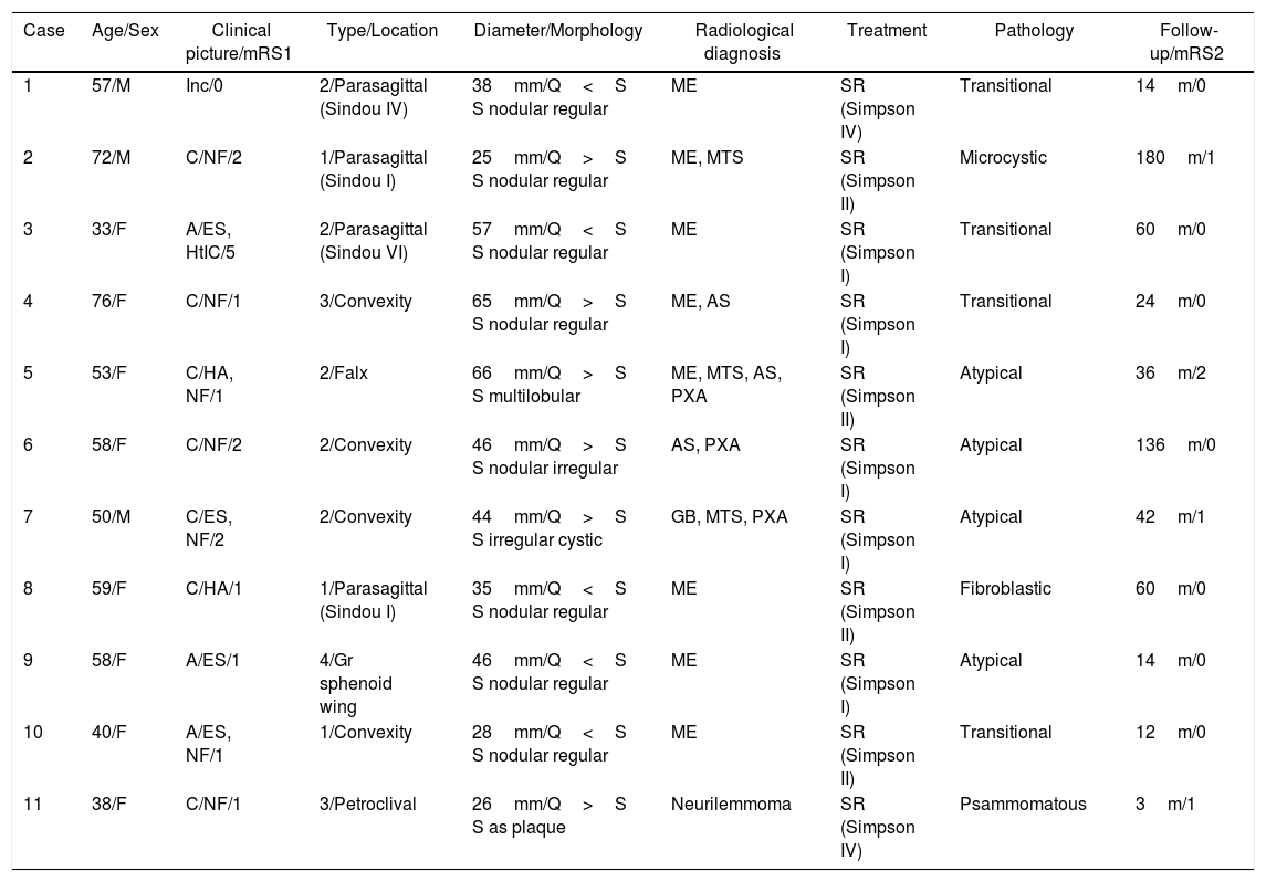
array:23 [
  "pii" => "S252984961830039X"
  "issn" => "25298496"
  "doi" => "10.1016/j.neucie.2018.10.001"
  "estado" => "S300"
  "fechaPublicacion" => "2019-01-01"
  "aid" => "346"
  "copyright" => "Sociedad Española de Neurocirugía"
  "copyrightAnyo" => "2018"
  "documento" => "article"
  "crossmark" => 1
  "subdocumento" => "fla"
  "cita" => "Neurocirugia. 2019;30:1-10"
  "abierto" => array:3 [
    "ES" => false
    "ES2" => false
    "LATM" => false
  ]
  "gratuito" => false
  "lecturas" => array:1 [
    "total" => 0
  ]
  "Traduccion" => array:1 [
    "es" => array:19 [
      "pii" => "S1130147318300861"
      "issn" => "11301473"
      "doi" => "10.1016/j.neucir.2018.08.002"
      "estado" => "S300"
      "fechaPublicacion" => "2019-01-01"
      "aid" => "346"
      "copyright" => "Sociedad Española de Neurocirugía"
      "documento" => "article"
      "crossmark" => 1
      "subdocumento" => "fla"
      "cita" => "Neurocirugia. 2019;30:1-10"
      "abierto" => array:3 [
        "ES" => false
        "ES2" => false
        "LATM" => false
      ]
      "gratuito" => false
      "lecturas" => array:2 [
        "total" => 27
        "formatos" => array:2 [
          "HTML" => 15
          "PDF" => 12
        ]
      ]
      "es" => array:13 [
        "idiomaDefecto" => true
        "cabecera" => "<span class="elsevierStyleTextfn">Investigaci&#243;n cl&#237;nica</span>"
        "titulo" => "Meningiomas qu&#237;sticos&#58; correlaci&#243;n radiol&#243;gica y patol&#243;gica con implicaciones quir&#250;rgicas"
        "tienePdf" => "es"
        "tieneTextoCompleto" => "es"
        "tieneResumen" => array:2 [
          0 => "es"
          1 => "en"
        ]
        "paginas" => array:1 [
          0 => array:2 [
            "paginaInicial" => "1"
            "paginaFinal" => "10"
          ]
        ]
        "titulosAlternativos" => array:1 [
          "en" => array:1 [
            "titulo" => "Cystic meningiomas&#58; Radiological and pathological correlation with surgical implications"
          ]
        ]
        "contieneResumen" => array:2 [
          "es" => true
          "en" => true
        ]
        "contieneTextoCompleto" => array:1 [
          "es" => true
        ]
        "contienePdf" => array:1 [
          "es" => true
        ]
        "resumenGrafico" => array:2 [
          "original" => 0
          "multimedia" => array:7 [
            "identificador" => "fig0035"
            "etiqueta" => "Figura 6"
            "tipo" => "MULTIMEDIAFIGURA"
            "mostrarFloat" => true
            "mostrarDisplay" => false
            "figura" => array:1 [
              0 => array:4 [
                "imagen" => "gr2.jpeg"
                "Alto" => 1205
                "Ancho" => 1250
                "Tamanyo" => 273766
              ]
            ]
            "descripcion" => array:1 [
              "es" => "<p id="spar0095" class="elsevierStyleSimplePara elsevierViewall">Aspectos hist&#243;ricos de los MQ&#46; A&#41; Seg&#250;n la literatura franc&#243;fona&#44; primer MQ de la historia&#46; Neoplasia leptomen&#237;ngea de base dural &#40;flecha negra&#41;&#44; con formaci&#243;n de una cavidad qu&#237;stica y atrofia-gliosis del par&#233;nquima encef&#225;lico adyacente &#40;flechas blancas&#41;&#59; coloraci&#243;n por el m&#233;todo de Loyez&#46; Extra&#237;do de Bouchut et al&#46;<a class="elsevierStyleCrossRef" href="#bib0180"><span class="elsevierStyleSup">8</span></a>&#44; con permiso&#46; B&#41; Atrapamiento a&#233;reo en el asta occipital &#40;flecha negra&#41; identificado durante una neumoencefalograf&#237;a&#44; dando lugar a una falsa imagen de quiste peritumoral en un meningioma intraventricular&#46; Reproducido de David et al&#46;<a class="elsevierStyleCrossRef" href="#bib0185"><span class="elsevierStyleSup">9</span></a>&#44; con permiso&#46; C&#41; Meningioma con voluminoso componente qu&#237;stico&#44; diagnosticado de forma incidental durante una neumoencefalograf&#237;a &#40;flecha blanca&#41;&#59; D&#41; la pieza de resecci&#243;n parece corresponder a un MQ de tipo 2 de la clasificaci&#243;n de Nauta &#40;flechas blancas&#41;&#46; Extra&#237;do de Cushing y Eisenhardt<a class="elsevierStyleCrossRef" href="#bib0190"><span class="elsevierStyleSup">10</span></a>&#44; con permiso&#46;</p>"
            ]
          ]
        ]
        "autores" => array:1 [
          0 => array:2 [
            "autoresLista" => "Rodrigo Carrasco Moro, Herbert D&#46; Jim&#233;nez Zapata, H&#233;ctor Pian Arias, Juan S&#46; Mart&#237;nez San Mill&#225;n, Mar&#237;a A&#46; Mart&#237;nez Rodrigo, Jos&#233; M&#46; Pascual Garvi"
            "autores" => array:6 [
              0 => array:2 [
                "nombre" => "Rodrigo"
                "apellidos" => "Carrasco Moro"
              ]
              1 => array:2 [
                "nombre" => "Herbert D&#46;"
                "apellidos" => "Jim&#233;nez Zapata"
              ]
              2 => array:2 [
                "nombre" => "H&#233;ctor"
                "apellidos" => "Pian Arias"
              ]
              3 => array:2 [
                "nombre" => "Juan S&#46;"
                "apellidos" => "Mart&#237;nez San Mill&#225;n"
              ]
              4 => array:2 [
                "nombre" => "Mar&#237;a A&#46;"
                "apellidos" => "Mart&#237;nez Rodrigo"
              ]
              5 => array:2 [
                "nombre" => "Jos&#233; M&#46;"
                "apellidos" => "Pascual Garvi"
              ]
            ]
          ]
        ]
      ]
      "idiomaDefecto" => "es"
      "Traduccion" => array:1 [
        "en" => array:9 [
          "pii" => "S252984961830039X"
          "doi" => "10.1016/j.neucie.2018.10.001"
          "estado" => "S300"
          "subdocumento" => ""
          "abierto" => array:3 [
            "ES" => false
            "ES2" => false
            "LATM" => false
          ]
          "gratuito" => false
          "lecturas" => array:1 [
            "total" => 0
          ]
          "idiomaDefecto" => "en"
          "EPUB" => "https://multimedia.elsevier.es/PublicationsMultimediaV1/item/epub/S252984961830039X?idApp=UINPBA00004B"
        ]
      ]
      "EPUB" => "https://multimedia.elsevier.es/PublicationsMultimediaV1/item/epub/S1130147318300861?idApp=UINPBA00004B"
      "url" => "/11301473/0000003000000001/v1_201901030610/S1130147318300861/v1_201901030610/es/main.assets"
    ]
  ]
  "itemSiguiente" => array:19 [
    "pii" => "S252984961830042X"
    "issn" => "25298496"
    "doi" => "10.1016/j.neucie.2018.06.001"
    "estado" => "S300"
    "fechaPublicacion" => "2019-01-01"
    "aid" => "342"
    "copyright" => "Sociedad Espa&#241;ola de Neurocirug&#237;a"
    "documento" => "article"
    "crossmark" => 1
    "subdocumento" => "fla"
    "cita" => "Neurocirugia. 2019;30:11-8"
    "abierto" => array:3 [
      "ES" => false
      "ES2" => false
      "LATM" => false
    ]
    "gratuito" => false
    "lecturas" => array:1 [
      "total" => 0
    ]
    "en" => array:13 [
      "idiomaDefecto" => true
      "cabecera" => "<span class="elsevierStyleTextfn">Clinical Research</span>"
      "titulo" => "Perfusion MRI grading diffuse gliomas&#58; Impact of permeability parameters on molecular biomarkers and survival"
      "tienePdf" => "en"
      "tieneTextoCompleto" => "en"
      "tieneResumen" => array:2 [
        0 => "en"
        1 => "es"
      ]
      "paginas" => array:1 [
        0 => array:2 [
          "paginaInicial" => "11"
          "paginaFinal" => "18"
        ]
      ]
      "titulosAlternativos" => array:1 [
        "es" => array:1 [
          "titulo" => "Utilidad de la RM perfusi&#243;n en la gradaci&#243;n de los gliomas difusos&#58; relaci&#243;n entre par&#225;metros de permeabilidad&#44; marcadores moleculares y supervivencia"
        ]
      ]
      "contieneResumen" => array:2 [
        "en" => true
        "es" => true
      ]
      "contieneTextoCompleto" => array:1 [
        "en" => true
      ]
      "contienePdf" => array:1 [
        "en" => true
      ]
      "resumenGrafico" => array:2 [
        "original" => 0
        "multimedia" => array:7 [
          "identificador" => "fig0005"
          "etiqueta" => "Fig&#46; 1"
          "tipo" => "MULTIMEDIAFIGURA"
          "mostrarFloat" => true
          "mostrarDisplay" => false
          "figura" => array:1 [
            0 => array:4 [
              "imagen" => "gr1.jpeg"
              "Alto" => 1452
              "Ancho" => 935
              "Tamanyo" => 264818
            ]
          ]
          "descripcion" => array:1 [
            "en" => "<p id="spar0045" class="elsevierStyleSimplePara elsevierViewall">Immunostaining for the mutated form of isocitrate dehydrogenase &#40;A&#41; and for the loss of ATRX protein expression &#40;B&#41;&#46;</p>"
          ]
        ]
      ]
      "autores" => array:1 [
        0 => array:2 [
          "autoresLista" => "Amaya Hilario, Aurelio Hernandez-Lain, Juan Manuel Sepulveda, Alfonso Lagares, Angel Perez-Nu&#241;ez, Ana Ramos"
          "autores" => array:6 [
            0 => array:2 [
              "nombre" => "Amaya"
              "apellidos" => "Hilario"
            ]
            1 => array:2 [
              "nombre" => "Aurelio"
              "apellidos" => "Hernandez-Lain"
            ]
            2 => array:2 [
              "nombre" => "Juan Manuel"
              "apellidos" => "Sepulveda"
            ]
            3 => array:2 [
              "nombre" => "Alfonso"
              "apellidos" => "Lagares"
            ]
            4 => array:2 [
              "nombre" => "Angel"
              "apellidos" => "Perez-Nu&#241;ez"
            ]
            5 => array:2 [
              "nombre" => "Ana"
              "apellidos" => "Ramos"
            ]
          ]
        ]
      ]
    ]
    "idiomaDefecto" => "en"
    "Traduccion" => array:1 [
      "en" => array:9 [
        "pii" => "S1130147318300678"
        "doi" => "10.1016/j.neucir.2018.06.004"
        "estado" => "S300"
        "subdocumento" => ""
        "abierto" => array:3 [
          "ES" => false
          "ES2" => false
          "LATM" => false
        ]
        "gratuito" => false
        "lecturas" => array:1 [
          "total" => 0
        ]
        "idiomaDefecto" => "en"
        "EPUB" => "https://multimedia.elsevier.es/PublicationsMultimediaV1/item/epub/S1130147318300678?idApp=UINPBA00004B"
      ]
    ]
    "EPUB" => "https://multimedia.elsevier.es/PublicationsMultimediaV1/item/epub/S252984961830042X?idApp=UINPBA00004B"
    "url" => "/25298496/0000003000000001/v1_201901030633/S252984961830042X/v1_201901030633/en/main.assets"
  ]
  "en" => array:21 [
    "idiomaDefecto" => true
    "cabecera" => "<span class="elsevierStyleTextfn">Clinical Research</span>"
    "titulo" => "Cystic meningiomas&#58; Radiological and pathological correlation with surgical implications"
    "tieneTextoCompleto" => true
    "paginas" => array:1 [
      0 => array:2 [
        "paginaInicial" => "1"
        "paginaFinal" => "10"
      ]
    ]
    "autores" => array:1 [
      0 => array:4 [
        "autoresLista" => "Rodrigo Carrasco Moro, Herbert D&#46; Jim&#233;nez Zapata, H&#233;ctor Pian Arias, Juan S&#46; Mart&#237;nez San Mill&#225;n, Mar&#237;a A&#46; Mart&#237;nez Rodrigo, Jos&#233; M&#46; Pascual Garvi"
        "autores" => array:6 [
          0 => array:4 [
            "nombre" => "Rodrigo"
            "apellidos" => "Carrasco Moro"
            "email" => array:1 [
              0 => "rocamo&#64;gmail&#46;com"
            ]
            "referencia" => array:2 [
              0 => array:2 [
                "etiqueta" => "<span class="elsevierStyleSup">a</span>"
                "identificador" => "aff0005"
              ]
              1 => array:2 [
                "etiqueta" => "<span class="elsevierStyleSup">&#42;</span>"
                "identificador" => "cor0005"
              ]
            ]
          ]
          1 => array:3 [
            "nombre" => "Herbert D&#46;"
            "apellidos" => "Jim&#233;nez Zapata"
            "referencia" => array:1 [
              0 => array:2 [
                "etiqueta" => "<span class="elsevierStyleSup">a</span>"
                "identificador" => "aff0005"
              ]
            ]
          ]
          2 => array:3 [
            "nombre" => "H&#233;ctor"
            "apellidos" => "Pian Arias"
            "referencia" => array:1 [
              0 => array:2 [
                "etiqueta" => "<span class="elsevierStyleSup">b</span>"
                "identificador" => "aff0010"
              ]
            ]
          ]
          3 => array:3 [
            "nombre" => "Juan S&#46;"
            "apellidos" => "Mart&#237;nez San Mill&#225;n"
            "referencia" => array:1 [
              0 => array:2 [
                "etiqueta" => "<span class="elsevierStyleSup">c</span>"
                "identificador" => "aff0015"
              ]
            ]
          ]
          4 => array:3 [
            "nombre" => "Mar&#237;a A&#46;"
            "apellidos" => "Mart&#237;nez Rodrigo"
            "referencia" => array:1 [
              0 => array:2 [
                "etiqueta" => "<span class="elsevierStyleSup">a</span>"
                "identificador" => "aff0005"
              ]
            ]
          ]
          5 => array:3 [
            "nombre" => "Jos&#233; M&#46;"
            "apellidos" => "Pascual Garvi"
            "referencia" => array:1 [
              0 => array:2 [
                "etiqueta" => "<span class="elsevierStyleSup">d</span>"
                "identificador" => "aff0020"
              ]
            ]
          ]
        ]
        "afiliaciones" => array:4 [
          0 => array:3 [
            "entidad" => "Servicio de Neurocirug&#237;a&#44; H&#46; U&#46; Ram&#243;n y Cajal&#44; Madrid&#44; Spain"
            "etiqueta" => "a"
            "identificador" => "aff0005"
          ]
          1 => array:3 [
            "entidad" => "Servicio de Anatom&#237;a Patol&#243;gica&#44; H&#46; U&#46; Ram&#243;n y Cajal&#44; Madrid&#44; Spain"
            "etiqueta" => "b"
            "identificador" => "aff0010"
          ]
          2 => array:3 [
            "entidad" => "Servicio de Radiodiagn&#243;stico&#44; H&#46; U&#46; Ram&#243;n y Cajal&#44; Madrid&#44; Spain"
            "etiqueta" => "c"
            "identificador" => "aff0015"
          ]
          3 => array:3 [
            "entidad" => "Servicio de Neurocirug&#237;a&#44; H&#46; U&#46; La Princesa&#44; Madrid&#44; Spain"
            "etiqueta" => "d"
            "identificador" => "aff0020"
          ]
        ]
        "correspondencia" => array:1 [
          0 => array:3 [
            "identificador" => "cor0005"
            "etiqueta" => "&#8270;"
            "correspondencia" => "<span class="elsevierStyleItalic">Corresponding author</span>&#46;"
          ]
        ]
      ]
    ]
    "titulosAlternativos" => array:1 [
      "es" => array:1 [
        "titulo" => "Meningiomas qu&#237;sticos&#58; correlaci&#243;n radiol&#243;gica y patol&#243;gica con implicaciones quir&#250;rgicas"
      ]
    ]
    "resumenGrafico" => array:2 [
      "original" => 0
      "multimedia" => array:7 [
        "identificador" => "fig0005"
        "etiqueta" => "Fig&#46; 1"
        "tipo" => "MULTIMEDIAFIGURA"
        "mostrarFloat" => true
        "mostrarDisplay" => false
        "figura" => array:1 [
          0 => array:4 [
            "imagen" => "gr1.jpeg"
            "Alto" => 1105
            "Ancho" => 1250
            "Tamanyo" => 155351
          ]
        ]
        "descripcion" => array:1 [
          "en" => "<p id="spar0050" class="elsevierStyleSimplePara elsevierViewall">Morphological classification of CMs based on the scheme by Nauta et al&#46; The following are differentiated among tumoural cysts in the strictest sense&#58; &#40;A&#41; those with a central location &#40;type 1&#41; from &#40;B&#41; those with an eccentric location &#40;type 2&#41;&#44; where the wall may in extreme cases be fibrotic with nodules of cancerous cells &#40;arrows&#41;&#46; The following are distinguished with regard to peri-tumoural cysts&#58; &#40;C&#41; those that form within the cerebral parenchyma&#44; giving rise to a parenchymatous pseudocapsule &#40;arrows&#41; of gliotic tissue without encompassing the leptomeninges &#40;type 3&#41;&#44; from &#40;D&#41; those formed through the trapping of CSF &#40;arrow&#41; in the subarachnoid space &#40;type 4&#41;&#46; Type 3 may contain proteinaceous material with shed cancer cells &#40;asterisk in C&#41;&#46; Q&#58; cyst&#59; the solid component of the tumour is shown in orange &#40;dark grey in black and white version&#41;&#46;</p>"
        ]
      ]
    ]
    "textoCompleto" => "<span class="elsevierStyleSections"><span id="sec0005" class="elsevierStyleSection elsevierViewall"><span class="elsevierStyleSectionTitle" id="sect0065">Introduction</span><p id="par0005" class="elsevierStylePara elsevierViewall">Meningiomas are the most common intracranial cancers&#46;<a class="elsevierStyleCrossRef" href="#bib0145"><span class="elsevierStyleSup">1</span></a> From a macroscopic perspective&#44; they usually present as extraparenchymal solid masses with a broad dural implantation base&#44; which facilitates their recognition in radiology and the planning of the most appropriate surgical strategy&#46; However&#44; it is estimated that some 15&#37; of meningiomas develop atypical morphological characteristics that can make their neuroradiological diagnosis more difficult&#46;<a class="elsevierStyleCrossRefs" href="#bib0150"><span class="elsevierStyleSup">2&#44;3</span></a> Among these&#44; the presence of multiple lesions&#44; growth in unusual locations&#44; the association of epiphenomena such as haemorrhage&#44; metaplasia and the development of cysts have been described&#46;<a class="elsevierStyleCrossRefs" href="#bib0150"><span class="elsevierStyleSup">2&#44;3</span></a> It is the last of these that we examine in this study&#44; in which we present our experience in the diagnostic and therapeutic management of a series of meningiomas with significant cystic changes &#40;CM&#41;&#44; discussing the results in the light of the most relevant scientific literature published to date&#46;</p></span><span id="sec0010" class="elsevierStyleSection elsevierViewall"><span class="elsevierStyleSectionTitle" id="sect0070">Material and methods</span><p id="par0010" class="elsevierStylePara elsevierViewall">A review of our surgical records was carried out in search of clinical cases of CM with pathological verification that were operated on in the last 15 years&#46; From each case selected&#44; the following variables were collected&#58; &#40;1&#41; <span class="elsevierStyleItalic">Epidemiological</span>&#58; age and sex&#59; &#40;2&#41; <span class="elsevierStyleItalic">Clinical</span>&#58; form of presentation and time of evolution of the symptoms&#59; &#40;3&#41; <span class="elsevierStyleItalic">Radiological</span>&#58; CT&#44; MRI and angiography findings and morphological classification using the scheme by Nauta et al&#46;<a class="elsevierStyleCrossRef" href="#bib0160"><span class="elsevierStyleSup">4</span></a> &#40;<a class="elsevierStyleCrossRef" href="#fig0005">Fig&#46; 1</a>&#41;&#59; in the event of invasion of the large venous sinuses&#44; this was classified using the model by Sindou and Alvernia<a class="elsevierStyleCrossRef" href="#bib0165"><span class="elsevierStyleSup">5</span></a>&#59; &#40;4&#41; <span class="elsevierStyleItalic">Pathological</span>&#58; WHO classification&#44;<a class="elsevierStyleCrossRef" href="#bib0145"><span class="elsevierStyleSup">1</span></a> cyst wall texture and cytology&#47;pathology of the cystic content&#59; &#40;5&#41; <span class="elsevierStyleItalic">Surgical</span>&#58; type of approach used&#44; degree of tumour resection using the Simpson classification<a class="elsevierStyleCrossRef" href="#bib0170"><span class="elsevierStyleSup">6</span></a> and complications of the surgical procedure&#59; and &#40;6&#41; <span class="elsevierStyleItalic">Prognostic</span>&#58; length of follow-up &#40;months&#41; and detection of recurrences&#46; The functional clinical situation of each patient was classified using the modified Rankin scale<a class="elsevierStyleCrossRef" href="#bib0175"><span class="elsevierStyleSup">7</span></a> at both the time of diagnosis and in the last clinical evaluation&#46;</p><elsevierMultimedia ident="fig0005"></elsevierMultimedia></span><span id="sec0015" class="elsevierStyleSection elsevierViewall"><span class="elsevierStyleSectionTitle" id="sect0075">Results</span><p id="par0015" class="elsevierStylePara elsevierViewall">We identified a total of 11 patients whose fundamental characteristics are described in <a class="elsevierStyleCrossRef" href="#tbl0005">Table 1</a>&#46; The ages of the patients varied between 33 and 76 years &#40;mean 54&#41;&#44; with a female majority &#40;72&#46;7&#37;&#41;&#46; The majority developed neurological symptoms over a period of more than one month&#44; including&#44; in order of frequency&#44; signs of focal involvement&#44; epileptic seizures and headaches&#59; only one patient presented a clinical picture of acute intracranial hypertension&#46; In case 1&#44; the intracranial lesion was an incidental radiological finding in the context of a systemic exploration of tumour extent in non-Hodgkin&#39;s lymphoma of the bone&#46;</p><elsevierMultimedia ident="tbl0005"></elsevierMultimedia><p id="par0020" class="elsevierStylePara elsevierViewall">All of the meningiomas were studied with cerebral CT and MRI &#40;<a class="elsevierStyleCrossRef" href="#tbl0005">Table 1</a>&#44; <a class="elsevierStyleCrossRefs" href="#fig0010">Figs&#46; 2&#8211;5</a>&#41;&#46; Case 1 was an exception in that the interior of the cyst presented a signal identical to that of CSF in all sequences&#44; as well as showing capsular enhancement with gadolinium&#46; In addition to conventional structural sequences&#44; diffusion studies were obtained in all cases &#40;with restriction to the solid component observed only in case 7&#41;&#46; In cases 7&#44; 10 and 11&#44; perfusion studies were performed&#44; revealing a significant increase in perfusion in case 7&#46; The spectroscopy pattern was not conclusive in either of the cases studied &#40;patients 4 and 5&#41;&#46; All of the tumours&#44; except in cases 3 and 5&#44; presented the &#8220;meningeal tail&#8221; sign&#46; In patient 10&#44; hyperostosis of the adjacent bone was also noted&#46; Cases 2&#44; 4&#8211;7&#44; 9 and 10 had associated prominent vasogenic oedema&#46; Cerebral angiography was only performed in patients 5 and 11&#44; with preoperative embolisation of the tumour in the first&#46;</p><elsevierMultimedia ident="fig0010"></elsevierMultimedia><elsevierMultimedia ident="fig0015"></elsevierMultimedia><elsevierMultimedia ident="fig0020"></elsevierMultimedia><elsevierMultimedia ident="fig0025"></elsevierMultimedia><p id="par0025" class="elsevierStylePara elsevierViewall">All patients were treated surgically &#40;<a class="elsevierStyleCrossRef" href="#tbl0005">Table 1</a>&#44; <a class="elsevierStyleCrossRefs" href="#fig0010">Figs&#46; 2&#8211;5</a>&#41;&#46; Type 1 and 2 CMs were resected en-bloc after partially draining the cyst by puncture&#44; followed by meticulous dissection and extirpation of the thin tumour capsule in type 2 cases&#46; Exceptionally&#44; in case 1&#44; the cystic cavity was used as a plane for dissection of the solid component&#44; without capsular resection&#44; since its contents were found to be identical in appearance to CSF&#46; This strategy was identical to that employed in type 3 CMs&#44; which included draining the contents of the cyst&#44; irrigating the resulting cavity with saline solution and taking a biopsy of the pseudocapsular tissue&#44; always with the result of reactive gliosis&#46; In type 4 CMs&#44; only the solid component of the lesion was resected&#46;</p><p id="par0030" class="elsevierStylePara elsevierViewall">The results of the pathological study of the tumours are summarised in <a class="elsevierStyleCrossRef" href="#tbl0005">Table 1</a>&#46; The cytological analysis of the cystic fluid in the type 2 and 3 CMs was negative&#59; however&#44; in case 4&#44; neoplastic cells could be identified within a proteinaceous material&#46;</p><p id="par0035" class="elsevierStylePara elsevierViewall">Follow-up time ranged from 3 to 180 months &#40;mean 52&#46;8&#41;&#44; and no cases of recurrence&#47;regrowth of the tumour were identified in the subsequent post-surgical imaging studies&#44; which were all performed using MRI&#46; The majority of the patients experienced a complete or partial resolution of their initial symptoms &#40;<a class="elsevierStyleCrossRef" href="#tbl0005">Table 1</a>&#41;&#46; As an exception&#44; case 5 suffered from partial retinal ischaemia due to occlusion of the ophthalmic nerve during the pre-operative endovascular procedure&#46;</p></span><span id="sec0020" class="elsevierStyleSection elsevierViewall"><span class="elsevierStyleSectionTitle" id="sect0080">Discussion</span><span id="sec0025" class="elsevierStyleSection elsevierViewall"><span class="elsevierStyleSectionTitle" id="sect0085">History&#44; classification and diagnosis</span><p id="par0040" class="elsevierStylePara elsevierViewall">Although older publications of isolated cases do exist&#44;<a class="elsevierStyleCrossRefs" href="#bib0180"><span class="elsevierStyleSup">8&#44;9</span></a> the first systematic discussion of cystic changes relating meningiomas in the celebrated monograph published by Cushing and Eisenhardt in 1938<a class="elsevierStyleCrossRef" href="#bib0190"><span class="elsevierStyleSup">10</span></a> &#40;<a class="elsevierStyleCrossRef" href="#fig0030">Fig&#46; 6</a>&#41;&#46; In that text&#44; the authors refer to the variability and occasional complexity of these changes&#44; including images of 7 illustrative cases&#46;<a class="elsevierStyleCrossRef" href="#bib0190"><span class="elsevierStyleSup">10</span></a> The cysts might be an accidental finding&#44; secondary to the incidental puncturing of the tumour while performing pneumoencephalography<a class="elsevierStyleCrossRef" href="#bib0190"><span class="elsevierStyleSup">10</span></a> &#40;<a class="elsevierStyleCrossRef" href="#fig0030">Fig&#46; 6</a>&#41;&#46; Given the limitations of the diagnostic technique available at the time&#44; other authors would later signal the importance of considering a presumptive CM diagnosis in this context&#44; given the possibility of confusion with other more common and potentially untreatable entities such as gliomas&#46;<a class="elsevierStyleCrossRefs" href="#bib0195"><span class="elsevierStyleSup">11&#44;12</span></a> The introduction of angiography led to a relevant advance in the diagnosis of these meningiomas&#44; identifying the typical finding of vascularisation of the solid neoplastic component originating from branches of the external carotid artery&#44; while the study of the internal carotid territory revealed phenomena such as angiographic gaps or vascular displacement&#44; with or without leptomeningeal parasitisation<a class="elsevierStyleCrossRefs" href="#bib0160"><span class="elsevierStyleSup">4&#44;11&#8211;13</span></a> &#40;<a class="elsevierStyleCrossRef" href="#fig0010">Fig&#46; 2</a>D&#8211;E&#41;&#46; Nevertheless&#44; in the case of CMs&#44; it can be seen that the rate of correct pre-operative diagnosis was below 20&#37;&#44; as a result of the morphological variability of these lesions&#46;<a class="elsevierStyleCrossRefs" href="#bib0200"><span class="elsevierStyleSup">12&#44;14</span></a></p><elsevierMultimedia ident="fig0030"></elsevierMultimedia><p id="par0045" class="elsevierStylePara elsevierViewall">The introduction of CT facilitated both the diagnosis of the meningiomas and the recognition of atypical changes in the same&#44; improving diagnostic performance to almost 40&#37; for CM specifically&#46;<a class="elsevierStyleCrossRefs" href="#bib0210"><span class="elsevierStyleSup">14&#44;15</span></a> Nowadays this rate is substantially higher thanks to the systematic used of MRI in the study of intracranial tumours&#46;<a class="elsevierStyleCrossRef" href="#bib0220"><span class="elsevierStyleSup">16</span></a> The arrival of modern neuroimaging techniques led to the first attempts to systematically correlate radiological and micro- and macroscopic morphological findings in CM&#46; In contrast to the traditional simplified classifications that merely considered the presence of intra- and&#47;or para-peri-tumoural cysts&#44;<a class="elsevierStyleCrossRefs" href="#bib0200"><span class="elsevierStyleSup">12&#44;13</span></a> other more complex systems were established that more closely reflected intra-operative findings&#46; This process culminated in the aforementioned scheme by Nauta et al&#46;<a class="elsevierStyleCrossRef" href="#bib0160"><span class="elsevierStyleSup">4</span></a> &#40;<a class="elsevierStyleCrossRef" href="#fig0005">Fig&#46; 1</a>&#41;&#44; later modified by Worthington et al&#46;&#44;<a class="elsevierStyleCrossRef" href="#bib0225"><span class="elsevierStyleSup">17</span></a> who added a fifth type of CM&#44; characterised by the presence of a fibrous peripheral cyst wall with nodules of tumour cells&#46; From our perspective&#44; this modification merely adds confusion to the previous classification&#44; since the additional category appears to constitute one extreme of the spectrum of type 2 cysts as described by Nauta et al&#46; More recent revisions&#44; on the other hand&#44; have proposed a return to more simplified schemes&#46;<a class="elsevierStyleCrossRef" href="#bib0230"><span class="elsevierStyleSup">18</span></a></p></span><span id="sec0030" class="elsevierStyleSection elsevierViewall"><span class="elsevierStyleSectionTitle" id="sect0090">Frequency and clinical picture</span><p id="par0050" class="elsevierStylePara elsevierViewall">Based on the series published&#44; it can be estimated that CMs account for 1&#46;7&#8211;11&#46;7&#37; of all meningiomas&#44; having been described in both the cranial and spinal compartments&#46;<a class="elsevierStyleCrossRefs" href="#bib0200"><span class="elsevierStyleSup">12&#44;16&#44;18&#44;19</span></a> They are diagnosed predominantly in middle-aged adult patients&#44; while the relative frequency of CMs in the paediatric population may be more than 10&#37;&#46;<a class="elsevierStyleCrossRef" href="#bib0220"><span class="elsevierStyleSup">16</span></a> Traditionally&#44; the presence of large-volume cysts has been associated with the development of symptoms of intracranial hypertension and a more rapid clinical evolution&#46;<a class="elsevierStyleCrossRefs" href="#bib0190"><span class="elsevierStyleSup">10&#44;13</span></a> Moreover&#44; various authors have demonstrated the particular tendency of type 2 and 3 CMs to produce greater vasogenic oedema&#44; and therefore a mass effect that is disproportionate to the size of the tumour&#46;<a class="elsevierStyleCrossRef" href="#bib0190"><span class="elsevierStyleSup">10</span></a> This presentation&#44; together with a radiological morphology indicative of malignancy&#44; often leads to an incorrect presumptive diagnosis&#46; However&#44; given the availability of advanced imaging techniques in our context&#44; which favours early diagnosis&#44; acute clinical presentation with severe neurological involvement should now be considered uncommon&#44; as we observed in our series&#44; except in those rare cases where it may be accompanied by epiphenomena such as tumoural bleeding&#46;<a class="elsevierStyleCrossRef" href="#bib0210"><span class="elsevierStyleSup">14</span></a></p></span><span id="sec0035" class="elsevierStyleSection elsevierViewall"><span class="elsevierStyleSectionTitle" id="sect0095">Pathogenesis</span><p id="par0055" class="elsevierStylePara elsevierViewall">The precise mechanisms that give rise to cystic changes in meningiomas have not yet been established&#46; Most authors propose the involvement of multiple phenomena&#44; including both sudden haemorrhage and&#47;or ischaemia within the tumour&#44;<a class="elsevierStyleCrossRefs" href="#bib0190"><span class="elsevierStyleSup">10&#44;12&#44;16&#44;19&#44;20</span></a> as well as processes that develop more slowly&#44; whether involutional &#40;vacuolar&#44; mucoid and&#47;or myxomatous degeneration&#41; or involving transudation and&#47;or secretion&#46;<a class="elsevierStyleCrossRefs" href="#bib0195"><span class="elsevierStyleSup">11&#44;14&#44;21</span></a> The second group may be implicated in particular in the genesis of type 2 and 3 CMs&#44; as the analysis of cyst content reveals a composition that is similar to that of cysts associated with other intracranial cancers&#44; ruling out the presence of detritus from prior necrosis or ischaemia&#46;<a class="elsevierStyleCrossRefs" href="#bib0160"><span class="elsevierStyleSup">4&#44;17&#44;22</span></a> The involvement of transudation from the parenchyma through the gliotic pseudocapsule has also been proposed for type 3 CMs&#44; given the absence of the leptomeninges&#46;<a class="elsevierStyleCrossRef" href="#bib0230"><span class="elsevierStyleSup">18</span></a> Finally&#44; in type 4 CMs&#44; the cysts could well be the result of a blockage in the circulation of CSF in the subarachnoid space&#44; or they may be true arachnoid cysts lying adjacent to the tumour&#46;<a class="elsevierStyleCrossRefs" href="#bib0210"><span class="elsevierStyleSup">14&#44;18</span></a></p><p id="par0060" class="elsevierStylePara elsevierViewall">Attempts have been made to correlate the presence of cystic changes with certain histological subtypes of meningioma&#46; Various authors mention a greater frequency among microcystic meningiomas&#44; also known in the literature as Masson&#39;s &#8220;forme humide&#8221;&#44; resulting in cysts of larger volume through the coalescence of smaller cysts&#44;<a class="elsevierStyleCrossRefs" href="#bib0235"><span class="elsevierStyleSup">19&#44;21</span></a> although we have not been able to find data that corroborate this hypothesis in our series or in any of those published previously&#46;<a class="elsevierStyleCrossRef" href="#bib0235"><span class="elsevierStyleSup">19</span></a> Finally&#44; it has also been proposed that cystic changes might reflect a greater histological aggressiveness of the cancer&#44; given the high frequency of atypical meningiomas in the series&#44; including our own&#46;<a class="elsevierStyleCrossRef" href="#bib0220"><span class="elsevierStyleSup">16</span></a> However&#44; we have observed that these changes are detected primarily in grade <span class="elsevierStyleSmallCaps">i</span> meningiomas&#44; with no specific association with any subtype&#44; and that their description in malignant meningiomas is extremely rare&#46;<a class="elsevierStyleCrossRefs" href="#bib0220"><span class="elsevierStyleSup">16&#44;19</span></a></p></span><span id="sec0040" class="elsevierStyleSection elsevierViewall"><span class="elsevierStyleSectionTitle" id="sect0100">Neuroradiological diagnosis and differential diagnosis</span><p id="par0065" class="elsevierStylePara elsevierViewall">The imaging technique of choice for the characterisation of CMs is MRI&#44; and together with CT this has been key to increasing the percentage of correct diagnoses of the lesions to 80&#37;&#44; especially in the presence of characteristic signs of meningiomas themselves&#46;<a class="elsevierStyleCrossRefs" href="#bib0155"><span class="elsevierStyleSup">3&#44;16&#44;23</span></a> Nevertheless&#44; it is estimated that some 10&#37; of CMs continue to be misinterpreted as malignant neoplasms&#44; especially those that also have disorganised morphological changes and prominent vasogenic oedema&#46;<a class="elsevierStyleCrossRef" href="#bib0220"><span class="elsevierStyleSup">16</span></a> In this context&#44; given that the majority of CMs are histologically benign&#44; the identification of restricted diffusion and increased perfusion in the solid component of the tumour will point towards the diagnosis of a cancer with more aggressive biological behaviour&#44; even though these findings do not rule out meningiomas of higher histological grades&#46;<a class="elsevierStyleCrossRefs" href="#bib0155"><span class="elsevierStyleSup">3&#44;16&#44;23</span></a> On the other hand&#44; the utility of spectroscopy for the diagnosis of CM is limited due to the non-homogeneous patterns obtained&#44; in which the characteristic alanine peak can be obscured by the lactate peak&#44; as we observed in the 2 cases in our series in which this sequence was obtained&#46;<a class="elsevierStyleCrossRef" href="#bib0230"><span class="elsevierStyleSup">18</span></a></p><p id="par0070" class="elsevierStylePara elsevierViewall">Type 1 CMs need to be differentiated from other lesions that produce a typical annular take-up image&#44; including primary high-grade tumours and secondary deposits&#44;<a class="elsevierStyleCrossRef" href="#bib0205"><span class="elsevierStyleSup">13</span></a> especially in the context of systemic disease&#46;<a class="elsevierStyleCrossRef" href="#bib0260"><span class="elsevierStyleSup">24</span></a> Type 2 and 3 CMs are the most polymorphic&#44; depending on the proportion of cyst to solid component&#44; the morphology of the latter and the presence of intracystic septa and&#47;or intense vasogenic oedema&#46;<a class="elsevierStyleCrossRefs" href="#bib0160"><span class="elsevierStyleSup">4&#44;17</span></a> Confusion most commonly results from cystic primary tumours with mural nodule with broad contact with the surface of the dura &#40;pleomorphic xanthoastrocytoma&#44; primary desmoplastic tumours&#41; and with haemangioblastomas&#44; where these develop in the posterior fossa&#46;<a class="elsevierStyleCrossRef" href="#bib0265"><span class="elsevierStyleSup">25</span></a> Conversely&#44; type 4 CMs are more easily identifiable due to the radiological behaviour of the cyst and its content&#46;<a class="elsevierStyleCrossRefs" href="#bib0230"><span class="elsevierStyleSup">18&#44;19&#44;24</span></a></p></span><span id="sec0045" class="elsevierStyleSection elsevierViewall"><span class="elsevierStyleSectionTitle" id="sect0105">Surgical treatment</span><p id="par0075" class="elsevierStylePara elsevierViewall">As in other meningiomas&#44; the treatment of choice in CM is surgical resection&#46; Nevertheless&#44; there may be exceptional contexts&#8212;difficulty of establishing an accurate pre-operative diagnosis&#44; difficult-to-access lesions&#44; or the generally unfavourable condition of the patient&#8212;in which it may be decided to perform a needle biopsy&#46; In this case&#44; it is advisable to obtain samples from the solid component of the tumour&#44; since a biopsy of the capsular region and&#47;or cystic content is associated with poor diagnostic returns&#59; findings may also be interpreted erroneously&#44; as has happened historically with the pseudocapsule in type 3 CMs&#44; which were often diagnosed as a cancer of astrocyte lineage&#46;<a class="elsevierStyleCrossRefs" href="#bib0160"><span class="elsevierStyleSup">4&#44;17&#44;22&#44;26</span></a></p><p id="par0080" class="elsevierStylePara elsevierViewall">Usually&#44; type 1 and 4 CMs can be differentiated during surgery if it is possible to define a leptomeningeal dissection plane&#46; However&#44; in the absence of such a plane&#44; intra-operative biopsy is necessary&#44; as they can be confused with tumours of similar radiological appearance&#44; especially secondary deposits and&#47;or primary tumours with leptomeningeal and even dural invasion with a high degree of consistency &#40;e&#46;g&#46; gliosarcomas&#44; metastasis&#44; cancers with associated desmoplasia&#41;&#44; giving rise to a false dissection plane and&#44; therefore&#44; the erroneous intraoperative impression that what is being resected is a meningioma&#46;<a class="elsevierStyleCrossRefs" href="#bib0160"><span class="elsevierStyleSup">4&#44;27</span></a></p><p id="par0085" class="elsevierStylePara elsevierViewall">On the other hand&#44; the presence of cysts facilitates the drainage of the tumour and&#44; as a result&#44; its subsequent peripheral dissection&#46;<a class="elsevierStyleCrossRef" href="#bib0280"><span class="elsevierStyleSup">28</span></a> The greatest problem is posed by those type 2 CMs with thin-walled cysts&#44; historically associated with a higher probability of local recurrence if the capsule is not completely removed&#44; since the cancer has invaded it in up to 60&#37; of cases&#46;<a class="elsevierStyleCrossRefs" href="#bib0210"><span class="elsevierStyleSup">14&#44;18&#44;19</span></a> In such CMs&#44; the recommendation is to perform partial drainage of the cyst and a meticulous dissection of the capsule&#44; which usually presents a good dissection plane with respect to the cerebral parenchyma&#44; as we observed in our series&#46;<a class="elsevierStyleCrossRef" href="#bib0280"><span class="elsevierStyleSup">28</span></a> In contrast&#44; in type 3 and 4 CMs&#44; the cystic cavity can be used as a dissection plane&#44;<a class="elsevierStyleCrossRef" href="#bib0200"><span class="elsevierStyleSup">12</span></a> although we must remember that in the former&#44; the content of the cyst must be fully evacuated due to the presence of cancer cells within it&#44; and the resulting cavity well-irrigated with saline&#46;<a class="elsevierStyleCrossRefs" href="#bib0220"><span class="elsevierStyleSup">16&#44;28</span></a></p></span></span><span id="sec0050" class="elsevierStyleSection elsevierViewall"><span class="elsevierStyleSectionTitle" id="sect0110">Conclusions</span><p id="par0090" class="elsevierStylePara elsevierViewall">CMs represent an uncommon variant on the morphological spectrum of meningiomas&#44; which&#44; due to their polymorphsim&#44; can give rise to serious difficulties in radiological differential diagnosis&#46; Although no association with any histological subtype has been demonstrated&#44; we observed a high frequency of atypical meningiomas in our series&#46; In all CMs&#44; especially types 2 and 3&#44; both biopsy of all tissue in which invasion by the cancer is suspected&#44; and thorough irrigation of the surgical field are recommended to avoid recurrences&#46;</p></span><span id="sec0055" class="elsevierStyleSection elsevierViewall"><span class="elsevierStyleSectionTitle" id="sect0115">Conflicts of interest</span><p id="par0095" class="elsevierStylePara elsevierViewall">The production of this manuscript does not give rise to any conflict of interest and has been conducted in accordance with current ethical standards&#44; with no financial assistance&#46;</p></span></span>"
    "textoCompletoSecciones" => array:1 [
      "secciones" => array:12 [
        0 => array:3 [
          "identificador" => "xres1133116"
          "titulo" => "Abstract"
          "secciones" => array:4 [
            0 => array:2 [
              "identificador" => "abst0005"
              "titulo" => "Introduction"
            ]
            1 => array:2 [
              "identificador" => "abst0010"
              "titulo" => "Material and methods"
            ]
            2 => array:2 [
              "identificador" => "abst0015"
              "titulo" => "Results"
            ]
            3 => array:2 [
              "identificador" => "abst0020"
              "titulo" => "Conclusions"
            ]
          ]
        ]
        1 => array:2 [
          "identificador" => "xpalclavsec1065524"
          "titulo" => "Keywords"
        ]
        2 => array:3 [
          "identificador" => "xres1133115"
          "titulo" => "Resumen"
          "secciones" => array:4 [
            0 => array:2 [
              "identificador" => "abst0025"
              "titulo" => "Introducci&#243;n"
            ]
            1 => array:2 [
              "identificador" => "abst0030"
              "titulo" => "Material y m&#233;todos"
            ]
            2 => array:2 [
              "identificador" => "abst0035"
              "titulo" => "Resultados"
            ]
            3 => array:2 [
              "identificador" => "abst0040"
              "titulo" => "Conclusiones"
            ]
          ]
        ]
        3 => array:2 [
          "identificador" => "xpalclavsec1065523"
          "titulo" => "Palabras clave"
        ]
        4 => array:2 [
          "identificador" => "sec0005"
          "titulo" => "Introduction"
        ]
        5 => array:2 [
          "identificador" => "sec0010"
          "titulo" => "Material and methods"
        ]
        6 => array:2 [
          "identificador" => "sec0015"
          "titulo" => "Results"
        ]
        7 => array:3 [
          "identificador" => "sec0020"
          "titulo" => "Discussion"
          "secciones" => array:5 [
            0 => array:2 [
              "identificador" => "sec0025"
              "titulo" => "History&#44; classification and diagnosis"
            ]
            1 => array:2 [
              "identificador" => "sec0030"
              "titulo" => "Frequency and clinical picture"
            ]
            2 => array:2 [
              "identificador" => "sec0035"
              "titulo" => "Pathogenesis"
            ]
            3 => array:2 [
              "identificador" => "sec0040"
              "titulo" => "Neuroradiological diagnosis and differential diagnosis"
            ]
            4 => array:2 [
              "identificador" => "sec0045"
              "titulo" => "Surgical treatment"
            ]
          ]
        ]
        8 => array:2 [
          "identificador" => "sec0050"
          "titulo" => "Conclusions"
        ]
        9 => array:2 [
          "identificador" => "sec0055"
          "titulo" => "Conflicts of interest"
        ]
        10 => array:2 [
          "identificador" => "xack385752"
          "titulo" => "Acknowledgements"
        ]
        11 => array:1 [
          "titulo" => "References"
        ]
      ]
    ]
    "pdfFichero" => "main.pdf"
    "tienePdf" => true
    "fechaRecibido" => "2018-03-23"
    "fechaAceptado" => "2018-08-18"
    "PalabrasClave" => array:2 [
      "en" => array:1 [
        0 => array:4 [
          "clase" => "keyword"
          "titulo" => "Keywords"
          "identificador" => "xpalclavsec1065524"
          "palabras" => array:3 [
            0 => "Meningioma"
            1 => "Cyst"
            2 => "Intracranial cystic tumours"
          ]
        ]
      ]
      "es" => array:1 [
        0 => array:4 [
          "clase" => "keyword"
          "titulo" => "Palabras clave"
          "identificador" => "xpalclavsec1065523"
          "palabras" => array:3 [
            0 => "Meningioma"
            1 => "Quiste"
            2 => "Tumores qu&#237;sticos intracraneales"
          ]
        ]
      ]
    ]
    "tieneResumen" => true
    "resumen" => array:2 [
      "en" => array:3 [
        "titulo" => "Abstract"
        "resumen" => "<span id="abst0005" class="elsevierStyleSection elsevierViewall"><span class="elsevierStyleSectionTitle" id="sect0010">Introduction</span><p id="spar0005" class="elsevierStyleSimplePara elsevierViewall">Meningiomas associating prominent cystic changes &#40;CM&#41; have challenged neurosurgeons since the beginning of this surgical discipline&#46; We present the experience in the diagnostic and therapeutic management of this entity in our institution&#46;</p></span> <span id="abst0010" class="elsevierStyleSection elsevierViewall"><span class="elsevierStyleSectionTitle" id="sect0015">Material and methods</span><p id="spar0010" class="elsevierStyleSimplePara elsevierViewall">A review of our patient database was carried out&#44; searching for those CM that were operated on in the last 15 years&#46; Relevant clinical data were recorded and analysed for each case&#44; with special emphasis in the correlation of radiological and pathological findings&#46; Cystic changes were classified according to the scheme proposed by Nauta et al&#46;</p></span> <span id="abst0015" class="elsevierStyleSection elsevierViewall"><span class="elsevierStyleSectionTitle" id="sect0020">Results</span><p id="spar0015" class="elsevierStyleSimplePara elsevierViewall">A total of 11 patients were gathered&#44; which represents 1&#46;8&#37; of the meningiomas operated on in our department during the period studied&#46; All were adults&#44; predominantly female patients &#40;8 cases&#41;&#46; Among the clinical symptoms a high rate of epileptic seizures was observed while only one patient developed acute intracranial hypertension&#46; Morphologically&#44; most tumours corresponded to type 2 CMs&#44; followed by types 1&#44; 3 and 4&#46; In only five patients an unequivocal radiological diagnosis of meningioma could be made&#46; All neoplasms were surgically removed and there were no records of recurrence &#40;mean follow-up&#58; 52&#46;8 months&#41;&#46; Microscopic findings were consistent with the pathological diagnosis of atypical meningioma in 4 cases&#44; while the remaining tumours corresponded to OMS grade I neoplasms with variable microscopic patterns&#46;</p></span> <span id="abst0020" class="elsevierStyleSection elsevierViewall"><span class="elsevierStyleSectionTitle" id="sect0025">Conclusions</span><p id="spar0020" class="elsevierStyleSimplePara elsevierViewall">Meningiomas can exceptionally associate cystic changes&#44; both intra and&#47;or extratumoral&#44; in variable number and size&#46; When cystic changes become too prominent &#40;a large number or big-sized cysts&#41;&#44; a serious preoperative diagnostic dilemma may arise&#46; The surgical management of those CMs displaying a peripheral&#44; thin-walled cyst &#40;types 2 and 3&#41; is especially complex&#44; as contrast enhancement of the tumour wall did not correlate strictly with neoplastic invasion&#59; even in the absence of this feature free floating islands of meningothelial cells intermixed with cyst fluid can be found&#46;</p><p id="spar0025" class="elsevierStyleSimplePara elsevierViewall">Consequently both surgical biopsy of every suspicious tissue and copious irrigation of the surgical cavity are strongly recommended for these CM types&#46;</p></span>"
        "secciones" => array:4 [
          0 => array:2 [
            "identificador" => "abst0005"
            "titulo" => "Introduction"
          ]
          1 => array:2 [
            "identificador" => "abst0010"
            "titulo" => "Material and methods"
          ]
          2 => array:2 [
            "identificador" => "abst0015"
            "titulo" => "Results"
          ]
          3 => array:2 [
            "identificador" => "abst0020"
            "titulo" => "Conclusions"
          ]
        ]
      ]
      "es" => array:3 [
        "titulo" => "Resumen"
        "resumen" => "<span id="abst0025" class="elsevierStyleSection elsevierViewall"><span class="elsevierStyleSectionTitle" id="sect0035">Introducci&#243;n</span><p id="spar0030" class="elsevierStyleSimplePara elsevierViewall">Los meningiomas que asocian cambios qu&#237;sticos prominentes &#40;MQ&#41; han representado un reto diagn&#243;stico y terap&#233;utico desde los or&#237;genes de la neurocirug&#237;a moderna&#46; Presentamos la experiencia en el manejo cl&#237;nico y quir&#250;rgico de esta entidad en nuestro Servicio&#46;</p></span> <span id="abst0030" class="elsevierStyleSection elsevierViewall"><span class="elsevierStyleSectionTitle" id="sect0040">Material y m&#233;todos</span><p id="spar0035" class="elsevierStyleSimplePara elsevierViewall">En este estudio descriptivo se incluyen los MQ intervenidos en nuestro Servicio en los &#250;ltimos 15 a&#241;os&#44; evaluando las variables cl&#237;nicas&#44; diagn&#243;sticas y terap&#233;uticas de este subgrupo de meningiomas&#44; con especial &#233;nfasis en la correlaci&#243;n de los hallazgos radiol&#243;gicos y patol&#243;gicos&#46; Los tumores se clasificaron siguiendo el esquema propuesto por Nauta y sus colaboradores&#46;</p></span> <span id="abst0035" class="elsevierStyleSection elsevierViewall"><span class="elsevierStyleSectionTitle" id="sect0045">Resultados</span><p id="spar0040" class="elsevierStyleSimplePara elsevierViewall">Se identificaron 11 casos&#44; lo que supone un 1&#44;8&#37; de los meningiomas intervenidos en ese periodo&#46; Todos los pacientes eran adultos y 8 de ellos&#44; mujeres&#46; Entre los s&#237;ntomas&#44; destac&#243; la alta frecuencia de crisis epil&#233;pticas&#44; mientras que solo un caso desarroll&#243; hipertensi&#243;n intracraneal aguda&#46; La morfolog&#237;a predominante fue el tipo 2&#44; seguida de los tipos 1&#44; 3 y 4&#46; En 5 casos pudo establecerse un diagn&#243;stico radiol&#243;gico inequ&#237;voco de meningioma&#46; Todos los pacientes fueron tratados mediante cirug&#237;a&#44; sin evidenciarse casos de recurrencia durante un seguimiento medio de 52&#44;8 meses&#46; Aunque la mayor&#237;a de las neoplasias correspondieron al grado <span class="elsevierStyleSmallCaps">i</span> de la OMS&#44; incluyendo subtipos variados&#44; 4 de ellas fueron diagnosticadas como meningiomas at&#237;picos&#46;</p></span> <span id="abst0040" class="elsevierStyleSection elsevierViewall"><span class="elsevierStyleSectionTitle" id="sect0050">Conclusiones</span><p id="spar0045" class="elsevierStyleSimplePara elsevierViewall">De forma excepcional&#44; los meningiomas pueden desarrollar cambios qu&#237;sticos&#44; intra- o extratumorales&#44; en n&#250;mero y tama&#241;o variable&#46; Estos constituyen hallazgos poco t&#237;picos que pueden dificultar seriamente su diagn&#243;stico diferencial radiol&#243;gico&#46; Los meningiomas con quistes perif&#233;ricos de c&#225;psula fina &#40;tipos 2 y 3&#41; suponen el escenario quir&#250;rgico m&#225;s complejo&#44; pues el realce capsular en las pruebas de imagen no traduce necesariamente infiltraci&#243;n tumoral&#44; mientras que el quiste puede contener c&#233;lulas neopl&#225;sicas&#46; Por lo tanto&#44; recomendamos realizar al menos biopsia de la c&#225;psula o seudoc&#225;psula&#44; y lavado minucioso de la cavidad quir&#250;rgica especialmente en estos subtipos&#46;</p></span>"
        "secciones" => array:4 [
          0 => array:2 [
            "identificador" => "abst0025"
            "titulo" => "Introducci&#243;n"
          ]
          1 => array:2 [
            "identificador" => "abst0030"
            "titulo" => "Material y m&#233;todos"
          ]
          2 => array:2 [
            "identificador" => "abst0035"
            "titulo" => "Resultados"
          ]
          3 => array:2 [
            "identificador" => "abst0040"
            "titulo" => "Conclusiones"
          ]
        ]
      ]
    ]
    "NotaPie" => array:2 [
      0 => array:2 [
        "etiqueta" => "&#9734;"
        "nota" => "<p class="elsevierStyleNotepara" id="npar0005">Please cite this article as&#58; Carrasco Moro R&#44; Jim&#233;nez Zapata HD&#44; Pian Arias H&#44; Mart&#237;nez San Mill&#225;n JS&#44; Mart&#237;nez Rodrigo MA&#44; Pascual Garvi JM&#46; Meningiomas qu&#237;sticos&#58; correlaci&#243;n radiol&#243;gica y patol&#243;gica con implicaciones quir&#250;rgicas&#46; Neurocirugia&#46; 2019&#59;30&#58;1&#8211;10&#46;</p>"
      ]
      1 => array:2 [
        "etiqueta" => "&#9734;&#9734;"
        "nota" => "<p class="elsevierStyleNotepara" id="npar0010">Some of the data contained in this document were presented as a poster communication during the Annual Congress of the European Association of Neurosurgical Societies &#40;EANS&#41;&#44; held in Venice in October 2017&#46;</p>"
      ]
    ]
    "multimedia" => array:7 [
      0 => array:7 [
        "identificador" => "fig0005"
        "etiqueta" => "Fig&#46; 1"
        "tipo" => "MULTIMEDIAFIGURA"
        "mostrarFloat" => true
        "mostrarDisplay" => false
        "figura" => array:1 [
          0 => array:4 [
            "imagen" => "gr1.jpeg"
            "Alto" => 1105
            "Ancho" => 1250
            "Tamanyo" => 155351
          ]
        ]
        "descripcion" => array:1 [
          "en" => "<p id="spar0050" class="elsevierStyleSimplePara elsevierViewall">Morphological classification of CMs based on the scheme by Nauta et al&#46; The following are differentiated among tumoural cysts in the strictest sense&#58; &#40;A&#41; those with a central location &#40;type 1&#41; from &#40;B&#41; those with an eccentric location &#40;type 2&#41;&#44; where the wall may in extreme cases be fibrotic with nodules of cancerous cells &#40;arrows&#41;&#46; The following are distinguished with regard to peri-tumoural cysts&#58; &#40;C&#41; those that form within the cerebral parenchyma&#44; giving rise to a parenchymatous pseudocapsule &#40;arrows&#41; of gliotic tissue without encompassing the leptomeninges &#40;type 3&#41;&#44; from &#40;D&#41; those formed through the trapping of CSF &#40;arrow&#41; in the subarachnoid space &#40;type 4&#41;&#46; Type 3 may contain proteinaceous material with shed cancer cells &#40;asterisk in C&#41;&#46; Q&#58; cyst&#59; the solid component of the tumour is shown in orange &#40;dark grey in black and white version&#41;&#46;</p>"
        ]
      ]
      1 => array:7 [
        "identificador" => "fig0010"
        "etiqueta" => "Fig&#46; 2"
        "tipo" => "MULTIMEDIAFIGURA"
        "mostrarFloat" => true
        "mostrarDisplay" => false
        "figura" => array:1 [
          0 => array:4 [
            "imagen" => "gr2.jpeg"
            "Alto" => 973
            "Ancho" => 1750
            "Tamanyo" => 259785
          ]
        ]
        "descripcion" => array:1 [
          "en" => "<p id="spar0055" class="elsevierStyleSimplePara elsevierViewall">Type 1 CM&#46; &#40;A&#8211;D&#41; Case 10&#46; Brain MRI&#44; coronal section in T1 weighted sequence after administration of paramagnetic contrast &#40;A&#41; showing an expansive process in the convexity of the left hemisphere&#44; with a broad base contacting the dura and &#8220;meningeal tail&#8221; sign&#44; as well as overlying hyperostosis in the CT with a bone window &#40;B&#41;&#46; The cross-section of the surgical resection specimen &#40;C&#41; shows a greyish peripheral component&#44; compatible from a macroscopic perspective with a transitional meningioma &#40;transparent arrow in C&#8211;D&#44; H-E&#44; 2&#46;5<span class="elsevierStyleHsp" style=""></span>&#215;&#41; and another central&#44; yellowish&#44; fibrotic component &#40;black arrow in C&#8211;D&#44; H&#8211;E 2&#46;5<span class="elsevierStyleHsp" style=""></span>&#215;&#41;&#46; E&#8211;F&#41; Case 8&#46; Tumour with behaviour similar to the previous case in the T1 weighted sequence with contrast &#40;E&#41;&#46; During surgery&#44; xanthochromic liquid content was identified inside it&#44; facilitating the drainage and subsequent resection of the lesion &#40;F&#41;&#46;</p>"
        ]
      ]
      2 => array:7 [
        "identificador" => "fig0015"
        "etiqueta" => "Fig&#46; 3"
        "tipo" => "MULTIMEDIAFIGURA"
        "mostrarFloat" => true
        "mostrarDisplay" => false
        "figura" => array:1 [
          0 => array:4 [
            "imagen" => "gr3.jpeg"
            "Alto" => 1827
            "Ancho" => 1250
            "Tamanyo" => 306054
          ]
        ]
        "descripcion" => array:1 [
          "en" => "<p id="spar0060" class="elsevierStyleSimplePara elsevierViewall">Type 2 CM &#40;case 5&#41;&#46; &#40;A&#41; Head CT with contrast&#46; An expansive process with a broad surface contacting the dura&#44; which includes a solid component with homogeneous contrast uptake&#44; and another larger&#44; septated&#44; hypodense peripheral component with contrast uptake in its periphery&#46; The lesion is associated with intense vasogenic oedema&#46; &#40;B&#41; Weighted T2 sequence MRI&#44; coronal section&#44; showing a hypointense signal from the solid component and a homogeneous hyperintense signal from the cystic component&#46; &#40;C&#41; FLAIR sequence MRI with gadolinium&#44; coronal projection&#59; disorganised uptake in the nodular portion&#44; as well as in the periphery and cystic septa&#44; with hypointense content in its interior&#46; &#40;D&#8211;E&#41; Cerebral angiography&#44; anteroposterior projection&#44; showing &#40;D&#41; arterial supply to the nodular portion from dural branches originating in the ophthalmic and middle meningeal arteries &#40;right external carotid axis&#41;&#44; and &#40;E&#41; vascular distortion of the left internal carotid arterial network&#46; &#40;F&#8211;I&#41; The following were identified during surgery&#58; &#40;H&#41; a thin wall&#44; &#40;I&#41; a fibrous membrane texture &#40;H&#8211;E 4&#215;&#41;&#44; dissectible from the surrounding encephalic parenchyma after partially draining its contents by puncture&#44; and &#40;F&#41; a deep nodular portion adhered to the falx cerebri&#44; corresponding to &#40;G&#41; a meningioma with an angiomatous pattern &#40;H&#8211;E 20&#215;&#41;&#44; eventually classified as atypical&#46;</p>"
        ]
      ]
      3 => array:7 [
        "identificador" => "fig0020"
        "etiqueta" => "Fig&#46; 4"
        "tipo" => "MULTIMEDIAFIGURA"
        "mostrarFloat" => true
        "mostrarDisplay" => false
        "figura" => array:1 [
          0 => array:4 [
            "imagen" => "gr4.jpeg"
            "Alto" => 1446
            "Ancho" => 1750
            "Tamanyo" => 407759
          ]
        ]
        "descripcion" => array:1 [
          "en" => "<p id="spar0065" class="elsevierStyleSimplePara elsevierViewall">Type 3 CM &#40;case 4&#41;&#46; An expansive process with a solid component with a broad dural base in the right frontal convexity&#44; and another cystic process posterior to the first&#44; hypodense in the CT &#40;A&#41;&#44; hyperintense in T2 &#40;B&#41; and hypointense in the FLAIR sequence&#44; without capsular enhancement after administering gadolinium &#40;D&#41;&#46; During the surgery&#44; the solid component &#40;E&#44; transparent arrow from D&#41; was resected en-bloc together with the overlying dura mater&#59; the microscopic study of this was compatible with the diagnosis of transitional meningioma &#40;F&#44; H&#8211;E 10&#215;&#41;&#46; The cystic portion &#40;G&#44; white arrow from D&#41; revealed an internal surface with a gliotic appearance&#44; verified in the pathological study &#40;H&#44; H&#8211;E 2&#46;5&#215;&#41;&#44; while the cystic content consisted of xanthochromic fluid and an amorphous material &#40;visible in the photograph between the ends of the forceps&#41;&#44; with a high protein content&#44; but with nodules of cancerous cells within it &#40;black arrow in I&#44; H&#8211;E 10&#215;&#41;&#46;</p>"
        ]
      ]
      4 => array:7 [
        "identificador" => "fig0025"
        "etiqueta" => "Fig&#46; 5"
        "tipo" => "MULTIMEDIAFIGURA"
        "mostrarFloat" => true
        "mostrarDisplay" => false
        "figura" => array:1 [
          0 => array:4 [
            "imagen" => "gr5.jpeg"
            "Alto" => 766
            "Ancho" => 1250
            "Tamanyo" => 183721
          ]
        ]
        "descripcion" => array:1 [
          "en" => "<p id="spar0070" class="elsevierStyleSimplePara elsevierViewall">Type 4 CM &#40;case 9&#41;&#46; &#40;A&#41; Cranial MRI&#44; T1 weighted sequence after administration of gadolinium&#44; axial cut&#44; showing a predominantly solid lesion with intense contrast uptake&#44; with a broad implantation base and &#8220;meningeal tail&#8221; sign&#46; In its anterior portion&#44; a small component is visible with a signal identical to CSF in all sequences &#40;transparent arrow&#41;&#44; without contrast uptake&#44; compatible with the diagnosis of a peri-tumoural arachnoid cyst&#44; as could be verified during surgery &#40;transparent arrow in B&#41;&#46;</p>"
        ]
      ]
      5 => array:7 [
        "identificador" => "fig0030"
        "etiqueta" => "Fig&#46; 6"
        "tipo" => "MULTIMEDIAFIGURA"
        "mostrarFloat" => true
        "mostrarDisplay" => false
        "figura" => array:1 [
          0 => array:4 [
            "imagen" => "gr6.jpeg"
            "Alto" => 1205
            "Ancho" => 1250
            "Tamanyo" => 273766
          ]
        ]
        "descripcion" => array:1 [
          "en" => "<p id="spar0075" class="elsevierStyleSimplePara elsevierViewall">Historical appearances of CMs&#46; &#40;A&#41; According to francophone literature&#44; the first CM in history&#46; Leptomeningeal tumour with a dural base &#40;black arrow&#41;&#44; with the formation of a cystic cavity and atrophy and gliosis of the adjacent encephalic parenchyma &#40;white arrows&#41;&#59; stained using the Loyez method&#46; Extract taken from Bouchut et al&#46;<a class="elsevierStyleCrossRef" href="#bib0180"><span class="elsevierStyleSup">8</span></a> with permission&#46; &#40;B&#41; Trapped air in the occipital horn &#40;black arrow&#41; identified during a pneumoencephalography&#44; producing a false image of a peri-tumoural cyst in an intraventricular meningioma&#46; Reproduced from David et al&#46;<a class="elsevierStyleCrossRef" href="#bib0185"><span class="elsevierStyleSup">9</span></a> with permission&#46; &#40;C&#41; Meningioma with a sizeable cystic component&#44; diagnosed incidentally during a pneumoencephalography &#40;white arrow&#41;&#59; &#40;D&#41; the resected specimen appears to be a type 2 CM based on Nauta&#39;s classification &#40;white arrows&#41;&#46; Extract taken from Cushing and Eisenhardt<a class="elsevierStyleCrossRef" href="#bib0190"><span class="elsevierStyleSup">10</span></a> with permission&#46;</p>"
        ]
      ]
      6 => array:8 [
        "identificador" => "tbl0005"
        "etiqueta" => "Table 1"
        "tipo" => "MULTIMEDIATABLA"
        "mostrarFloat" => true
        "mostrarDisplay" => false
        "detalles" => array:1 [
          0 => array:3 [
            "identificador" => "at1"
            "detalle" => "Table "
            "rol" => "short"
          ]
        ]
        "tabla" => array:2 [
          "leyenda" => "<p id="spar0085" class="elsevierStyleSimplePara elsevierViewall">A&#58; acute&#59; Age&#58; age in years&#59; C&#58; chronic&#59; ES&#58; epileptic seizure&#59; F&#58; female&#59; HA&#58; headache&#59; Inc&#58; incidental&#59; M&#58; male&#59; mRS&#58; modified Rankin scale&#59; mRS1&#58; pre-operative value&#59; mRS2 post-operative value&#59; NF&#58; signs of neurological focal involvement&#59; S&#58; sex&#46;</p><p id="spar0090" class="elsevierStyleSimplePara elsevierViewall">Type&#58; CM according to classification by Nauta et al&#46;&#59; AS&#58; astrocytoma&#59; Follow-up&#58; follow-up time in months &#40;m&#41;&#59; Gr&#58; greater&#59; ME&#58; meningioma&#59; MTS&#58; metastasis&#59; PXA&#58; pleomorphic xanthoastrocytoma&#59; Q&#58; cystic portion&#59; S&#58; solid portion&#59; SR&#58; surgical resection&#46;</p>"
          "tablatextoimagen" => array:1 [
            0 => array:2 [
              "tabla" => array:1 [
                0 => """
                  <table border="0" frame="\n
                  \t\t\t\t\tvoid\n
                  \t\t\t\t" class=""><thead title="thead"><tr title="table-row"><th class="td" title="table-head  " align="left" valign="top" scope="col" style="border-bottom: 2px solid black">Case&nbsp;\t\t\t\t\t\t\n
                  \t\t\t\t</th><th class="td" title="table-head  " align="center" valign="top" scope="col" style="border-bottom: 2px solid black">Age&#47;Sex&nbsp;\t\t\t\t\t\t\n
                  \t\t\t\t</th><th class="td" title="table-head  " align="center" valign="top" scope="col" style="border-bottom: 2px solid black">Clinical picture&#47;mRS1&nbsp;\t\t\t\t\t\t\n
                  \t\t\t\t</th><th class="td" title="table-head  " align="center" valign="top" scope="col" style="border-bottom: 2px solid black">Type&#47;Location&nbsp;\t\t\t\t\t\t\n
                  \t\t\t\t</th><th class="td" title="table-head  " align="center" valign="top" scope="col" style="border-bottom: 2px solid black">Diameter&#47;Morphology&nbsp;\t\t\t\t\t\t\n
                  \t\t\t\t</th><th class="td" title="table-head  " align="center" valign="top" scope="col" style="border-bottom: 2px solid black">Radiological diagnosis&nbsp;\t\t\t\t\t\t\n
                  \t\t\t\t</th><th class="td" title="table-head  " align="center" valign="top" scope="col" style="border-bottom: 2px solid black">Treatment&nbsp;\t\t\t\t\t\t\n
                  \t\t\t\t</th><th class="td" title="table-head  " align="center" valign="top" scope="col" style="border-bottom: 2px solid black">Pathology&nbsp;\t\t\t\t\t\t\n
                  \t\t\t\t</th><th class="td" title="table-head  " align="center" valign="top" scope="col" style="border-bottom: 2px solid black">Follow-up&#47;mRS2&nbsp;\t\t\t\t\t\t\n
                  \t\t\t\t</th></tr></thead><tbody title="tbody"><tr title="table-row"><td class="td-with-role" title="table-entry ; entry_with_role_rowhead " align="left" valign="top">1&nbsp;\t\t\t\t\t\t\n
                  \t\t\t\t</td><td class="td" title="table-entry  " align="left" valign="top">57&#47;M&nbsp;\t\t\t\t\t\t\n
                  \t\t\t\t</td><td class="td" title="table-entry  " align="left" valign="top">Inc&#47;0&nbsp;\t\t\t\t\t\t\n
                  \t\t\t\t</td><td class="td" title="table-entry  " align="left" valign="top">2&#47;Parasagittal &#40;Sindou IV&#41;&nbsp;\t\t\t\t\t\t\n
                  \t\t\t\t</td><td class="td" title="table-entry  " align="left" valign="top">38<span class="elsevierStyleHsp" style=""></span>mm&#47;Q<span class="elsevierStyleHsp" style=""></span>&#60;<span class="elsevierStyleHsp" style=""></span>S<br>S nodular regular&nbsp;\t\t\t\t\t\t\n
                  \t\t\t\t</td><td class="td" title="table-entry  " align="left" valign="top">ME&nbsp;\t\t\t\t\t\t\n
                  \t\t\t\t</td><td class="td" title="table-entry  " align="left" valign="top">SR &#40;Simpson IV&#41;&nbsp;\t\t\t\t\t\t\n
                  \t\t\t\t</td><td class="td" title="table-entry  " align="left" valign="top">Transitional&nbsp;\t\t\t\t\t\t\n
                  \t\t\t\t</td><td class="td" title="table-entry  " align="left" valign="top">14<span class="elsevierStyleHsp" style=""></span>m&#47;0&nbsp;\t\t\t\t\t\t\n
                  \t\t\t\t</td></tr><tr title="table-row"><td class="td-with-role" title="table-entry ; entry_with_role_rowhead " align="left" valign="top">2&nbsp;\t\t\t\t\t\t\n
                  \t\t\t\t</td><td class="td" title="table-entry  " align="left" valign="top">72&#47;M&nbsp;\t\t\t\t\t\t\n
                  \t\t\t\t</td><td class="td" title="table-entry  " align="left" valign="top">C&#47;NF&#47;2&nbsp;\t\t\t\t\t\t\n
                  \t\t\t\t</td><td class="td" title="table-entry  " align="left" valign="top">1&#47;Parasagittal &#40;Sindou I&#41;&nbsp;\t\t\t\t\t\t\n
                  \t\t\t\t</td><td class="td" title="table-entry  " align="left" valign="top">25<span class="elsevierStyleHsp" style=""></span>mm&#47;Q<span class="elsevierStyleHsp" style=""></span>&#62;<span class="elsevierStyleHsp" style=""></span>S<br>S nodular regular&nbsp;\t\t\t\t\t\t\n
                  \t\t\t\t</td><td class="td" title="table-entry  " align="left" valign="top">ME&#44; MTS&nbsp;\t\t\t\t\t\t\n
                  \t\t\t\t</td><td class="td" title="table-entry  " align="left" valign="top">SR &#40;Simpson II&#41;&nbsp;\t\t\t\t\t\t\n
                  \t\t\t\t</td><td class="td" title="table-entry  " align="left" valign="top">Microcystic&nbsp;\t\t\t\t\t\t\n
                  \t\t\t\t</td><td class="td" title="table-entry  " align="left" valign="top">180<span class="elsevierStyleHsp" style=""></span>m&#47;1&nbsp;\t\t\t\t\t\t\n
                  \t\t\t\t</td></tr><tr title="table-row"><td class="td-with-role" title="table-entry ; entry_with_role_rowhead " align="left" valign="top">3&nbsp;\t\t\t\t\t\t\n
                  \t\t\t\t</td><td class="td" title="table-entry  " align="left" valign="top">33&#47;F&nbsp;\t\t\t\t\t\t\n
                  \t\t\t\t</td><td class="td" title="table-entry  " align="left" valign="top">A&#47;ES&#44; HtIC&#47;5&nbsp;\t\t\t\t\t\t\n
                  \t\t\t\t</td><td class="td" title="table-entry  " align="left" valign="top">2&#47;Parasagittal &#40;Sindou VI&#41;&nbsp;\t\t\t\t\t\t\n
                  \t\t\t\t</td><td class="td" title="table-entry  " align="left" valign="top">57<span class="elsevierStyleHsp" style=""></span>mm&#47;Q<span class="elsevierStyleHsp" style=""></span>&#60;<span class="elsevierStyleHsp" style=""></span>S<br>S nodular regular&nbsp;\t\t\t\t\t\t\n
                  \t\t\t\t</td><td class="td" title="table-entry  " align="left" valign="top">ME&nbsp;\t\t\t\t\t\t\n
                  \t\t\t\t</td><td class="td" title="table-entry  " align="left" valign="top">SR &#40;Simpson I&#41;&nbsp;\t\t\t\t\t\t\n
                  \t\t\t\t</td><td class="td" title="table-entry  " align="left" valign="top">Transitional&nbsp;\t\t\t\t\t\t\n
                  \t\t\t\t</td><td class="td" title="table-entry  " align="left" valign="top">60<span class="elsevierStyleHsp" style=""></span>m&#47;0&nbsp;\t\t\t\t\t\t\n
                  \t\t\t\t</td></tr><tr title="table-row"><td class="td-with-role" title="table-entry ; entry_with_role_rowhead " align="left" valign="top">4&nbsp;\t\t\t\t\t\t\n
                  \t\t\t\t</td><td class="td" title="table-entry  " align="left" valign="top">76&#47;F&nbsp;\t\t\t\t\t\t\n
                  \t\t\t\t</td><td class="td" title="table-entry  " align="left" valign="top">C&#47;NF&#47;1&nbsp;\t\t\t\t\t\t\n
                  \t\t\t\t</td><td class="td" title="table-entry  " align="left" valign="top">3&#47;Convexity&nbsp;\t\t\t\t\t\t\n
                  \t\t\t\t</td><td class="td" title="table-entry  " align="left" valign="top">65<span class="elsevierStyleHsp" style=""></span>mm&#47;Q<span class="elsevierStyleHsp" style=""></span>&#62;<span class="elsevierStyleHsp" style=""></span>S<br>S nodular regular&nbsp;\t\t\t\t\t\t\n
                  \t\t\t\t</td><td class="td" title="table-entry  " align="left" valign="top">ME&#44; AS&nbsp;\t\t\t\t\t\t\n
                  \t\t\t\t</td><td class="td" title="table-entry  " align="left" valign="top">SR &#40;Simpson I&#41;&nbsp;\t\t\t\t\t\t\n
                  \t\t\t\t</td><td class="td" title="table-entry  " align="left" valign="top">Transitional&nbsp;\t\t\t\t\t\t\n
                  \t\t\t\t</td><td class="td" title="table-entry  " align="left" valign="top">24<span class="elsevierStyleHsp" style=""></span>m&#47;0&nbsp;\t\t\t\t\t\t\n
                  \t\t\t\t</td></tr><tr title="table-row"><td class="td-with-role" title="table-entry ; entry_with_role_rowhead " align="left" valign="top">5&nbsp;\t\t\t\t\t\t\n
                  \t\t\t\t</td><td class="td" title="table-entry  " align="left" valign="top">53&#47;F&nbsp;\t\t\t\t\t\t\n
                  \t\t\t\t</td><td class="td" title="table-entry  " align="left" valign="top">C&#47;HA&#44; NF&#47;1&nbsp;\t\t\t\t\t\t\n
                  \t\t\t\t</td><td class="td" title="table-entry  " align="left" valign="top">2&#47;Falx&nbsp;\t\t\t\t\t\t\n
                  \t\t\t\t</td><td class="td" title="table-entry  " align="left" valign="top">66<span class="elsevierStyleHsp" style=""></span>mm&#47;Q<span class="elsevierStyleHsp" style=""></span>&#62;<span class="elsevierStyleHsp" style=""></span>S<br>S multilobular&nbsp;\t\t\t\t\t\t\n
                  \t\t\t\t</td><td class="td" title="table-entry  " align="left" valign="top">ME&#44; MTS&#44; AS&#44; PXA&nbsp;\t\t\t\t\t\t\n
                  \t\t\t\t</td><td class="td" title="table-entry  " align="left" valign="top">SR &#40;Simpson II&#41;&nbsp;\t\t\t\t\t\t\n
                  \t\t\t\t</td><td class="td" title="table-entry  " align="left" valign="top">Atypical&nbsp;\t\t\t\t\t\t\n
                  \t\t\t\t</td><td class="td" title="table-entry  " align="left" valign="top">36<span class="elsevierStyleHsp" style=""></span>m&#47;2&nbsp;\t\t\t\t\t\t\n
                  \t\t\t\t</td></tr><tr title="table-row"><td class="td-with-role" title="table-entry ; entry_with_role_rowhead " align="left" valign="top">6&nbsp;\t\t\t\t\t\t\n
                  \t\t\t\t</td><td class="td" title="table-entry  " align="left" valign="top">58&#47;F&nbsp;\t\t\t\t\t\t\n
                  \t\t\t\t</td><td class="td" title="table-entry  " align="left" valign="top">C&#47;NF&#47;2&nbsp;\t\t\t\t\t\t\n
                  \t\t\t\t</td><td class="td" title="table-entry  " align="left" valign="top">2&#47;Convexity&nbsp;\t\t\t\t\t\t\n
                  \t\t\t\t</td><td class="td" title="table-entry  " align="left" valign="top">46<span class="elsevierStyleHsp" style=""></span>mm&#47;Q<span class="elsevierStyleHsp" style=""></span>&#62;<span class="elsevierStyleHsp" style=""></span>S<br>S nodular irregular&nbsp;\t\t\t\t\t\t\n
                  \t\t\t\t</td><td class="td" title="table-entry  " align="left" valign="top">AS&#44; PXA&nbsp;\t\t\t\t\t\t\n
                  \t\t\t\t</td><td class="td" title="table-entry  " align="left" valign="top">SR &#40;Simpson I&#41;&nbsp;\t\t\t\t\t\t\n
                  \t\t\t\t</td><td class="td" title="table-entry  " align="left" valign="top">Atypical&nbsp;\t\t\t\t\t\t\n
                  \t\t\t\t</td><td class="td" title="table-entry  " align="left" valign="top">136<span class="elsevierStyleHsp" style=""></span>m&#47;0&nbsp;\t\t\t\t\t\t\n
                  \t\t\t\t</td></tr><tr title="table-row"><td class="td-with-role" title="table-entry ; entry_with_role_rowhead " align="left" valign="top">7&nbsp;\t\t\t\t\t\t\n
                  \t\t\t\t</td><td class="td" title="table-entry  " align="left" valign="top">50&#47;M&nbsp;\t\t\t\t\t\t\n
                  \t\t\t\t</td><td class="td" title="table-entry  " align="left" valign="top">C&#47;ES&#44; NF&#47;2&nbsp;\t\t\t\t\t\t\n
                  \t\t\t\t</td><td class="td" title="table-entry  " align="left" valign="top">2&#47;Convexity&nbsp;\t\t\t\t\t\t\n
                  \t\t\t\t</td><td class="td" title="table-entry  " align="left" valign="top">44<span class="elsevierStyleHsp" style=""></span>mm&#47;Q<span class="elsevierStyleHsp" style=""></span>&#62;<span class="elsevierStyleHsp" style=""></span>S<br>S irregular cystic&nbsp;\t\t\t\t\t\t\n
                  \t\t\t\t</td><td class="td" title="table-entry  " align="left" valign="top">GB&#44; MTS&#44; PXA&nbsp;\t\t\t\t\t\t\n
                  \t\t\t\t</td><td class="td" title="table-entry  " align="left" valign="top">SR &#40;Simpson I&#41;&nbsp;\t\t\t\t\t\t\n
                  \t\t\t\t</td><td class="td" title="table-entry  " align="left" valign="top">Atypical&nbsp;\t\t\t\t\t\t\n
                  \t\t\t\t</td><td class="td" title="table-entry  " align="left" valign="top">42<span class="elsevierStyleHsp" style=""></span>m&#47;1&nbsp;\t\t\t\t\t\t\n
                  \t\t\t\t</td></tr><tr title="table-row"><td class="td-with-role" title="table-entry ; entry_with_role_rowhead " align="left" valign="top">8&nbsp;\t\t\t\t\t\t\n
                  \t\t\t\t</td><td class="td" title="table-entry  " align="left" valign="top">59&#47;F&nbsp;\t\t\t\t\t\t\n
                  \t\t\t\t</td><td class="td" title="table-entry  " align="left" valign="top">C&#47;HA&#47;1&nbsp;\t\t\t\t\t\t\n
                  \t\t\t\t</td><td class="td" title="table-entry  " align="left" valign="top">1&#47;Parasagittal &#40;Sindou I&#41;&nbsp;\t\t\t\t\t\t\n
                  \t\t\t\t</td><td class="td" title="table-entry  " align="left" valign="top">35<span class="elsevierStyleHsp" style=""></span>mm&#47;Q<span class="elsevierStyleHsp" style=""></span>&#60;<span class="elsevierStyleHsp" style=""></span>S<br>S nodular regular&nbsp;\t\t\t\t\t\t\n
                  \t\t\t\t</td><td class="td" title="table-entry  " align="left" valign="top">ME&nbsp;\t\t\t\t\t\t\n
                  \t\t\t\t</td><td class="td" title="table-entry  " align="left" valign="top">SR &#40;Simpson II&#41;&nbsp;\t\t\t\t\t\t\n
                  \t\t\t\t</td><td class="td" title="table-entry  " align="left" valign="top">Fibroblastic&nbsp;\t\t\t\t\t\t\n
                  \t\t\t\t</td><td class="td" title="table-entry  " align="left" valign="top">60<span class="elsevierStyleHsp" style=""></span>m&#47;0&nbsp;\t\t\t\t\t\t\n
                  \t\t\t\t</td></tr><tr title="table-row"><td class="td-with-role" title="table-entry ; entry_with_role_rowhead " align="left" valign="top">9&nbsp;\t\t\t\t\t\t\n
                  \t\t\t\t</td><td class="td" title="table-entry  " align="left" valign="top">58&#47;F&nbsp;\t\t\t\t\t\t\n
                  \t\t\t\t</td><td class="td" title="table-entry  " align="left" valign="top">A&#47;ES&#47;1&nbsp;\t\t\t\t\t\t\n
                  \t\t\t\t</td><td class="td" title="table-entry  " align="left" valign="top">4&#47;Gr sphenoid wing&nbsp;\t\t\t\t\t\t\n
                  \t\t\t\t</td><td class="td" title="table-entry  " align="left" valign="top">46<span class="elsevierStyleHsp" style=""></span>mm&#47;Q<span class="elsevierStyleHsp" style=""></span>&#60;<span class="elsevierStyleHsp" style=""></span>S<br>S nodular regular&nbsp;\t\t\t\t\t\t\n
                  \t\t\t\t</td><td class="td" title="table-entry  " align="left" valign="top">ME&nbsp;\t\t\t\t\t\t\n
                  \t\t\t\t</td><td class="td" title="table-entry  " align="left" valign="top">SR &#40;Simpson I&#41;&nbsp;\t\t\t\t\t\t\n
                  \t\t\t\t</td><td class="td" title="table-entry  " align="left" valign="top">Atypical&nbsp;\t\t\t\t\t\t\n
                  \t\t\t\t</td><td class="td" title="table-entry  " align="left" valign="top">14<span class="elsevierStyleHsp" style=""></span>m&#47;0&nbsp;\t\t\t\t\t\t\n
                  \t\t\t\t</td></tr><tr title="table-row"><td class="td-with-role" title="table-entry ; entry_with_role_rowhead " align="left" valign="top">10&nbsp;\t\t\t\t\t\t\n
                  \t\t\t\t</td><td class="td" title="table-entry  " align="left" valign="top">40&#47;F&nbsp;\t\t\t\t\t\t\n
                  \t\t\t\t</td><td class="td" title="table-entry  " align="left" valign="top">A&#47;ES&#44; NF&#47;1&nbsp;\t\t\t\t\t\t\n
                  \t\t\t\t</td><td class="td" title="table-entry  " align="left" valign="top">1&#47;Convexity&nbsp;\t\t\t\t\t\t\n
                  \t\t\t\t</td><td class="td" title="table-entry  " align="left" valign="top">28<span class="elsevierStyleHsp" style=""></span>mm&#47;Q<span class="elsevierStyleHsp" style=""></span>&#60;<span class="elsevierStyleHsp" style=""></span>S<br>S nodular regular&nbsp;\t\t\t\t\t\t\n
                  \t\t\t\t</td><td class="td" title="table-entry  " align="left" valign="top">ME&nbsp;\t\t\t\t\t\t\n
                  \t\t\t\t</td><td class="td" title="table-entry  " align="left" valign="top">SR &#40;Simpson II&#41;&nbsp;\t\t\t\t\t\t\n
                  \t\t\t\t</td><td class="td" title="table-entry  " align="left" valign="top">Transitional&nbsp;\t\t\t\t\t\t\n
                  \t\t\t\t</td><td class="td" title="table-entry  " align="left" valign="top">12<span class="elsevierStyleHsp" style=""></span>m&#47;0&nbsp;\t\t\t\t\t\t\n
                  \t\t\t\t</td></tr><tr title="table-row"><td class="td-with-role" title="table-entry ; entry_with_role_rowhead " align="left" valign="top">11&nbsp;\t\t\t\t\t\t\n
                  \t\t\t\t</td><td class="td" title="table-entry  " align="left" valign="top">38&#47;F&nbsp;\t\t\t\t\t\t\n
                  \t\t\t\t</td><td class="td" title="table-entry  " align="left" valign="top">C&#47;NF&#47;1&nbsp;\t\t\t\t\t\t\n
                  \t\t\t\t</td><td class="td" title="table-entry  " align="left" valign="top">3&#47;Petroclival&nbsp;\t\t\t\t\t\t\n
                  \t\t\t\t</td><td class="td" title="table-entry  " align="left" valign="top">26<span class="elsevierStyleHsp" style=""></span>mm&#47;Q<span class="elsevierStyleHsp" style=""></span>&#62;<span class="elsevierStyleHsp" style=""></span>S<br>S as plaque&nbsp;\t\t\t\t\t\t\n
                  \t\t\t\t</td><td class="td" title="table-entry  " align="left" valign="top">Neurilemmoma&nbsp;\t\t\t\t\t\t\n
                  \t\t\t\t</td><td class="td" title="table-entry  " align="left" valign="top">SR &#40;Simpson IV&#41;&nbsp;\t\t\t\t\t\t\n
                  \t\t\t\t</td><td class="td" title="table-entry  " align="left" valign="top">Psammomatous&nbsp;\t\t\t\t\t\t\n
                  \t\t\t\t</td><td class="td" title="table-entry  " align="left" valign="top">3<span class="elsevierStyleHsp" style=""></span>m&#47;1&nbsp;\t\t\t\t\t\t\n
                  \t\t\t\t</td></tr></tbody></table>
                  """
              ]
              "imagenFichero" => array:1 [
                0 => "xTab1933268.png"
              ]
            ]
          ]
        ]
        "descripcion" => array:1 [
          "en" => "<p id="spar0080" class="elsevierStyleSimplePara elsevierViewall">CM cases included in our series&#46;</p>"
        ]
      ]
    ]
    "bibliografia" => array:2 [
      "titulo" => "References"
      "seccion" => array:1 [
        0 => array:2 [
          "identificador" => "bibs0015"
          "bibliografiaReferencia" => array:28 [
            0 => array:3 [
              "identificador" => "bib0145"
              "etiqueta" => "1"
              "referencia" => array:1 [
                0 => array:2 [
                  "contribucion" => array:1 [
                    0 => array:2 [
                      "titulo" => "Meningioma variants"
                      "autores" => array:1 [
                        0 => array:2 [
                          "etal" => true
                          "autores" => array:6 [
                            0 => "A&#46; Perry"
                            1 => "D&#46;N&#46; Louis"
                            2 => "H&#46; Budka"
                            3 => "A&#46; von Deimling"
                            4 => "F&#46; Sahm"
                            5 => "C&#46; Mawrin"
                          ]
                        ]
                      ]
                    ]
                  ]
                  "host" => array:1 [
                    0 => array:1 [
                      "LibroEditado" => array:4 [
                        "titulo" => "WHO classification of tumors of the central nervous system"
                        "paginaInicial" => "237"
                        "paginaFinal" => "245"
                        "serieFecha" => "2016"
                      ]
                    ]
                  ]
                ]
              ]
            ]
            1 => array:3 [
              "identificador" => "bib0150"
              "etiqueta" => "2"
              "referencia" => array:1 [
                0 => array:2 [
                  "contribucion" => array:1 [
                    0 => array:2 [
                      "titulo" => "Typical&#44; atypical and misleading features in meningioma"
                      "autores" => array:1 [
                        0 => array:2 [
                          "etal" => false
                          "autores" => array:3 [
                            0 => "M&#46;P&#46; Buetow"
                            1 => "P&#46;C&#46; Buetow"
                            2 => "J&#46;G&#46; Smirniotopoulos"
                          ]
                        ]
                      ]
                    ]
                  ]
                  "host" => array:1 [
                    0 => array:2 [
                      "doi" => "10.1148/radiographics.11.6.1749851"
                      "Revista" => array:6 [
                        "tituloSerie" => "Radiographics"
                        "fecha" => "1991"
                        "volumen" => "11"
                        "paginaInicial" => "1087"
                        "paginaFinal" => "1106"
                        "link" => array:1 [
                          0 => array:2 [
                            "url" => "https://www.ncbi.nlm.nih.gov/pubmed/1749851"
                            "web" => "Medline"
                          ]
                        ]
                      ]
                    ]
                  ]
                ]
              ]
            ]
            2 => array:3 [
              "identificador" => "bib0155"
              "etiqueta" => "3"
              "referencia" => array:1 [
                0 => array:2 [
                  "contribucion" => array:1 [
                    0 => array:2 [
                      "titulo" => "Uncommon cranial meningioma&#58; key imaging features on conventional and advanced imaging"
                      "autores" => array:1 [
                        0 => array:2 [
                          "etal" => false
                          "autores" => array:4 [
                            0 => "N&#46; Zakhari"
                            1 => "C&#46; Torres"
                            2 => "M&#46; Castillo"
                            3 => "T&#46;B&#46; Nguyen"
                          ]
                        ]
                      ]
                    ]
                  ]
                  "host" => array:1 [
                    0 => array:2 [
                      "doi" => "10.1007/s00062-017-0583-y"
                      "Revista" => array:6 [
                        "tituloSerie" => "Clin Neuroradiol"
                        "fecha" => "2017"
                        "volumen" => "27"
                        "paginaInicial" => "135"
                        "paginaFinal" => "144"
                        "link" => array:1 [
                          0 => array:2 [
                            "url" => "https://www.ncbi.nlm.nih.gov/pubmed/28466126"
                            "web" => "Medline"
                          ]
                        ]
                      ]
                    ]
                  ]
                ]
              ]
            ]
            3 => array:3 [
              "identificador" => "bib0160"
              "etiqueta" => "4"
              "referencia" => array:1 [
                0 => array:2 [
                  "contribucion" => array:1 [
                    0 => array:2 [
                      "titulo" => "Xanthocromic cysts associated with meningioma"
                      "autores" => array:1 [
                        0 => array:2 [
                          "etal" => false
                          "autores" => array:5 [
                            0 => "H&#46;J&#46;W&#46; Nauta"
                            1 => "W&#46;S&#46; Tucker"
                            2 => "W&#46;J&#46; Horsey"
                            3 => "J&#46;M&#46; Bilbao"
                            4 => "C&#46; Gonsalves"
                          ]
                        ]
                      ]
                    ]
                  ]
                  "host" => array:1 [
                    0 => array:1 [
                      "Revista" => array:6 [
                        "tituloSerie" => "J Neurol Neurosurg Psychiatry"
                        "fecha" => "1979"
                        "volumen" => "42"
                        "paginaInicial" => "529"
                        "paginaFinal" => "535"
                        "link" => array:1 [
                          0 => array:2 [
                            "url" => "https://www.ncbi.nlm.nih.gov/pubmed/469560"
                            "web" => "Medline"
                          ]
                        ]
                      ]
                    ]
                  ]
                ]
              ]
            ]
            4 => array:3 [
              "identificador" => "bib0165"
              "etiqueta" => "5"
              "referencia" => array:1 [
                0 => array:2 [
                  "contribucion" => array:1 [
                    0 => array:2 [
                      "titulo" => "Results of attempted radical tumor removal and venous repair in 100 consecutive meningiomas involving the major dural sinuses"
                      "autores" => array:1 [
                        0 => array:2 [
                          "etal" => false
                          "autores" => array:2 [
                            0 => "M&#46;P&#46; Sindou"
                            1 => "J&#46;E&#46; Alvernia"
                          ]
                        ]
                      ]
                    ]
                  ]
                  "host" => array:1 [
                    0 => array:2 [
                      "doi" => "10.3171/jns.2006.105.4.514"
                      "Revista" => array:6 [
                        "tituloSerie" => "J Neurosurg"
                        "fecha" => "2006"
                        "volumen" => "105"
                        "paginaInicial" => "514"
                        "paginaFinal" => "525"
                        "link" => array:1 [
                          0 => array:2 [
                            "url" => "https://www.ncbi.nlm.nih.gov/pubmed/17044551"
                            "web" => "Medline"
                          ]
                        ]
                      ]
                    ]
                  ]
                ]
              ]
            ]
            5 => array:3 [
              "identificador" => "bib0170"
              "etiqueta" => "6"
              "referencia" => array:1 [
                0 => array:2 [
                  "contribucion" => array:1 [
                    0 => array:2 [
                      "titulo" => "The recurrence of meningiomas after surgical treatment"
                      "autores" => array:1 [
                        0 => array:2 [
                          "etal" => false
                          "autores" => array:1 [
                            0 => "D&#46; Simpson"
                          ]
                        ]
                      ]
                    ]
                  ]
                  "host" => array:1 [
                    0 => array:1 [
                      "Revista" => array:5 [
                        "tituloSerie" => "J Neurol Neurosurg Psych"
                        "fecha" => "1957"
                        "volumen" => "20"
                        "paginaInicial" => "22"
                        "paginaFinal" => "39"
                      ]
                    ]
                  ]
                ]
              ]
            ]
            6 => array:3 [
              "identificador" => "bib0175"
              "etiqueta" => "7"
              "referencia" => array:1 [
                0 => array:2 [
                  "contribucion" => array:1 [
                    0 => array:2 [
                      "titulo" => "Patient satisfaction and short-term outcome in elective cranial neurosurgery"
                      "autores" => array:1 [
                        0 => array:2 [
                          "etal" => false
                          "autores" => array:4 [
                            0 => "E&#46; Reponen"
                            1 => "H&#46; Tuominen"
                            2 => "J&#46; Hernesniemi"
                            3 => "M&#46; Korja"
                          ]
                        ]
                      ]
                    ]
                  ]
                  "host" => array:1 [
                    0 => array:2 [
                      "doi" => "10.1227/NEU.0000000000000931"
                      "Revista" => array:6 [
                        "tituloSerie" => "Neurosurgery"
                        "fecha" => "2015"
                        "volumen" => "77"
                        "paginaInicial" => "769"
                        "paginaFinal" => "775"
                        "link" => array:1 [
                          0 => array:2 [
                            "url" => "https://www.ncbi.nlm.nih.gov/pubmed/26244270"
                            "web" => "Medline"
                          ]
                        ]
                      ]
                    ]
                  ]
                ]
              ]
            ]
            7 => array:3 [
              "identificador" => "bib0180"
              "etiqueta" => "8"
              "referencia" => array:1 [
                0 => array:3 [
                  "comentario" => "&#91;in Franch&#93;"
                  "contribucion" => array:1 [
                    0 => array:2 [
                      "titulo" => "Tumeurs kystiques des m&#233;ninges&#46; &#201;tude histologique des accidents &#233;volutifs des m&#233;ningoblastomas"
                      "autores" => array:1 [
                        0 => array:2 [
                          "etal" => false
                          "autores" => array:3 [
                            0 => "M&#46; Bouchut"
                            1 => "J&#46; Dechaume"
                            2 => "J&#46; Barbier"
                          ]
                        ]
                      ]
                    ]
                  ]
                  "host" => array:1 [
                    0 => array:1 [
                      "Revista" => array:5 [
                        "tituloSerie" => "J Med Lyon"
                        "fecha" => "1928"
                        "volumen" => "9"
                        "paginaInicial" => "81"
                        "paginaFinal" => "86"
                      ]
                    ]
                  ]
                ]
              ]
            ]
            8 => array:3 [
              "identificador" => "bib0185"
              "etiqueta" => "9"
              "referencia" => array:1 [
                0 => array:3 [
                  "comentario" => "&#91;in French&#93;"
                  "contribucion" => array:1 [
                    0 => array:2 [
                      "titulo" => "Intraventricular meningioma"
                      "autores" => array:1 [
                        0 => array:2 [
                          "etal" => false
                          "autores" => array:3 [
                            0 => "M&#46; David"
                            1 => "L&#46; Guillaumat"
                            2 => "H&#46; Ask&#233;nasy"
                          ]
                        ]
                      ]
                    ]
                  ]
                  "host" => array:1 [
                    0 => array:1 [
                      "Revista" => array:5 [
                        "tituloSerie" => "Rev Neurol"
                        "fecha" => "1937"
                        "volumen" => "67"
                        "paginaInicial" => "504"
                        "paginaFinal" => "514"
                      ]
                    ]
                  ]
                ]
              ]
            ]
            9 => array:3 [
              "identificador" => "bib0190"
              "etiqueta" => "10"
              "referencia" => array:1 [
                0 => array:2 [
                  "contribucion" => array:1 [
                    0 => array:2 [
                      "titulo" => "Gross and microscopical pathology"
                      "autores" => array:1 [
                        0 => array:2 [
                          "etal" => false
                          "autores" => array:2 [
                            0 => "H&#46; Cushing"
                            1 => "L&#46; Eisenhardt"
                          ]
                        ]
                      ]
                    ]
                  ]
                  "host" => array:1 [
                    0 => array:1 [
                      "LibroEditado" => array:4 [
                        "titulo" => "Meningiomas&#46; Their classification&#44; regional behavior&#44; life history&#44; and surgical end results"
                        "paginaInicial" => "26"
                        "paginaFinal" => "27"
                        "serieFecha" => "1938"
                      ]
                    ]
                  ]
                ]
              ]
            ]
            10 => array:3 [
              "identificador" => "bib0195"
              "etiqueta" => "11"
              "referencia" => array:1 [
                0 => array:3 [
                  "comentario" => "&#91;in Spanish&#93;"
                  "contribucion" => array:1 [
                    0 => array:2 [
                      "titulo" => "Two cases of cystic meningioma of the parietal region"
                      "autores" => array:1 [
                        0 => array:2 [
                          "etal" => false
                          "autores" => array:2 [
                            0 => "W&#46; Calvo"
                            1 => "J&#46;L&#46; Barcia-Salorio"
                          ]
                        ]
                      ]
                    ]
                  ]
                  "host" => array:1 [
                    0 => array:1 [
                      "Revista" => array:6 [
                        "tituloSerie" => "Rev Esp Otoneurooftalmol Neurocir"
                        "fecha" => "1956"
                        "volumen" => "15"
                        "paginaInicial" => "547"
                        "paginaFinal" => "554"
                        "link" => array:1 [
                          0 => array:2 [
                            "url" => "https://www.ncbi.nlm.nih.gov/pubmed/13408689"
                            "web" => "Medline"
                          ]
                        ]
                      ]
                    ]
                  ]
                ]
              ]
            ]
            11 => array:3 [
              "identificador" => "bib0200"
              "etiqueta" => "12"
              "referencia" => array:1 [
                0 => array:3 [
                  "comentario" => "&#91;in Franch&#93;"
                  "contribucion" => array:1 [
                    0 => array:2 [
                      "titulo" => "Supratentorial meningiomas with cysts"
                      "autores" => array:1 [
                        0 => array:2 [
                          "etal" => false
                          "autores" => array:2 [
                            0 => "J&#46; Le Beau"
                            1 => "J&#46; Cophignon"
                          ]
                        ]
                      ]
                    ]
                  ]
                  "host" => array:1 [
                    0 => array:1 [
                      "Revista" => array:6 [
                        "tituloSerie" => "Neurochirurgie"
                        "fecha" => "1963"
                        "volumen" => "9"
                        "paginaInicial" => "81"
                        "paginaFinal" => "87"
                        "link" => array:1 [
                          0 => array:2 [
                            "url" => "https://www.ncbi.nlm.nih.gov/pubmed/13928773"
                            "web" => "Medline"
                          ]
                        ]
                      ]
                    ]
                  ]
                ]
              ]
            ]
            12 => array:3 [
              "identificador" => "bib0205"
              "etiqueta" => "13"
              "referencia" => array:1 [
                0 => array:2 [
                  "contribucion" => array:1 [
                    0 => array:2 [
                      "titulo" => "Cystic lesions associated with intracranial meningiomas"
                      "autores" => array:1 [
                        0 => array:2 [
                          "etal" => false
                          "autores" => array:6 [
                            0 => "S&#46; Rengachary"
                            1 => "S&#46; Batnitzky"
                            2 => "J&#46;J&#46; Kepes"
                            3 => "R&#46;A&#46; Morantz"
                            4 => "P&#46; O&#8217;Boynick"
                            5 => "I&#46; Watanabe"
                          ]
                        ]
                      ]
                    ]
                  ]
                  "host" => array:1 [
                    0 => array:1 [
                      "Revista" => array:6 [
                        "tituloSerie" => "Neurosurgery"
                        "fecha" => "1979"
                        "volumen" => "4"
                        "paginaInicial" => "107"
                        "paginaFinal" => "114"
                        "link" => array:1 [
                          0 => array:2 [
                            "url" => "https://www.ncbi.nlm.nih.gov/pubmed/440541"
                            "web" => "Medline"
                          ]
                        ]
                      ]
                    ]
                  ]
                ]
              ]
            ]
            13 => array:3 [
              "identificador" => "bib0210"
              "etiqueta" => "14"
              "referencia" => array:1 [
                0 => array:2 [
                  "contribucion" => array:1 [
                    0 => array:2 [
                      "titulo" => "Cystic meningiomas"
                      "autores" => array:1 [
                        0 => array:2 [
                          "etal" => false
                          "autores" => array:5 [
                            0 => "A&#46; Fortuna"
                            1 => "M&#46; Ferrante"
                            2 => "M&#46; Acqui"
                            3 => "G&#46; Guglielmi"
                            4 => "L&#46; Mastronardi"
                          ]
                        ]
                      ]
                    ]
                  ]
                  "host" => array:1 [
                    0 => array:1 [
                      "Revista" => array:6 [
                        "tituloSerie" => "Acta Neurochir"
                        "fecha" => "1988"
                        "volumen" => "90"
                        "paginaInicial" => "23"
                        "paginaFinal" => "30"
                        "link" => array:1 [
                          0 => array:2 [
                            "url" => "https://www.ncbi.nlm.nih.gov/pubmed/3344623"
                            "web" => "Medline"
                          ]
                        ]
                      ]
                    ]
                  ]
                ]
              ]
            ]
            14 => array:3 [
              "identificador" => "bib0215"
              "etiqueta" => "15"
              "referencia" => array:1 [
                0 => array:2 [
                  "contribucion" => array:1 [
                    0 => array:2 [
                      "titulo" => "Atypical computed tomographic features of intracranial meningiomas&#46; Radiological-pathological correlation in a series of 131 consecutive cases"
                      "autores" => array:1 [
                        0 => array:2 [
                          "etal" => false
                          "autores" => array:4 [
                            0 => "E&#46;J&#46; Russell"
                            1 => "A&#46;E&#46; George"
                            2 => "I&#46;I&#46; Kricheff"
                            3 => "G&#46; Budzilovich"
                          ]
                        ]
                      ]
                    ]
                  ]
                  "host" => array:1 [
                    0 => array:2 [
                      "doi" => "10.1148/radiology.135.3.7384454"
                      "Revista" => array:6 [
                        "tituloSerie" => "Radiology"
                        "fecha" => "1980"
                        "volumen" => "135"
                        "paginaInicial" => "673"
                        "paginaFinal" => "682"
                        "link" => array:1 [
                          0 => array:2 [
                            "url" => "https://www.ncbi.nlm.nih.gov/pubmed/7384454"
                            "web" => "Medline"
                          ]
                        ]
                      ]
                    ]
                  ]
                ]
              ]
            ]
            15 => array:3 [
              "identificador" => "bib0220"
              "etiqueta" => "16"
              "referencia" => array:1 [
                0 => array:2 [
                  "contribucion" => array:1 [
                    0 => array:2 [
                      "titulo" => "Clinical and hitopathological analysis of cystic meningiomas"
                      "autores" => array:1 [
                        0 => array:2 [
                          "etal" => true
                          "autores" => array:6 [
                            0 => "T&#46;Y&#46; Jung"
                            1 => "S&#46; Jung"
                            2 => "S&#46;R&#46; Shin"
                            3 => "K&#46;S&#46; Moon"
                            4 => "I&#46;Y&#46; Kim"
                            5 => "S&#46;J&#46; Park"
                          ]
                        ]
                      ]
                    ]
                  ]
                  "host" => array:1 [
                    0 => array:2 [
                      "doi" => "10.1016/j.jocn.2004.09.020"
                      "Revista" => array:6 [
                        "tituloSerie" => "J Clin Neurosci"
                        "fecha" => "2005"
                        "volumen" => "12"
                        "paginaInicial" => "651"
                        "paginaFinal" => "655"
                        "link" => array:1 [
                          0 => array:2 [
                            "url" => "https://www.ncbi.nlm.nih.gov/pubmed/16098756"
                            "web" => "Medline"
                          ]
                        ]
                      ]
                    ]
                  ]
                ]
              ]
            ]
            16 => array:3 [
              "identificador" => "bib0225"
              "etiqueta" => "17"
              "referencia" => array:1 [
                0 => array:2 [
                  "contribucion" => array:1 [
                    0 => array:2 [
                      "titulo" => "Meningioma cysts"
                      "autores" => array:1 [
                        0 => array:2 [
                          "etal" => false
                          "autores" => array:4 [
                            0 => "C&#46; Worthington"
                            1 => "J&#46;L&#46; Caron"
                            2 => "D&#46; Melanson"
                            3 => "R&#46; Leblanc"
                          ]
                        ]
                      ]
                    ]
                  ]
                  "host" => array:1 [
                    0 => array:1 [
                      "Revista" => array:6 [
                        "tituloSerie" => "Neurology"
                        "fecha" => "1985"
                        "volumen" => "35"
                        "paginaInicial" => "1720"
                        "paginaFinal" => "1724"
                        "link" => array:1 [
                          0 => array:2 [
                            "url" => "https://www.ncbi.nlm.nih.gov/pubmed/4069363"
                            "web" => "Medline"
                          ]
                        ]
                      ]
                    ]
                  ]
                ]
              ]
            ]
            17 => array:3 [
              "identificador" => "bib0230"
              "etiqueta" => "18"
              "referencia" => array:1 [
                0 => array:2 [
                  "contribucion" => array:1 [
                    0 => array:2 [
                      "titulo" => "Intracranial cystic meningiomas&#58; a rare type of tumors"
                      "autores" => array:1 [
                        0 => array:2 [
                          "etal" => false
                          "autores" => array:2 [
                            0 => "E&#46; Ghani"
                            1 => "M&#46; Al-Yamani"
                          ]
                        ]
                      ]
                    ]
                  ]
                  "host" => array:1 [
                    0 => array:2 [
                      "doi" => "10.3109/02688697.2014.997673"
                      "Revista" => array:6 [
                        "tituloSerie" => "Br J Neurosurg"
                        "fecha" => "2015"
                        "volumen" => "29"
                        "paginaInicial" => "396"
                        "paginaFinal" => "400"
                        "link" => array:1 [
                          0 => array:2 [
                            "url" => "https://www.ncbi.nlm.nih.gov/pubmed/25619978"
                            "web" => "Medline"
                          ]
                        ]
                      ]
                    ]
                  ]
                ]
              ]
            ]
            18 => array:3 [
              "identificador" => "bib0235"
              "etiqueta" => "19"
              "referencia" => array:1 [
                0 => array:2 [
                  "contribucion" => array:1 [
                    0 => array:2 [
                      "titulo" => "Cystic meningiomas&#58; radiological&#44; histological&#44; and surgical particularities in 43 patients"
                      "autores" => array:1 [
                        0 => array:2 [
                          "etal" => true
                          "autores" => array:6 [
                            0 => "M&#46; Boukobza"
                            1 => "H&#46; Cebula"
                            2 => "R&#46; Pop"
                            3 => "F&#46; Kouakou"
                            4 => "A&#46; Sadoun"
                            5 => "H&#46;A&#46; Coca"
                          ]
                        ]
                      ]
                    ]
                  ]
                  "host" => array:1 [
                    0 => array:2 [
                      "doi" => "10.1007/s00701-016-2898-x"
                      "Revista" => array:6 [
                        "tituloSerie" => "Acta Neurochir"
                        "fecha" => "2016"
                        "volumen" => "158"
                        "paginaInicial" => "1955"
                        "paginaFinal" => "1964"
                        "link" => array:1 [
                          0 => array:2 [
                            "url" => "https://www.ncbi.nlm.nih.gov/pubmed/27510826"
                            "web" => "Medline"
                          ]
                        ]
                      ]
                    ]
                  ]
                ]
              ]
            ]
            19 => array:3 [
              "identificador" => "bib0240"
              "etiqueta" => "20"
              "referencia" => array:1 [
                0 => array:3 [
                  "comentario" => "&#91;in Italian&#93;"
                  "contribucion" => array:1 [
                    0 => array:2 [
                      "titulo" => "Association of multiple meningiomas and pachymeningitis haemorrhagica interna"
                      "autores" => array:1 [
                        0 => array:2 [
                          "etal" => false
                          "autores" => array:1 [
                            0 => "A&#46; Pacifico"
                          ]
                        ]
                      ]
                    ]
                  ]
                  "host" => array:1 [
                    0 => array:1 [
                      "Revista" => array:5 [
                        "tituloSerie" => "Riv Patol Nerv Ment"
                        "fecha" => "1937"
                        "volumen" => "5"
                        "paginaInicial" => "299"
                        "paginaFinal" => "324"
                      ]
                    ]
                  ]
                ]
              ]
            ]
            20 => array:3 [
              "identificador" => "bib0245"
              "etiqueta" => "21"
              "referencia" => array:1 [
                0 => array:3 [
                  "comentario" => "&#91;in Franch&#93;"
                  "contribucion" => array:1 [
                    0 => array:2 [
                      "titulo" => "The cystic meningiomas&#58; an aspect of &#8220;forme humide&#8221; of Masson"
                      "autores" => array:1 [
                        0 => array:2 [
                          "etal" => false
                          "autores" => array:2 [
                            0 => "A&#46; Moraci"
                            1 => "F&#46; Cioffi"
                          ]
                        ]
                      ]
                    ]
                  ]
                  "host" => array:1 [
                    0 => array:1 [
                      "Revista" => array:6 [
                        "tituloSerie" => "Neurochirurgie"
                        "fecha" => "1976"
                        "volumen" => "22"
                        "paginaInicial" => "701"
                        "paginaFinal" => "710"
                        "link" => array:1 [
                          0 => array:2 [
                            "url" => "https://www.ncbi.nlm.nih.gov/pubmed/1031465"
                            "web" => "Medline"
                          ]
                        ]
                      ]
                    ]
                  ]
                ]
              ]
            ]
            21 => array:3 [
              "identificador" => "bib0250"
              "etiqueta" => "22"
              "referencia" => array:1 [
                0 => array:2 [
                  "contribucion" => array:1 [
                    0 => array:2 [
                      "titulo" => "The diagnostic value of the chemical examination of cerebral cyst fluids"
                      "autores" => array:1 [
                        0 => array:2 [
                          "etal" => false
                          "autores" => array:2 [
                            0 => "H&#46;B&#46; Szliwowski"
                            1 => "J&#46;N&#46; Cumings"
                          ]
                        ]
                      ]
                    ]
                  ]
                  "host" => array:1 [
                    0 => array:1 [
                      "Revista" => array:6 [
                        "tituloSerie" => "Brain"
                        "fecha" => "1961"
                        "volumen" => "84"
                        "paginaInicial" => "204"
                        "paginaFinal" => "211"
                        "link" => array:1 [
                          0 => array:2 [
                            "url" => "https://www.ncbi.nlm.nih.gov/pubmed/13774627"
                            "web" => "Medline"
                          ]
                        ]
                      ]
                    ]
                  ]
                ]
              ]
            ]
            22 => array:3 [
              "identificador" => "bib0255"
              "etiqueta" => "23"
              "referencia" => array:1 [
                0 => array:2 [
                  "contribucion" => array:1 [
                    0 => array:2 [
                      "titulo" => "Meningioma mimics&#58; five key imaging features to differenciate them from meningiomas"
                      "autores" => array:1 [
                        0 => array:2 [
                          "etal" => false
                          "autores" => array:2 [
                            0 => "C&#46;J&#46; Starr"
                            1 => "S&#46; Cha"
                          ]
                        ]
                      ]
                    ]
                  ]
                  "host" => array:1 [
                    0 => array:2 [
                      "doi" => "10.1016/j.crad.2017.05.002"
                      "Revista" => array:6 [
                        "tituloSerie" => "Clin Radiol"
                        "fecha" => "2017"
                        "volumen" => "72"
                        "paginaInicial" => "722"
                        "paginaFinal" => "728"
                        "link" => array:1 [
                          0 => array:2 [
                            "url" => "https://www.ncbi.nlm.nih.gov/pubmed/28554578"
                            "web" => "Medline"
                          ]
                        ]
                      ]
                    ]
                  ]
                ]
              ]
            ]
            23 => array:3 [
              "identificador" => "bib0260"
              "etiqueta" => "24"
              "referencia" => array:1 [
                0 => array:2 [
                  "contribucion" => array:1 [
                    0 => array:2 [
                      "titulo" => "Computed tomography and pathological correlation in cystic meningiomas&#46; Report of two cases"
                      "autores" => array:1 [
                        0 => array:2 [
                          "etal" => false
                          "autores" => array:3 [
                            0 => "D&#46; Becker"
                            1 => "D&#46; Norman"
                            2 => "C&#46;B&#46; Wilson"
                          ]
                        ]
                      ]
                    ]
                  ]
                  "host" => array:1 [
                    0 => array:2 [
                      "doi" => "10.3171/jns.1979.50.1.0103"
                      "Revista" => array:6 [
                        "tituloSerie" => "J Neurosurg"
                        "fecha" => "1979"
                        "volumen" => "50"
                        "paginaInicial" => "103"
                        "paginaFinal" => "105"
                        "link" => array:1 [
                          0 => array:2 [
                            "url" => "https://www.ncbi.nlm.nih.gov/pubmed/758370"
                            "web" => "Medline"
                          ]
                        ]
                      ]
                    ]
                  ]
                ]
              ]
            ]
            24 => array:3 [
              "identificador" => "bib0265"
              "etiqueta" => "25"
              "referencia" => array:1 [
                0 => array:2 [
                  "contribucion" => array:1 [
                    0 => array:2 [
                      "titulo" => "Cystic meningioma of posterior fossa"
                      "autores" => array:1 [
                        0 => array:2 [
                          "etal" => false
                          "autores" => array:4 [
                            0 => "J&#46;R&#46; Boixad&#243;s"
                            1 => "R&#46;D&#46; Lobato"
                            2 => "J&#46; Est&#233;vez"
                            3 => "A&#46; C&#243;rdoba"
                          ]
                        ]
                      ]
                    ]
                  ]
                  "host" => array:1 [
                    0 => array:1 [
                      "Revista" => array:5 [
                        "tituloSerie" => "Surg Neurol"
                        "fecha" => "1974"
                        "volumen" => "2"
                        "paginaInicial" => "289"
                        "paginaFinal" => "291"
                      ]
                    ]
                  ]
                ]
              ]
            ]
            25 => array:3 [
              "identificador" => "bib0270"
              "etiqueta" => "26"
              "referencia" => array:1 [
                0 => array:2 [
                  "contribucion" => array:1 [
                    0 => array:2 [
                      "titulo" => "Cystic meningioma in a 10-month-old infant&#46; Case report"
                      "autores" => array:1 [
                        0 => array:2 [
                          "etal" => true
                          "autores" => array:6 [
                            0 => "K&#46; Amano"
                            1 => "N&#46; Miura"
                            2 => "Y&#46; Tajika"
                            3 => "K&#46; Matsumori"
                            4 => "O&#46; Kubo"
                            5 => "N&#46; Kobayashi"
                          ]
                        ]
                      ]
                    ]
                  ]
                  "host" => array:1 [
                    0 => array:2 [
                      "doi" => "10.3171/jns.1980.52.6.0829"
                      "Revista" => array:6 [
                        "tituloSerie" => "J Neurosurg"
                        "fecha" => "1980"
                        "volumen" => "52"
                        "paginaInicial" => "829"
                        "paginaFinal" => "833"
                        "link" => array:1 [
                          0 => array:2 [
                            "url" => "https://www.ncbi.nlm.nih.gov/pubmed/7381541"
                            "web" => "Medline"
                          ]
                        ]
                      ]
                    ]
                  ]
                ]
              ]
            ]
            26 => array:3 [
              "identificador" => "bib0275"
              "etiqueta" => "27"
              "referencia" => array:1 [
                0 => array:2 [
                  "contribucion" => array:1 [
                    0 => array:2 [
                      "titulo" => "Cystic meningiomas simulating astrocytomas&#46; Report of three cases"
                      "autores" => array:1 [
                        0 => array:2 [
                          "etal" => false
                          "autores" => array:4 [
                            0 => "J&#46;H&#46; Henry"
                            1 => "F&#46;T&#46; Schwartz"
                            2 => "M&#46;A&#46; Sartawi"
                            3 => "J&#46;L&#46; Fox"
                          ]
                        ]
                      ]
                    ]
                  ]
                  "host" => array:1 [
                    0 => array:1 [
                      "Revista" => array:5 [
                        "tituloSerie" => "J Neurosurg"
                        "fecha" => "1974"
                        "volumen" => "40"
                        "paginaInicial" => "647"
                        "paginaFinal" => "650"
                      ]
                    ]
                  ]
                ]
              ]
            ]
            27 => array:3 [
              "identificador" => "bib0280"
              "etiqueta" => "28"
              "referencia" => array:1 [
                0 => array:2 [
                  "contribucion" => array:1 [
                    0 => array:2 [
                      "titulo" => "Intraoperative management of cystic meningiomas"
                      "autores" => array:1 [
                        0 => array:2 [
                          "etal" => false
                          "autores" => array:5 [
                            0 => "J&#46; Weber"
                            1 => "A&#46;M&#46; Gassel"
                            2 => "A&#46; Hoch"
                            3 => "L&#46; Kilisek"
                            4 => "A&#46; Spring"
                          ]
                        ]
                      ]
                    ]
                  ]
                  "host" => array:1 [
                    0 => array:2 [
                      "doi" => "10.1007/s10143-002-0228-7"
                      "Revista" => array:6 [
                        "tituloSerie" => "Neurosurg Rev"
                        "fecha" => "2003"
                        "volumen" => "26"
                        "paginaInicial" => "62"
                        "paginaFinal" => "66"
                        "link" => array:1 [
                          0 => array:2 [
                            "url" => "https://www.ncbi.nlm.nih.gov/pubmed/12520319"
                            "web" => "Medline"
                          ]
                        ]
                      ]
                    ]
                  ]
                ]
              ]
            ]
          ]
        ]
      ]
    ]
    "agradecimientos" => array:1 [
      0 => array:4 [
        "identificador" => "xack385752"
        "titulo" => "Acknowledgements"
        "texto" => "<p id="par0100" class="elsevierStylePara elsevierViewall">We would like to acknowledge and thank the extraordinary work of the staff of our hospital&#39;s medical library during the search and bibliographical documentation stage&#44; without which the production of this manuscript would not have been possible&#46;</p>"
        "vista" => "all"
      ]
    ]
  ]
  "idiomaDefecto" => "en"
  "url" => "/25298496/0000003000000001/v1_201901030633/S252984961830039X/v1_201901030633/en/main.assets"
  "Apartado" => array:4 [
    "identificador" => "65581"
    "tipo" => "SECCION"
    "en" => array:2 [
      "titulo" => "Clinical research"
      "idiomaDefecto" => true
    ]
    "idiomaDefecto" => "en"
  ]
  "PDF" => "https://static.elsevier.es/multimedia/25298496/0000003000000001/v1_201901030633/S252984961830039X/v1_201901030633/en/main.pdf?idApp=UINPBA00004B&text.app=https://revistaneurocirugia.com/"
  "EPUB" => "https://multimedia.elsevier.es/PublicationsMultimediaV1/item/epub/S252984961830039X?idApp=UINPBA00004B"
]
Share
Journal Information
Vol. 30. Issue 1.
Pages 1-10 (January - February 2019)
Share
Share
Download PDF
More article options
Visits
8
Vol. 30. Issue 1.
Pages 1-10 (January - February 2019)
Clinical Research
Cystic meningiomas: Radiological and pathological correlation with surgical implications
Meningiomas quísticos: correlación radiológica y patológica con implicaciones quirúrgicas
Visits
8
Rodrigo Carrasco Moroa,
Corresponding author
rocamo@gmail.com

Corresponding author.
, Herbert D. Jiménez Zapataa, Héctor Pian Ariasb, Juan S. Martínez San Millánc, María A. Martínez Rodrigoa, José M. Pascual Garvid
a Servicio de Neurocirugía, H. U. Ramón y Cajal, Madrid, Spain
b Servicio de Anatomía Patológica, H. U. Ramón y Cajal, Madrid, Spain
c Servicio de Radiodiagnóstico, H. U. Ramón y Cajal, Madrid, Spain
d Servicio de Neurocirugía, H. U. La Princesa, Madrid, Spain
This item has received
Article information
Abstract
Full Text
Bibliography
Download PDF
Statistics
Figures (6)
Show moreShow less
Tables (1)
Table 1. CM cases included in our series.
Abstract
Introduction

Meningiomas associating prominent cystic changes (CM) have challenged neurosurgeons since the beginning of this surgical discipline. We present the experience in the diagnostic and therapeutic management of this entity in our institution.

Material and methods

A review of our patient database was carried out, searching for those CM that were operated on in the last 15 years. Relevant clinical data were recorded and analysed for each case, with special emphasis in the correlation of radiological and pathological findings. Cystic changes were classified according to the scheme proposed by Nauta et al.

Results

A total of 11 patients were gathered, which represents 1.8% of the meningiomas operated on in our department during the period studied. All were adults, predominantly female patients (8 cases). Among the clinical symptoms a high rate of epileptic seizures was observed while only one patient developed acute intracranial hypertension. Morphologically, most tumours corresponded to type 2 CMs, followed by types 1, 3 and 4. In only five patients an unequivocal radiological diagnosis of meningioma could be made. All neoplasms were surgically removed and there were no records of recurrence (mean follow-up: 52.8 months). Microscopic findings were consistent with the pathological diagnosis of atypical meningioma in 4 cases, while the remaining tumours corresponded to OMS grade I neoplasms with variable microscopic patterns.

Conclusions

Meningiomas can exceptionally associate cystic changes, both intra and/or extratumoral, in variable number and size. When cystic changes become too prominent (a large number or big-sized cysts), a serious preoperative diagnostic dilemma may arise. The surgical management of those CMs displaying a peripheral, thin-walled cyst (types 2 and 3) is especially complex, as contrast enhancement of the tumour wall did not correlate strictly with neoplastic invasion; even in the absence of this feature free floating islands of meningothelial cells intermixed with cyst fluid can be found.

Consequently both surgical biopsy of every suspicious tissue and copious irrigation of the surgical cavity are strongly recommended for these CM types.

Keywords:
Meningioma
Cyst
Intracranial cystic tumours
Resumen
Introducción

Los meningiomas que asocian cambios quísticos prominentes (MQ) han representado un reto diagnóstico y terapéutico desde los orígenes de la neurocirugía moderna. Presentamos la experiencia en el manejo clínico y quirúrgico de esta entidad en nuestro Servicio.

Material y métodos

En este estudio descriptivo se incluyen los MQ intervenidos en nuestro Servicio en los últimos 15 años, evaluando las variables clínicas, diagnósticas y terapéuticas de este subgrupo de meningiomas, con especial énfasis en la correlación de los hallazgos radiológicos y patológicos. Los tumores se clasificaron siguiendo el esquema propuesto por Nauta y sus colaboradores.

Resultados

Se identificaron 11 casos, lo que supone un 1,8% de los meningiomas intervenidos en ese periodo. Todos los pacientes eran adultos y 8 de ellos, mujeres. Entre los síntomas, destacó la alta frecuencia de crisis epilépticas, mientras que solo un caso desarrolló hipertensión intracraneal aguda. La morfología predominante fue el tipo 2, seguida de los tipos 1, 3 y 4. En 5 casos pudo establecerse un diagnóstico radiológico inequívoco de meningioma. Todos los pacientes fueron tratados mediante cirugía, sin evidenciarse casos de recurrencia durante un seguimiento medio de 52,8 meses. Aunque la mayoría de las neoplasias correspondieron al grado i de la OMS, incluyendo subtipos variados, 4 de ellas fueron diagnosticadas como meningiomas atípicos.

Conclusiones

De forma excepcional, los meningiomas pueden desarrollar cambios quísticos, intra- o extratumorales, en número y tamaño variable. Estos constituyen hallazgos poco típicos que pueden dificultar seriamente su diagnóstico diferencial radiológico. Los meningiomas con quistes periféricos de cápsula fina (tipos 2 y 3) suponen el escenario quirúrgico más complejo, pues el realce capsular en las pruebas de imagen no traduce necesariamente infiltración tumoral, mientras que el quiste puede contener células neoplásicas. Por lo tanto, recomendamos realizar al menos biopsia de la cápsula o seudocápsula, y lavado minucioso de la cavidad quirúrgica especialmente en estos subtipos.

Palabras clave:
Meningioma
Quiste
Tumores quísticos intracraneales

Article

These are the options to access the full texts of the publication Neurocirugía (English edition)
Member
Member of the Sociedad Española de Neurocirugía

If it is the first time you have accessed you can obtain your credentials by contacting Elsevier Spain in suscripciones@elsevier.com or by calling our Customer Service at902 88 87 40 if you are calling from Spain or at +34 932 418 800 (from 9 to 18h., GMT + 1) if you are calling outside of Spain.

If you already have your login data, please click here .

If you have forgotten your password you can you can recover it by clicking here and selecting the option ¿I have forgotten my password¿.

Subscriber
Subscriber

If you already have your login data, please click here .

If you have forgotten your password you can you can recover it by clicking here and selecting the option “I have forgotten my password”
Subscribe
Subscribe to

Neurocirugía (English edition)

Purchase
Purchase article

Purchasing article the PDF version will be downloaded

Price 19.34 €

Purchase now
Contact
Phone for subscriptions and reporting of errors
From Monday to Friday from 9 a.m. to 6 p.m. (GMT + 1) except for the months of July and August which will be from 9 a.m. to 3 p.m.
Calls from Spain
932 415 960
Calls from outside Spain
+34 932 415 960
Email
Idiomas
Neurocirugía (English edition)
Article options
Tools
es en

¿Es usted profesional sanitario apto para prescribir o dispensar medicamentos?

Are you a health professional able to prescribe or dispense drugs?