Compartir
array:23 [
"pii" => "S1130147318300290"
"issn" => "11301473"
"doi" => "10.1016/j.neucir.2018.03.003"
"estado" => "S300"
"fechaPublicacion" => "2019-01-01"
"aid" => "328"
"copyright" => "Sociedad Española de Neurocirugía"
"copyrightAnyo" => "2018"
"documento" => "simple-article"
"crossmark" => 1
"subdocumento" => "crp"
"cita" => "Neurocirugia. 2019;30:38-43"
"abierto" => array:3 [
"ES" => false
"ES2" => false
"LATM" => false
]
"gratuito" => false
"lecturas" => array:2 [
"total" => 19
"formatos" => array:2 [
"HTML" => 10
"PDF" => 9
]
]
"itemSiguiente" => array:18 [
"pii" => "S1130147318300289"
"issn" => "11301473"
"doi" => "10.1016/j.neucir.2018.03.002"
"estado" => "S300"
"fechaPublicacion" => "2019-01-01"
"aid" => "327"
"copyright" => "Sociedad Española de Neurocirugía"
"documento" => "simple-article"
"crossmark" => 1
"subdocumento" => "crp"
"cita" => "Neurocirugia. 2019;30:44-9"
"abierto" => array:3 [
"ES" => false
"ES2" => false
"LATM" => false
]
"gratuito" => false
"lecturas" => array:2 [
"total" => 20
"formatos" => array:2 [
"HTML" => 16
"PDF" => 4
]
]
"es" => array:13 [
"idiomaDefecto" => true
"cabecera" => "<span class="elsevierStyleTextfn">Caso clínico</span>"
"titulo" => "Resección completa de neurocitoma extraventricular hipervascularizado tras embolización preoperatoria"
"tienePdf" => "es"
"tieneTextoCompleto" => "es"
"tieneResumen" => array:2 [
0 => "es"
1 => "en"
]
"paginas" => array:1 [
0 => array:2 [
"paginaInicial" => "44"
"paginaFinal" => "49"
]
]
"titulosAlternativos" => array:1 [
"en" => array:1 [
"titulo" => "Complete resection of hypervascularised extraventricular neurocytoma after preoperative embolisation"
]
]
"contieneResumen" => array:2 [
"es" => true
"en" => true
]
"contieneTextoCompleto" => array:1 [
"es" => true
]
"contienePdf" => array:1 [
"es" => true
]
"resumenGrafico" => array:2 [
"original" => 0
"multimedia" => array:7 [
"identificador" => "fig0010"
"etiqueta" => "Figura 2"
"tipo" => "MULTIMEDIAFIGURA"
"mostrarFloat" => true
"mostrarDisplay" => false
"figura" => array:1 [
0 => array:4 [
"imagen" => "gr2.jpeg"
"Alto" => 1342
"Ancho" => 1200
"Tamanyo" => 132928
]
]
"descripcion" => array:1 [
"es" => "<p id="spar0020" class="elsevierStyleSimplePara elsevierViewall">A y B) Imágenes de angiografía pre y postembolización parcial de la lesión respectivamente. C y D) Imágenes de secuencias T1 de RM con Gd axial y coronal, respectivamente, tras la segunda cirugía con la resección de la lesión de forma completa.</p>"
]
]
]
"autores" => array:1 [
0 => array:2 [
"autoresLista" => "Majed Katati, Isabel Ortiz García, Clara Isabel Chamorro, Ángel Horcajadas, Alicia Hurtado, Carlos Sánchez, Benjamín Iañez, Enrique Saura, Ernesto García, Heriberto Busquier"
"autores" => array:10 [
0 => array:2 [
"nombre" => "Majed"
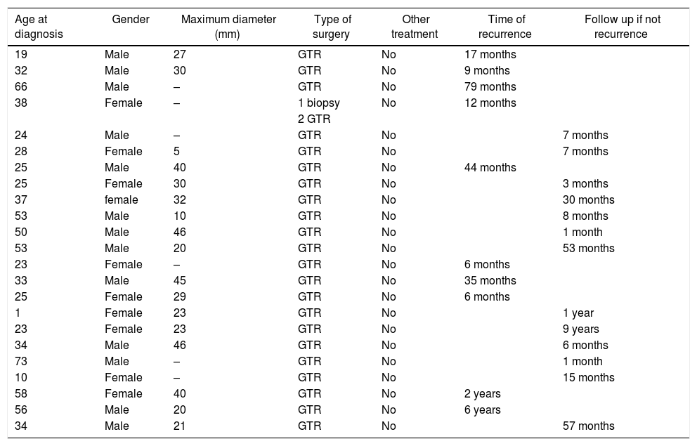
"apellidos" => "Katati"
]
1 => array:2 [
"nombre" => "Isabel"
"apellidos" => "Ortiz García"
]
2 => array:2 [
"nombre" => "Clara Isabel"
"apellidos" => "Chamorro"
]
3 => array:2 [
"nombre" => "Ángel"
"apellidos" => "Horcajadas"
]
4 => array:2 [
"nombre" => "Alicia"
"apellidos" => "Hurtado"
]
5 => array:2 [
"nombre" => "Carlos"
"apellidos" => "Sánchez"
]
6 => array:2 [
"nombre" => "Benjamín"
"apellidos" => "Iañez"
]
7 => array:2 [
"nombre" => "Enrique"
"apellidos" => "Saura"
]
8 => array:2 [
"nombre" => "Ernesto"
"apellidos" => "García"
]
9 => array:2 [
"nombre" => "Heriberto"
"apellidos" => "Busquier"
]
]
]
]
]
"idiomaDefecto" => "es"
"EPUB" => "https://multimedia.elsevier.es/PublicationsMultimediaV1/item/epub/S1130147318300289?idApp=UINPBA00004B"
"url" => "/11301473/0000003000000001/v1_201901030610/S1130147318300289/v1_201901030610/es/main.assets"
]
"itemAnterior" => array:18 [
"pii" => "S1130147318300034"
"issn" => "11301473"
"doi" => "10.1016/j.neucir.2018.01.003"
"estado" => "S300"
"fechaPublicacion" => "2019-01-01"
"aid" => "315"
"copyright" => "Sociedad Española de Neurocirugía"
"documento" => "simple-article"
"crossmark" => 1
"subdocumento" => "crp"
"cita" => "Neurocirugia. 2019;30:33-7"
"abierto" => array:3 [
"ES" => false
"ES2" => false
"LATM" => false
]
"gratuito" => false
"lecturas" => array:2 [
"total" => 12
"formatos" => array:2 [
"HTML" => 6
"PDF" => 6
]
]
"es" => array:13 [
"idiomaDefecto" => true
"cabecera" => "<span class="elsevierStyleTextfn">Caso clínico</span>"
"titulo" => "Tumor fibroso solitario extra-intramedular de localización dorsal"
"tienePdf" => "es"
"tieneTextoCompleto" => "es"
"tieneResumen" => array:2 [
0 => "es"
1 => "en"
]
"paginas" => array:1 [
0 => array:2 [
"paginaInicial" => "33"
"paginaFinal" => "37"
]
]
"titulosAlternativos" => array:1 [
"en" => array:1 [
"titulo" => "Solitary fibrous tumor: A clinical case"
]
]
"contieneResumen" => array:2 [
"es" => true
"en" => true
]
"contieneTextoCompleto" => array:1 [
"es" => true
]
"contienePdf" => array:1 [
"es" => true
]
"resumenGrafico" => array:2 [
"original" => 0
"multimedia" => array:7 [
"identificador" => "fig0010"
"etiqueta" => "Figura 2"
"tipo" => "MULTIMEDIAFIGURA"
"mostrarFloat" => true
"mostrarDisplay" => false
"figura" => array:1 [
0 => array:4 [
"imagen" => "gr2.jpeg"
"Alto" => 1573
"Ancho" => 2133
"Tamanyo" => 685453
]
]
"descripcion" => array:1 [
"es" => "<p id="spar0050" class="elsevierStyleSimplePara elsevierViewall">Inmunohistoquímica característica del tumor fibroso solitario: STAT-6 positivo a ×20 (A) y CD34 positivo, a ×20 (B) hematoxilina-eosina ×20 (C) observamos una proliferación mesenquimal de haces de células fusiformes estructuradas.</p>"
]
]
]
"autores" => array:1 [
0 => array:2 [
"autoresLista" => "Beatriz Mansilla Fernández, María Román de Aragón, José Francisco Paz Solís, Pablo García Feijoo, José Roda Frade, María Rita Regojo Zapata"
"autores" => array:6 [
0 => array:2 [
"nombre" => "Beatriz"
"apellidos" => "Mansilla Fernández"
]
1 => array:2 [
"nombre" => "María"
"apellidos" => "Román de Aragón"
]
2 => array:2 [
"nombre" => "José Francisco"
"apellidos" => "Paz Solís"
]
3 => array:2 [
"nombre" => "Pablo"
"apellidos" => "García Feijoo"
]
4 => array:2 [
"nombre" => "José"
"apellidos" => "Roda Frade"
]
5 => array:2 [
"nombre" => "María Rita"
"apellidos" => "Regojo Zapata"
]
]
]
]
]
"idiomaDefecto" => "es"
"EPUB" => "https://multimedia.elsevier.es/PublicationsMultimediaV1/item/epub/S1130147318300034?idApp=UINPBA00004B"
"url" => "/11301473/0000003000000001/v1_201901030610/S1130147318300034/v1_201901030610/es/main.assets"
]
"en" => array:19 [
"idiomaDefecto" => true
"cabecera" => "<span class="elsevierStyleTextfn">Case Report</span>"
"titulo" => "Papillary tumor of the pineal region: Case report and review of the literature"
"tieneTextoCompleto" => true
"paginas" => array:1 [
0 => array:2 [
"paginaInicial" => "38"
"paginaFinal" => "43"
]
]
"autores" => array:1 [
0 => array:4 [
"autoresLista" => "María de los Ángeles Cañizares Méndez, Manuel Amosa Delgado, Juan Antonio Álvarez Salgado, Jorge Javier Villaseñor Ledezma, Elena Capilla Cabezuelo, Francisco Díaz Crespo"
"autores" => array:6 [
0 => array:4 [
"nombre" => "María de los Ángeles"
"apellidos" => "Cañizares Méndez"
"email" => array:1 [
0 => "angeles.ca.me@gmail.com"
]
"referencia" => array:2 [
0 => array:2 [
"etiqueta" => "<span class="elsevierStyleSup">a</span>"
"identificador" => "aff0005"
]
1 => array:2 [
"etiqueta" => "<span class="elsevierStyleSup">*</span>"
"identificador" => "cor0005"
]
]
]
1 => array:3 [
"nombre" => "Manuel"
"apellidos" => "Amosa Delgado"
"referencia" => array:1 [
0 => array:2 [
"etiqueta" => "<span class="elsevierStyleSup">a</span>"
"identificador" => "aff0005"
]
]
]
2 => array:3 [
"nombre" => "Juan Antonio"
"apellidos" => "Álvarez Salgado"
"referencia" => array:1 [
0 => array:2 [
"etiqueta" => "<span class="elsevierStyleSup">a</span>"
"identificador" => "aff0005"
]
]
]
3 => array:3 [
"nombre" => "Jorge Javier"
"apellidos" => "Villaseñor Ledezma"
"referencia" => array:1 [
0 => array:2 [
"etiqueta" => "<span class="elsevierStyleSup">a</span>"
"identificador" => "aff0005"
]
]
]
4 => array:3 [
"nombre" => "Elena"
"apellidos" => "Capilla Cabezuelo"
"referencia" => array:1 [
0 => array:2 [
"etiqueta" => "<span class="elsevierStyleSup">b</span>"
"identificador" => "aff0010"
]
]
]
5 => array:3 [
"nombre" => "Francisco"
"apellidos" => "Díaz Crespo"
"referencia" => array:1 [
0 => array:2 [
"etiqueta" => "<span class="elsevierStyleSup">c</span>"
"identificador" => "aff0015"
]
]
]
]
"afiliaciones" => array:3 [
0 => array:3 [
"entidad" => "Department of Neurosurgery, Complejo Hospitalario de Toledo (CHT), Spain"
"etiqueta" => "a"
"identificador" => "aff0005"
]
1 => array:3 [
"entidad" => "Department of Radiology, Complejo Hospitalario de Toledo (CHT), Spain"
"etiqueta" => "b"
"identificador" => "aff0010"
]
2 => array:3 [
"entidad" => "Department of Pathology, Complejo Hospitalario de Toledo (CHT), Spain"
"etiqueta" => "c"
"identificador" => "aff0015"
]
]
"correspondencia" => array:1 [
0 => array:3 [
"identificador" => "cor0005"
"etiqueta" => "⁎"
"correspondencia" => "<span class="elsevierStyleItalic">Corresponding author</span>."
]
]
]
]
"titulosAlternativos" => array:1 [
"es" => array:1 [
"titulo" => "Tumor papilar de la región pineal, caso clínico y revisión de la literatura"
]
]
"resumenGrafico" => array:2 [
"original" => 0
"multimedia" => array:7 [
"identificador" => "fig0005"
"etiqueta" => "Fig. 1"
"tipo" => "MULTIMEDIAFIGURA"
"mostrarFloat" => true
"mostrarDisplay" => false
"figura" => array:1 [
0 => array:4 [
"imagen" => "gr1.jpeg"
"Alto" => 2334
"Ancho" => 1200
"Tamanyo" => 339768
]
]
"descripcion" => array:1 [
"en" => "<p id="spar0015" class="elsevierStyleSimplePara elsevierViewall">(a–d) Radiological images from the Radiology Department of Complejo Hospitalario de Toledo. A nodular lesion with a cystic component and peripheral enhancement in the pineal region can be seen in the MRI. (a) T1 axial MRI. (b) T1 Sagital MRI with gadolinium. (c) T2 coronal MRI. (d) Spectroscopy shows an increase in membrane replacement rates as well as internal lactate spikes. (e) Image of the surgical field from the Neurosurgical Department of Complejo Hospitalario de Toledo. (e) Image of the surgical field. Supracerebellar approach with the pineal lesion in the center of the image. (f–h) Histological images from the Anatomic Pathology Department of Complejo Hospitalario de Toledo. (f) Hematoxylin and eosin stain with papillary tumor structure. (g) Immunohistochemistry: CKAE1/AE3+, S100+, EMA−, Synaptophysin−. (h) Immunohistochemistry: Chromogranin−, P53−, PGFA−, and showing a low proliferative rate.</p>"
]
]
]
"textoCompleto" => "<span class="elsevierStyleSections"><span id="sec0005" class="elsevierStyleSection elsevierViewall"><span class="elsevierStyleSectionTitle" id="sect0030">Introduction</span><p id="par0005" class="elsevierStylePara elsevierViewall">Papillary tumor of the pineal region (PTPR) is a rare neuroepithelial tumor characterized by papillary architecture and epithelial cytology, cytokeratin-positive, and ependymal cell differentiation. It is considered World Health Organization (WHO) grade II–III, according to its biological behavior. It was first described by Jouvet in 2003,<a class="elsevierStyleCrossRef" href="#bib0080"><span class="elsevierStyleSup">1</span></a> and was included in the 2007 WHO classification.<a class="elsevierStyleCrossRef" href="#bib0085"><span class="elsevierStyleSup">2</span></a></p></span><span id="sec0010" class="elsevierStyleSection elsevierViewall"><span class="elsevierStyleSectionTitle" id="sect0035">Case report</span><p id="par0010" class="elsevierStylePara elsevierViewall">A 34-year-old Caucasian male with history of migraines arrived at the emergency room for episodes of blurry vision followed by holocranial headache which occurred 2–3 times a week, occasionally accompanied by nausea, and progressed over 15 days. Neurological examination was normal.</p><p id="par0015" class="elsevierStylePara elsevierViewall">An emergent cranial computerized tomography (CT) scan was performed. A nodulocystic lesion was found in the pineal region accompanied by triventricular ventriculomegaly.</p><p id="par0020" class="elsevierStylePara elsevierViewall">He was admitted for study and the cranial magnetic resonance imaging (MRI) showed a solid cystic mass with well-defined edges in the pineal region, measuring 21<span class="elsevierStyleHsp" style=""></span>mm<span class="elsevierStyleHsp" style=""></span>×<span class="elsevierStyleHsp" style=""></span>24<span class="elsevierStyleHsp" style=""></span>mm<span class="elsevierStyleHsp" style=""></span>×<span class="elsevierStyleHsp" style=""></span>23<span class="elsevierStyleHsp" style=""></span>mm, which caudally compressed the quadrigeminal plate and aqueduct, noting a slight increase in the size of the supratentorial ventricular system without signs of active hydrocephalus. The lesion was predominantly cystic, T1-hypointense and T2-hyperintense, and showed a solid posterior and outer zone with a maximum width of 5<span class="elsevierStyleHsp" style=""></span>mm which was homogenously enhanced with the administration of contrast. This lesion presented restriction in the diffusion study and an increase in cerebral blood volume in the solid region during the perfusion scan. Spectroscopy study showed an increase in membrane replacement rates as well as in internal lactate spikes. The initial presumptive diagnosis was pineocytoma (<a class="elsevierStyleCrossRef" href="#fig0005">Fig. 1</a>a–d).</p><elsevierMultimedia ident="fig0005"></elsevierMultimedia><p id="par0025" class="elsevierStylePara elsevierViewall">The patient underwent a planned procedure without requiring cerebral spinal fluid (CSF) shunt, performing a supra/infratentorial craniotomy and infratentorial supracerebellar approach. The presence of a well vascularized, predominantly cystic rosy tumor was observed in the quadrigeminal cistern and was resected completely (<a class="elsevierStyleCrossRef" href="#fig0005">Fig. 1</a>e).</p><p id="par0030" class="elsevierStylePara elsevierViewall">Ventriculomegaly was completely eliminated after removal of the tumor and there were no post-surgical complications.</p><p id="par0035" class="elsevierStylePara elsevierViewall">Histological cross-sections showed a neoplastic proliferation that formed papillary structures and perivascular pseudorosettes. Cells were polygonal with extensive eosinophils cytoplasms which were clear in some areas. Isolated mitosis were observed. Pineal gland parenchyma with calcifications, reactive gliosis and Rosenthal fibers were focally observed. Immunohistochemistry of the tumor was cytokeratin (CK) AE1/AE3+, glial fibrillary acidic protein (GFAP)−, S100+, epithelial membrane antigen (EMA)−, Chromogranin−, Synaptophysin−, P53− and showed a low proliferative rate, Ki67 5% and 1 mitosis per 10 high-power. Definitive diagnosis was PTPR (<a class="elsevierStyleCrossRef" href="#fig0005">Fig. 1</a>f–h).</p><p id="par0040" class="elsevierStylePara elsevierViewall">The hospital's neuro-oncology committee decided to discharge the patient without adjuvant therapy, undergoing periodical clinical and radiological follow-up.</p><p id="par0045" class="elsevierStylePara elsevierViewall">The patient showed good recovery without adjuvant therapy and after 57 months of follow-up he remained asymptomatic, showing no radiological or clinical signs of tumor recurrence.</p></span><span id="sec0015" class="elsevierStyleSection elsevierViewall"><span class="elsevierStyleSectionTitle" id="sect0040">Discussion</span><p id="par0050" class="elsevierStylePara elsevierViewall">Papillary tumor of the pineal region is a rare neuroepithelial tumor but description is increasing since its discovery. It is characterized by papillary architecture, epithelial cytology, and ependymal cell differentiation. PTPR derives from specialized ependymal cells in the subcomissural organ,<a class="elsevierStyleCrossRef" href="#bib0080"><span class="elsevierStyleSup">1</span></a> a small gland located in the third ventricle under the posterior commissure and just behind the pineal gland, which secretes glycoproteins into the CSF. This hypothesis justifies the neuroendocrine origin and the ependymal morphology.</p><p id="par0055" class="elsevierStylePara elsevierViewall">It is considered to be grade II–III, according to its biological behavior. On average, it presents around the age of 32, although it can appear at any age including childhood.<a class="elsevierStyleCrossRef" href="#bib0090"><span class="elsevierStyleSup">3</span></a> There has been no evidence of gender predilection and it usually presents as a headache caused by non-communicating hydrocephalus.<a class="elsevierStyleCrossRef" href="#bib0095"><span class="elsevierStyleSup">4</span></a></p><p id="par0060" class="elsevierStylePara elsevierViewall">Upon the discovery of lesions in the pineal region the radiological differential diagnosis should mind all solid-cystic lesions that could be found in this area, especially pineal parenchymal tumors and germ cell tumors (GCT), although lesions from others origins can be also found (<a class="elsevierStyleCrossRef" href="#fig0010">Fig. 2</a>a). An intrinsic characteristic which suggests diagnosis of PTPR for a mass in the pineal gland region and may help to differentiate it from other lesions in the same area is that PTPR presents as a solid-cystic lesion hyperintense in T2-weighted MRI sequences in the absence of fat, hemorrhage, melanin or calcifications.<a class="elsevierStyleCrossRef" href="#bib0100"><span class="elsevierStyleSup">5</span></a> This is due to the high concentrations of proteins in the small cysts that these lesions contain.<a class="elsevierStyleCrossRef" href="#bib0105"><span class="elsevierStyleSup">6</span></a> Enhancement is moderate after contrast administration.</p><elsevierMultimedia ident="fig0010"></elsevierMultimedia><p id="par0065" class="elsevierStylePara elsevierViewall">After radiological findings, CSF and serum markers could be used although they are not adequate enough for diagnosis. The most useful markers for pineal area are alpha fetoprotein (AFP) and beta human chorionic gonadotropin (β-hCG), which are negative for PTPR and other pineal parenchymal tumors but positives for GCT<a class="elsevierStyleCrossRef" href="#bib0110"><span class="elsevierStyleSup">7</span></a> (<a class="elsevierStyleCrossRef" href="#fig0010">Fig. 2</a>b). Results of serum or CSF markers may help the treatment decision, if they are elevated, a biopsy, that could be endoscopic and combined if necessary with third ventriculostomy,<a class="elsevierStyleCrossRef" href="#bib0115"><span class="elsevierStyleSup">8</span></a> should be performed because the most probable diagnosis will be any GCT, needing complementary treatment instead of surgery. If CSF or serum makers are not elevated total resection surgery could be performed although some authors defend endoscopic biopsy with third ventriculostomy as the first step in all the cases to get a biopsy for diagnosis and solve hydrocephalus without implanting a shunt.</p><p id="par0070" class="elsevierStylePara elsevierViewall">The definitive diagnosis always depends on histopathological analysis. The main pathological differential diagnosis is between lesions with papillary histology,<a class="elsevierStyleCrossRef" href="#bib0105"><span class="elsevierStyleSup">6</span></a> specially papillary ependymoma, papillary meningioma, choroid plexus papilloma or metastatic papillary carcinomas.</p><p id="par0075" class="elsevierStylePara elsevierViewall">Tumor cells resemble ependymal cells and display microvilli as well as adherens junctions. They are also similar to choroid plexus cells which suggests reticulum secretory function. In contrast, the presence of annulate lamellae, dense core vesicles and microtubules suggest neuronal or neuroendocrine differentiation.<a class="elsevierStyleCrossRef" href="#bib0120"><span class="elsevierStyleSup">9</span></a> A PTPR is histologically less papillary than a choroid plexus papilloma, more epithelial than an ependymoma and does not have the fibrillary background.<a class="elsevierStyleCrossRef" href="#bib0080"><span class="elsevierStyleSup">1</span></a> These histological similarities of PTPR have infradiagnosed it against these entities.<a class="elsevierStyleCrossRef" href="#bib0090"><span class="elsevierStyleSup">3</span></a> The histopathological definitive diagnosis will be the result of the combination of morphological and immunohistochemical studies (<a class="elsevierStyleCrossRef" href="#fig0010">Fig. 2</a>c).</p><p id="par0080" class="elsevierStylePara elsevierViewall">Clinical course and management of PTPR have been partially described in the literature.</p><p id="par0085" class="elsevierStylePara elsevierViewall">Clinical behavior of PTPR is characterized by progressive growth and frequent local relapse, even after the complete resection and has potential for CSF dissemination.<a class="elsevierStyleCrossRef" href="#bib0125"><span class="elsevierStyleSup">10</span></a> Its ability to differentiate into a more malignant histology is still unknown. Incomplete resection or elevated mitotic activity (3 or more mitosis per 10 high-power fields or Ki 67/MIB1 proliferative activity greater or equal to 10%) tend to be associated with a lower survival rate.<a class="elsevierStyleCrossRef" href="#bib0130"><span class="elsevierStyleSup">11</span></a></p><p id="par0090" class="elsevierStylePara elsevierViewall">In 2011 Poulgrain published the last systematic review of the literature,<a class="elsevierStyleCrossRef" href="#bib0105"><span class="elsevierStyleSup">6</span></a> which includes all the 70 cases published plus her 2 cases.</p><p id="par0095" class="elsevierStylePara elsevierViewall">In 2013 Fauchon published a multi-center study which collected the most extensive clinical series to date, with a total of 44 patients. It established a recurrence rate of 58% after 5 years and up to 70% at 6 years, without noting any conclusive results on the benefits of radiotherapy or chemotherapy adjuvant to surgery.<a class="elsevierStyleCrossRef" href="#bib0115"><span class="elsevierStyleSup">8</span></a></p><p id="par0100" class="elsevierStylePara elsevierViewall">The optimal initial management of this pathology has not been well established,<a class="elsevierStyleCrossRef" href="#bib0135"><span class="elsevierStyleSup">12</span></a> although complete surgical excision seems to be the goal.<a class="elsevierStyleCrossRef" href="#bib0135"><span class="elsevierStyleSup">12</span></a> The value of chemotherapy and radiotherapy in terms of tumor recurrence has not been defined,<a class="elsevierStyleCrossRef" href="#bib0115"><span class="elsevierStyleSup">8</span></a> therefore it is necessary to continue studying the need for radiotherapy after complete surgical excision in non-aggressive histology.<a class="elsevierStyleCrossRef" href="#bib0135"><span class="elsevierStyleSup">12</span></a> Some authors defend the use of biopsy and radiotherapy, and in some of cases positron emission tomography<a class="elsevierStyleCrossRef" href="#bib0135"><span class="elsevierStyleSup">12</span></a> or gamma knife radiosurgery<a class="elsevierStyleCrossRef" href="#bib0130"><span class="elsevierStyleSup">11</span></a> are also used.</p><p id="par0105" class="elsevierStylePara elsevierViewall">For recurrent tumors after surgery, radiotherapy and chemotherapy are used. Recently the administration of an antibody targeting the endothelial growth factor (Bevacizumab) has been proposed as an alternative based on the similarities this tumor shares with ependymoma.<a class="elsevierStyleCrossRef" href="#bib0140"><span class="elsevierStyleSup">13</span></a></p><p id="par0110" class="elsevierStylePara elsevierViewall">Due to the high variability of treatments published for PTPR we decided to evaluate the recurrence and survival of patients with surgical gross total resection (GTR), without complementary treatment and with some follow up. A systematic review of the literature was performed on Pubmed including all papers related to PTPR published after Poulgrain's (2011) work to February 2018. A total of 23 patients meet the requirements including 14 patients of 10 Pubmed papers,<a class="elsevierStyleCrossRefs" href="#bib0090"><span class="elsevierStyleSup">3–5,8–12,14,15</span></a> 8 patients of Poulgrain's<a class="elsevierStyleCrossRef" href="#bib0105"><span class="elsevierStyleSup">6</span></a> paper, and our patient (<a class="elsevierStyleCrossRef" href="#tbl0005">Table 1</a>).</p><elsevierMultimedia ident="tbl0005"></elsevierMultimedia><p id="par0115" class="elsevierStylePara elsevierViewall">Given these 23 patients, the average age at diagnosis was 35.65 years old, 13 males and 10 females, a maximal diameter average of 28.6<span class="elsevierStyleHsp" style=""></span>mm. Seven patients were followed for less than one year, 10 of the patients had recurrence, 4 of them during the first year after surgery. The earliest recurrence was 6 months after surgery and the latest 79 months after surgery. Our patient with 57 months is the second longest disease-free period in the literature, after the patient with the recurrence at 79 months.</p></span><span id="sec0020" class="elsevierStyleSection elsevierViewall"><span class="elsevierStyleSectionTitle" id="sect0045">Conclusion</span><p id="par0120" class="elsevierStylePara elsevierViewall">This case shows typical characteristics of PTPR in epidemiology and histopathology.</p><p id="par0125" class="elsevierStylePara elsevierViewall">After 57 months of post-surgical follow-up and without having received adjuvant therapy, the patient remained asymptomatic without clinical or radiological signs of tumor recurrence, the second longest disease-free period in the literature.</p><p id="par0130" class="elsevierStylePara elsevierViewall">With increased diagnoses since its inclusion in the WHO classification in 2007, there is much that is still unknown about the pathogenesis, prognosis and management of this tumor and thus it is necessary to continue collecting information and following up cases.</p></span><span id="sec0025" class="elsevierStyleSection elsevierViewall"><span class="elsevierStyleSectionTitle" id="sect0050">Conflicts of interest</span><p id="par0135" class="elsevierStylePara elsevierViewall">The authors report no conflict of interest.</p></span><span id="sec0030" class="elsevierStyleSection elsevierViewall"><span class="elsevierStyleSectionTitle" id="sect0055">Authors contributions</span><p id="par0140" class="elsevierStylePara elsevierViewall">Conception and design: Cañizares and Amosa.</p><p id="par0145" class="elsevierStylePara elsevierViewall">Acquisition of data: all authors.</p><p id="par0150" class="elsevierStylePara elsevierViewall">Analysis and interpretation of data: all authors.</p><p id="par0155" class="elsevierStylePara elsevierViewall">Drafting the article: Cañizares.</p><p id="par0160" class="elsevierStylePara elsevierViewall">Critically revising the article: all authors.</p><p id="par0165" class="elsevierStylePara elsevierViewall">Reviewed submitted version of manuscript: all authors.</p></span></span>"
"textoCompletoSecciones" => array:1 [
"secciones" => array:12 [
0 => array:3 [
"identificador" => "xres1132653"
"titulo" => "Abstract"
"secciones" => array:1 [
0 => array:1 [
"identificador" => "abst0005"
]
]
]
1 => array:2 [
"identificador" => "xpalclavsec1065250"
"titulo" => "Keywords"
]
2 => array:2 [
"identificador" => "xpalclavsec1065249"
"titulo" => "Abbreviations"
]
3 => array:3 [
"identificador" => "xres1132652"
"titulo" => "Resumen"
"secciones" => array:1 [
0 => array:1 [
"identificador" => "abst0010"
]
]
]
4 => array:2 [
"identificador" => "xpalclavsec1065248"
"titulo" => "Palabras clave"
]
5 => array:2 [
"identificador" => "sec0005"
"titulo" => "Introduction"
]
6 => array:2 [
"identificador" => "sec0010"
"titulo" => "Case report"
]
7 => array:2 [
"identificador" => "sec0015"
"titulo" => "Discussion"
]
8 => array:2 [
"identificador" => "sec0020"
"titulo" => "Conclusion"
]
9 => array:2 [
"identificador" => "sec0025"
"titulo" => "Conflicts of interest"
]
10 => array:2 [
"identificador" => "sec0030"
"titulo" => "Authors contributions"
]
11 => array:1 [
"titulo" => "References"
]
]
]
"pdfFichero" => "main.pdf"
"tienePdf" => true
"fechaRecibido" => "2017-10-31"
"fechaAceptado" => "2018-03-15"
"PalabrasClave" => array:2 [
"en" => array:2 [
0 => array:4 [
"clase" => "keyword"
"titulo" => "Keywords"
"identificador" => "xpalclavsec1065250"
"palabras" => array:4 [
0 => "Hydrocephalus"
1 => "Pineal gland"
2 => "Pineal gland tumor"
3 => "Immunohistochemistry"
]
]
1 => array:4 [
"clase" => "abr"
"titulo" => "Abbreviations"
"identificador" => "xpalclavsec1065249"
"palabras" => array:13 [
0 => "PTPR"
1 => "WHO"
2 => "CT"
3 => "MRI"
4 => "CBV"
5 => "CSF"
6 => "CK"
7 => "GFAP"
8 => "EMA"
9 => "GCT"
10 => "AFP"
11 => "β-hCG"
12 => "GTR"
]
]
]
"es" => array:1 [
0 => array:4 [
"clase" => "keyword"
"titulo" => "Palabras clave"
"identificador" => "xpalclavsec1065248"
"palabras" => array:4 [
0 => "Hidrocefalia"
1 => "Glándula pineal"
2 => "Tumor de la glándula pineal"
3 => "Inmunohistoquímica"
]
]
]
]
"tieneResumen" => true
"resumen" => array:2 [
"en" => array:2 [
"titulo" => "Abstract"
"resumen" => "<span id="abst0005" class="elsevierStyleSection elsevierViewall"><p id="spar0005" class="elsevierStyleSimplePara elsevierViewall">Papillary tumor of the pineal region is a rare neuroepithelial tumor characterized by papillary architecture and epithelial cytology, immunopositivity for cytokeratin and ependymal differentiation. It is considered grade II–III by the World Health Organization and was first described by Jouvet in 2003. We present a 34-year-old male with headaches, blurred vision and normal examination. Radiological study showed a nodulocystic lesion in the pineal region compatible with pineocytoma. Surgery was performed using an infratentorial supracerebellar approach, finding a cystic tumor in the quadrigeminal cistern which was completely resected. Histopathology reported a papillary tumor of the pineal region. The patient made good progress without adjuvant therapy, and after 57 months of follow-up he remained asymptomatic and free of recurrence. A review of the literature was performed to collect all the cases published with gross total resection and no complementary treatment. In conclusion, there is still much to be learned about the pathogenesis, prognosis and management of this tumor.</p></span>"
]
"es" => array:2 [
"titulo" => "Resumen"
"resumen" => "<span id="abst0010" class="elsevierStyleSection elsevierViewall"><p id="spar0010" class="elsevierStyleSimplePara elsevierViewall">El tumor papilar de la región pineal es un infrecuente tumor neuroepitelial con arquitectura y citología epitelial, citoqueratina positiva y signos de diferenciación ependimaria. Descrito por Jouvet en 2003 es considerado grado II-III de la Organización Mundial de la Salud. Presentamos el caso de un varón de 34 años con cefalea, visión borrosa y exploración normal. El estudio radiológico muestra una lesión noduloquística en región pineal compatible con pineocitoma. Es intervenido mediante abordaje supracerebeloso infratentorial apreciándose lesión de aspecto quístico en cisterna cuadrigémina que es resecada por completo. El resultado histopatológico es tumor papilar de la región pineal. El paciente presenta buena evolución sin tratamiento adyuvante y tras 57 meses de seguimiento permanece asintomático y sin recidiva. Realizamos una revisión sistemática sobre la evolución de los casos publicados tratados mediante resección completa y sin tratamiento complementario, concluyendo que continuamos necesitando aprender sobre la patogénesis, pronóstico y manejo de este tumor.</p></span>"
]
]
"multimedia" => array:3 [
0 => array:7 [
"identificador" => "fig0005"
"etiqueta" => "Fig. 1"
"tipo" => "MULTIMEDIAFIGURA"
"mostrarFloat" => true
"mostrarDisplay" => false
"figura" => array:1 [
0 => array:4 [
"imagen" => "gr1.jpeg"
"Alto" => 2334
"Ancho" => 1200
"Tamanyo" => 339768
]
]
"descripcion" => array:1 [
"en" => "<p id="spar0015" class="elsevierStyleSimplePara elsevierViewall">(a–d) Radiological images from the Radiology Department of Complejo Hospitalario de Toledo. A nodular lesion with a cystic component and peripheral enhancement in the pineal region can be seen in the MRI. (a) T1 axial MRI. (b) T1 Sagital MRI with gadolinium. (c) T2 coronal MRI. (d) Spectroscopy shows an increase in membrane replacement rates as well as internal lactate spikes. (e) Image of the surgical field from the Neurosurgical Department of Complejo Hospitalario de Toledo. (e) Image of the surgical field. Supracerebellar approach with the pineal lesion in the center of the image. (f–h) Histological images from the Anatomic Pathology Department of Complejo Hospitalario de Toledo. (f) Hematoxylin and eosin stain with papillary tumor structure. (g) Immunohistochemistry: CKAE1/AE3+, S100+, EMA−, Synaptophysin−. (h) Immunohistochemistry: Chromogranin−, P53−, PGFA−, and showing a low proliferative rate.</p>"
]
]
1 => array:7 [
"identificador" => "fig0010"
"etiqueta" => "Fig. 2"
"tipo" => "MULTIMEDIAFIGURA"
"mostrarFloat" => true
"mostrarDisplay" => false
"figura" => array:1 [
0 => array:4 [
"imagen" => "gr2.jpeg"
"Alto" => 4364
"Ancho" => 2583
"Tamanyo" => 516928
]
]
"descripcion" => array:1 [
"en" => "<p id="spar0020" class="elsevierStyleSimplePara elsevierViewall">Differential diagnosis process. A. Radiological differential diagnosis of solid cystic pineal lesions. B. Biochemical differential diagnosis according to tumor markers in CSF. C. Immunohistochemical differential diagnosis.</p>"
]
]
2 => array:8 [
"identificador" => "tbl0005"
"etiqueta" => "Table 1"
"tipo" => "MULTIMEDIATABLA"
"mostrarFloat" => true
"mostrarDisplay" => false
"detalles" => array:1 [
0 => array:3 [
"identificador" => "at1"
"detalle" => "Table "
"rol" => "short"
]
]
"tabla" => array:1 [
"tablatextoimagen" => array:1 [
0 => array:2 [
"tabla" => array:1 [
0 => """
<table border="0" frame="\n
\t\t\t\t\tvoid\n
\t\t\t\t" class=""><thead title="thead"><tr title="table-row"><th class="td" title="table-head " align="left" valign="top" scope="col" style="border-bottom: 2px solid black">Age at diagnosis \t\t\t\t\t\t\n
\t\t\t\t</th><th class="td" title="table-head " align="center" valign="top" scope="col" style="border-bottom: 2px solid black">Gender \t\t\t\t\t\t\n
\t\t\t\t</th><th class="td" title="table-head " align="center" valign="top" scope="col" style="border-bottom: 2px solid black">Maximum diameter (mm) \t\t\t\t\t\t\n
\t\t\t\t</th><th class="td" title="table-head " align="center" valign="top" scope="col" style="border-bottom: 2px solid black">Type of surgery \t\t\t\t\t\t\n
\t\t\t\t</th><th class="td" title="table-head " align="center" valign="top" scope="col" style="border-bottom: 2px solid black">Other treatment \t\t\t\t\t\t\n
\t\t\t\t</th><th class="td" title="table-head " align="center" valign="top" scope="col" style="border-bottom: 2px solid black">Time of recurrence \t\t\t\t\t\t\n
\t\t\t\t</th><th class="td" title="table-head " align="center" valign="top" scope="col" style="border-bottom: 2px solid black">Follow up if not recurrence \t\t\t\t\t\t\n
\t\t\t\t</th></tr></thead><tbody title="tbody"><tr title="table-row"><td class="td-with-role" title="table-entry ; entry_with_role_rowhead " align="left" valign="top">19 \t\t\t\t\t\t\n
\t\t\t\t</td><td class="td" title="table-entry " align="left" valign="top">Male \t\t\t\t\t\t\n
\t\t\t\t</td><td class="td" title="table-entry " align="left" valign="top">27 \t\t\t\t\t\t\n
\t\t\t\t</td><td class="td" title="table-entry " align="left" valign="top">GTR \t\t\t\t\t\t\n
\t\t\t\t</td><td class="td" title="table-entry " align="left" valign="top">No \t\t\t\t\t\t\n
\t\t\t\t</td><td class="td" title="table-entry " align="left" valign="top">17 months \t\t\t\t\t\t\n
\t\t\t\t</td><td class="td" title="table-entry " align="" valign="top"> \t\t\t\t\t\t\n
\t\t\t\t</td></tr><tr title="table-row"><td class="td-with-role" title="table-entry ; entry_with_role_rowhead " align="left" valign="top">32 \t\t\t\t\t\t\n
\t\t\t\t</td><td class="td" title="table-entry " align="left" valign="top">Male \t\t\t\t\t\t\n
\t\t\t\t</td><td class="td" title="table-entry " align="left" valign="top">30 \t\t\t\t\t\t\n
\t\t\t\t</td><td class="td" title="table-entry " align="left" valign="top">GTR \t\t\t\t\t\t\n
\t\t\t\t</td><td class="td" title="table-entry " align="left" valign="top">No \t\t\t\t\t\t\n
\t\t\t\t</td><td class="td" title="table-entry " align="left" valign="top">9 months \t\t\t\t\t\t\n
\t\t\t\t</td><td class="td" title="table-entry " align="" valign="top"> \t\t\t\t\t\t\n
\t\t\t\t</td></tr><tr title="table-row"><td class="td-with-role" title="table-entry ; entry_with_role_rowhead " align="left" valign="top">66 \t\t\t\t\t\t\n
\t\t\t\t</td><td class="td" title="table-entry " align="left" valign="top">Male \t\t\t\t\t\t\n
\t\t\t\t</td><td class="td" title="table-entry " align="left" valign="top">– \t\t\t\t\t\t\n
\t\t\t\t</td><td class="td" title="table-entry " align="left" valign="top">GTR \t\t\t\t\t\t\n
\t\t\t\t</td><td class="td" title="table-entry " align="left" valign="top">No \t\t\t\t\t\t\n
\t\t\t\t</td><td class="td" title="table-entry " align="left" valign="top">79 months \t\t\t\t\t\t\n
\t\t\t\t</td><td class="td" title="table-entry " align="" valign="top"> \t\t\t\t\t\t\n
\t\t\t\t</td></tr><tr title="table-row"><td class="td-with-role" title="table-entry ; entry_with_role_rowhead " align="left" valign="top">38 \t\t\t\t\t\t\n
\t\t\t\t</td><td class="td" title="table-entry " align="left" valign="top">Female \t\t\t\t\t\t\n
\t\t\t\t</td><td class="td" title="table-entry " align="left" valign="top">– \t\t\t\t\t\t\n
\t\t\t\t</td><td class="td" title="table-entry " align="left" valign="top">1 biopsy \t\t\t\t\t\t\n
\t\t\t\t</td><td class="td" title="table-entry " align="left" valign="top">No \t\t\t\t\t\t\n
\t\t\t\t</td><td class="td" title="table-entry " align="left" valign="top">12 months \t\t\t\t\t\t\n
\t\t\t\t</td><td class="td" title="table-entry " align="" valign="top"> \t\t\t\t\t\t\n
\t\t\t\t</td></tr><tr title="table-row"><td class="td" title="table-entry " align="" valign="top"> \t\t\t\t\t\t\n
\t\t\t\t</td><td class="td" title="table-entry " align="" valign="top"> \t\t\t\t\t\t\n
\t\t\t\t</td><td class="td" title="table-entry " align="left" valign="top">2 GTR \t\t\t\t\t\t\n
\t\t\t\t</td><td class="td" title="table-entry " align="" valign="top"> \t\t\t\t\t\t\n
\t\t\t\t</td></tr><tr title="table-row"><td class="td-with-role" title="table-entry ; entry_with_role_rowhead " align="left" valign="top">24 \t\t\t\t\t\t\n
\t\t\t\t</td><td class="td" title="table-entry " align="left" valign="top">Male \t\t\t\t\t\t\n
\t\t\t\t</td><td class="td" title="table-entry " align="left" valign="top">– \t\t\t\t\t\t\n
\t\t\t\t</td><td class="td" title="table-entry " align="left" valign="top">GTR \t\t\t\t\t\t\n
\t\t\t\t</td><td class="td" title="table-entry " align="left" valign="top">No \t\t\t\t\t\t\n
\t\t\t\t</td><td class="td" title="table-entry " align="" valign="top"> \t\t\t\t\t\t\n
\t\t\t\t</td><td class="td" title="table-entry " align="left" valign="top">7 months \t\t\t\t\t\t\n
\t\t\t\t</td></tr><tr title="table-row"><td class="td-with-role" title="table-entry ; entry_with_role_rowhead " align="left" valign="top">28 \t\t\t\t\t\t\n
\t\t\t\t</td><td class="td" title="table-entry " align="left" valign="top">Female \t\t\t\t\t\t\n
\t\t\t\t</td><td class="td" title="table-entry " align="left" valign="top">5 \t\t\t\t\t\t\n
\t\t\t\t</td><td class="td" title="table-entry " align="left" valign="top">GTR \t\t\t\t\t\t\n
\t\t\t\t</td><td class="td" title="table-entry " align="left" valign="top">No \t\t\t\t\t\t\n
\t\t\t\t</td><td class="td" title="table-entry " align="" valign="top"> \t\t\t\t\t\t\n
\t\t\t\t</td><td class="td" title="table-entry " align="left" valign="top">7 months \t\t\t\t\t\t\n
\t\t\t\t</td></tr><tr title="table-row"><td class="td-with-role" title="table-entry ; entry_with_role_rowhead " align="left" valign="top">25 \t\t\t\t\t\t\n
\t\t\t\t</td><td class="td" title="table-entry " align="left" valign="top">Male \t\t\t\t\t\t\n
\t\t\t\t</td><td class="td" title="table-entry " align="left" valign="top">40 \t\t\t\t\t\t\n
\t\t\t\t</td><td class="td" title="table-entry " align="left" valign="top">GTR \t\t\t\t\t\t\n
\t\t\t\t</td><td class="td" title="table-entry " align="left" valign="top">No \t\t\t\t\t\t\n
\t\t\t\t</td><td class="td" title="table-entry " align="left" valign="top">44 months \t\t\t\t\t\t\n
\t\t\t\t</td><td class="td" title="table-entry " align="" valign="top"> \t\t\t\t\t\t\n
\t\t\t\t</td></tr><tr title="table-row"><td class="td-with-role" title="table-entry ; entry_with_role_rowhead " align="left" valign="top">25 \t\t\t\t\t\t\n
\t\t\t\t</td><td class="td" title="table-entry " align="left" valign="top">Female \t\t\t\t\t\t\n
\t\t\t\t</td><td class="td" title="table-entry " align="left" valign="top">30 \t\t\t\t\t\t\n
\t\t\t\t</td><td class="td" title="table-entry " align="left" valign="top">GTR \t\t\t\t\t\t\n
\t\t\t\t</td><td class="td" title="table-entry " align="left" valign="top">No \t\t\t\t\t\t\n
\t\t\t\t</td><td class="td" title="table-entry " align="" valign="top"> \t\t\t\t\t\t\n
\t\t\t\t</td><td class="td" title="table-entry " align="left" valign="top">3 months \t\t\t\t\t\t\n
\t\t\t\t</td></tr><tr title="table-row"><td class="td-with-role" title="table-entry ; entry_with_role_rowhead " align="left" valign="top">37 \t\t\t\t\t\t\n
\t\t\t\t</td><td class="td" title="table-entry " align="left" valign="top">female \t\t\t\t\t\t\n
\t\t\t\t</td><td class="td" title="table-entry " align="left" valign="top">32 \t\t\t\t\t\t\n
\t\t\t\t</td><td class="td" title="table-entry " align="left" valign="top">GTR \t\t\t\t\t\t\n
\t\t\t\t</td><td class="td" title="table-entry " align="left" valign="top">No \t\t\t\t\t\t\n
\t\t\t\t</td><td class="td" title="table-entry " align="" valign="top"> \t\t\t\t\t\t\n
\t\t\t\t</td><td class="td" title="table-entry " align="left" valign="top">30 months \t\t\t\t\t\t\n
\t\t\t\t</td></tr><tr title="table-row"><td class="td-with-role" title="table-entry ; entry_with_role_rowhead " align="left" valign="top">53 \t\t\t\t\t\t\n
\t\t\t\t</td><td class="td" title="table-entry " align="left" valign="top">Male \t\t\t\t\t\t\n
\t\t\t\t</td><td class="td" title="table-entry " align="left" valign="top">10 \t\t\t\t\t\t\n
\t\t\t\t</td><td class="td" title="table-entry " align="left" valign="top">GTR \t\t\t\t\t\t\n
\t\t\t\t</td><td class="td" title="table-entry " align="left" valign="top">No \t\t\t\t\t\t\n
\t\t\t\t</td><td class="td" title="table-entry " align="" valign="top"> \t\t\t\t\t\t\n
\t\t\t\t</td><td class="td" title="table-entry " align="left" valign="top">8 months \t\t\t\t\t\t\n
\t\t\t\t</td></tr><tr title="table-row"><td class="td-with-role" title="table-entry ; entry_with_role_rowhead " align="left" valign="top">50 \t\t\t\t\t\t\n
\t\t\t\t</td><td class="td" title="table-entry " align="left" valign="top">Male \t\t\t\t\t\t\n
\t\t\t\t</td><td class="td" title="table-entry " align="left" valign="top">46 \t\t\t\t\t\t\n
\t\t\t\t</td><td class="td" title="table-entry " align="left" valign="top">GTR \t\t\t\t\t\t\n
\t\t\t\t</td><td class="td" title="table-entry " align="left" valign="top">No \t\t\t\t\t\t\n
\t\t\t\t</td><td class="td" title="table-entry " align="" valign="top"> \t\t\t\t\t\t\n
\t\t\t\t</td><td class="td" title="table-entry " align="left" valign="top">1 month \t\t\t\t\t\t\n
\t\t\t\t</td></tr><tr title="table-row"><td class="td-with-role" title="table-entry ; entry_with_role_rowhead " align="left" valign="top">53 \t\t\t\t\t\t\n
\t\t\t\t</td><td class="td" title="table-entry " align="left" valign="top">Male \t\t\t\t\t\t\n
\t\t\t\t</td><td class="td" title="table-entry " align="left" valign="top">20 \t\t\t\t\t\t\n
\t\t\t\t</td><td class="td" title="table-entry " align="left" valign="top">GTR \t\t\t\t\t\t\n
\t\t\t\t</td><td class="td" title="table-entry " align="left" valign="top">No \t\t\t\t\t\t\n
\t\t\t\t</td><td class="td" title="table-entry " align="" valign="top"> \t\t\t\t\t\t\n
\t\t\t\t</td><td class="td" title="table-entry " align="left" valign="top">53 months \t\t\t\t\t\t\n
\t\t\t\t</td></tr><tr title="table-row"><td class="td-with-role" title="table-entry ; entry_with_role_rowhead " align="left" valign="top">23 \t\t\t\t\t\t\n
\t\t\t\t</td><td class="td" title="table-entry " align="left" valign="top">Female \t\t\t\t\t\t\n
\t\t\t\t</td><td class="td" title="table-entry " align="left" valign="top">– \t\t\t\t\t\t\n
\t\t\t\t</td><td class="td" title="table-entry " align="left" valign="top">GTR \t\t\t\t\t\t\n
\t\t\t\t</td><td class="td" title="table-entry " align="left" valign="top">No \t\t\t\t\t\t\n
\t\t\t\t</td><td class="td" title="table-entry " align="left" valign="top">6 months \t\t\t\t\t\t\n
\t\t\t\t</td><td class="td" title="table-entry " align="" valign="top"> \t\t\t\t\t\t\n
\t\t\t\t</td></tr><tr title="table-row"><td class="td-with-role" title="table-entry ; entry_with_role_rowhead " align="left" valign="top">33 \t\t\t\t\t\t\n
\t\t\t\t</td><td class="td" title="table-entry " align="left" valign="top">Male \t\t\t\t\t\t\n
\t\t\t\t</td><td class="td" title="table-entry " align="left" valign="top">45 \t\t\t\t\t\t\n
\t\t\t\t</td><td class="td" title="table-entry " align="left" valign="top">GTR \t\t\t\t\t\t\n
\t\t\t\t</td><td class="td" title="table-entry " align="left" valign="top">No \t\t\t\t\t\t\n
\t\t\t\t</td><td class="td" title="table-entry " align="left" valign="top">35 months \t\t\t\t\t\t\n
\t\t\t\t</td><td class="td" title="table-entry " align="" valign="top"> \t\t\t\t\t\t\n
\t\t\t\t</td></tr><tr title="table-row"><td class="td-with-role" title="table-entry ; entry_with_role_rowhead " align="left" valign="top">25 \t\t\t\t\t\t\n
\t\t\t\t</td><td class="td" title="table-entry " align="left" valign="top">Female \t\t\t\t\t\t\n
\t\t\t\t</td><td class="td" title="table-entry " align="left" valign="top">29 \t\t\t\t\t\t\n
\t\t\t\t</td><td class="td" title="table-entry " align="left" valign="top">GTR \t\t\t\t\t\t\n
\t\t\t\t</td><td class="td" title="table-entry " align="left" valign="top">No \t\t\t\t\t\t\n
\t\t\t\t</td><td class="td" title="table-entry " align="left" valign="top">6 months \t\t\t\t\t\t\n
\t\t\t\t</td><td class="td" title="table-entry " align="" valign="top"> \t\t\t\t\t\t\n
\t\t\t\t</td></tr><tr title="table-row"><td class="td-with-role" title="table-entry ; entry_with_role_rowhead " align="left" valign="top">1 \t\t\t\t\t\t\n
\t\t\t\t</td><td class="td" title="table-entry " align="left" valign="top">Female \t\t\t\t\t\t\n
\t\t\t\t</td><td class="td" title="table-entry " align="left" valign="top">23 \t\t\t\t\t\t\n
\t\t\t\t</td><td class="td" title="table-entry " align="left" valign="top">GTR \t\t\t\t\t\t\n
\t\t\t\t</td><td class="td" title="table-entry " align="left" valign="top">No \t\t\t\t\t\t\n
\t\t\t\t</td><td class="td" title="table-entry " align="" valign="top"> \t\t\t\t\t\t\n
\t\t\t\t</td><td class="td" title="table-entry " align="left" valign="top">1 year \t\t\t\t\t\t\n
\t\t\t\t</td></tr><tr title="table-row"><td class="td-with-role" title="table-entry ; entry_with_role_rowhead " align="left" valign="top">23 \t\t\t\t\t\t\n
\t\t\t\t</td><td class="td" title="table-entry " align="left" valign="top">Female \t\t\t\t\t\t\n
\t\t\t\t</td><td class="td" title="table-entry " align="left" valign="top">23 \t\t\t\t\t\t\n
\t\t\t\t</td><td class="td" title="table-entry " align="left" valign="top">GTR \t\t\t\t\t\t\n
\t\t\t\t</td><td class="td" title="table-entry " align="left" valign="top">No \t\t\t\t\t\t\n
\t\t\t\t</td><td class="td" title="table-entry " align="" valign="top"> \t\t\t\t\t\t\n
\t\t\t\t</td><td class="td" title="table-entry " align="left" valign="top">9 years \t\t\t\t\t\t\n
\t\t\t\t</td></tr><tr title="table-row"><td class="td-with-role" title="table-entry ; entry_with_role_rowhead " align="left" valign="top">34 \t\t\t\t\t\t\n
\t\t\t\t</td><td class="td" title="table-entry " align="left" valign="top">Male \t\t\t\t\t\t\n
\t\t\t\t</td><td class="td" title="table-entry " align="left" valign="top">46 \t\t\t\t\t\t\n
\t\t\t\t</td><td class="td" title="table-entry " align="left" valign="top">GTR \t\t\t\t\t\t\n
\t\t\t\t</td><td class="td" title="table-entry " align="left" valign="top">No \t\t\t\t\t\t\n
\t\t\t\t</td><td class="td" title="table-entry " align="" valign="top"> \t\t\t\t\t\t\n
\t\t\t\t</td><td class="td" title="table-entry " align="left" valign="top">6 months \t\t\t\t\t\t\n
\t\t\t\t</td></tr><tr title="table-row"><td class="td-with-role" title="table-entry ; entry_with_role_rowhead " align="left" valign="top">73 \t\t\t\t\t\t\n
\t\t\t\t</td><td class="td" title="table-entry " align="left" valign="top">Male \t\t\t\t\t\t\n
\t\t\t\t</td><td class="td" title="table-entry " align="left" valign="top">– \t\t\t\t\t\t\n
\t\t\t\t</td><td class="td" title="table-entry " align="left" valign="top">GTR \t\t\t\t\t\t\n
\t\t\t\t</td><td class="td" title="table-entry " align="left" valign="top">No \t\t\t\t\t\t\n
\t\t\t\t</td><td class="td" title="table-entry " align="" valign="top"> \t\t\t\t\t\t\n
\t\t\t\t</td><td class="td" title="table-entry " align="left" valign="top">1 month \t\t\t\t\t\t\n
\t\t\t\t</td></tr><tr title="table-row"><td class="td-with-role" title="table-entry ; entry_with_role_rowhead " align="left" valign="top">10 \t\t\t\t\t\t\n
\t\t\t\t</td><td class="td" title="table-entry " align="left" valign="top">Female \t\t\t\t\t\t\n
\t\t\t\t</td><td class="td" title="table-entry " align="left" valign="top">– \t\t\t\t\t\t\n
\t\t\t\t</td><td class="td" title="table-entry " align="left" valign="top">GTR \t\t\t\t\t\t\n
\t\t\t\t</td><td class="td" title="table-entry " align="left" valign="top">No \t\t\t\t\t\t\n
\t\t\t\t</td><td class="td" title="table-entry " align="" valign="top"> \t\t\t\t\t\t\n
\t\t\t\t</td><td class="td" title="table-entry " align="left" valign="top">15 months \t\t\t\t\t\t\n
\t\t\t\t</td></tr><tr title="table-row"><td class="td-with-role" title="table-entry ; entry_with_role_rowhead " align="left" valign="top">58 \t\t\t\t\t\t\n
\t\t\t\t</td><td class="td" title="table-entry " align="left" valign="top">Female \t\t\t\t\t\t\n
\t\t\t\t</td><td class="td" title="table-entry " align="left" valign="top">40 \t\t\t\t\t\t\n
\t\t\t\t</td><td class="td" title="table-entry " align="left" valign="top">GTR \t\t\t\t\t\t\n
\t\t\t\t</td><td class="td" title="table-entry " align="left" valign="top">No \t\t\t\t\t\t\n
\t\t\t\t</td><td class="td" title="table-entry " align="left" valign="top">2 years \t\t\t\t\t\t\n
\t\t\t\t</td><td class="td" title="table-entry " align="" valign="top"> \t\t\t\t\t\t\n
\t\t\t\t</td></tr><tr title="table-row"><td class="td-with-role" title="table-entry ; entry_with_role_rowhead " align="left" valign="top">56 \t\t\t\t\t\t\n
\t\t\t\t</td><td class="td" title="table-entry " align="left" valign="top">Male \t\t\t\t\t\t\n
\t\t\t\t</td><td class="td" title="table-entry " align="left" valign="top">20 \t\t\t\t\t\t\n
\t\t\t\t</td><td class="td" title="table-entry " align="left" valign="top">GTR \t\t\t\t\t\t\n
\t\t\t\t</td><td class="td" title="table-entry " align="left" valign="top">No \t\t\t\t\t\t\n
\t\t\t\t</td><td class="td" title="table-entry " align="left" valign="top">6 years \t\t\t\t\t\t\n
\t\t\t\t</td><td class="td" title="table-entry " align="" valign="top"> \t\t\t\t\t\t\n
\t\t\t\t</td></tr><tr title="table-row"><td class="td-with-role" title="table-entry ; entry_with_role_rowhead " align="left" valign="top">34 \t\t\t\t\t\t\n
\t\t\t\t</td><td class="td" title="table-entry " align="left" valign="top">Male \t\t\t\t\t\t\n
\t\t\t\t</td><td class="td" title="table-entry " align="left" valign="top">21 \t\t\t\t\t\t\n
\t\t\t\t</td><td class="td" title="table-entry " align="left" valign="top">GTR \t\t\t\t\t\t\n
\t\t\t\t</td><td class="td" title="table-entry " align="left" valign="top">No \t\t\t\t\t\t\n
\t\t\t\t</td><td class="td" title="table-entry " align="" valign="top"> \t\t\t\t\t\t\n
\t\t\t\t</td><td class="td" title="table-entry " align="left" valign="top">57 months \t\t\t\t\t\t\n
\t\t\t\t</td></tr></tbody></table>
"""
]
"imagenFichero" => array:1 [
0 => "xTab1932604.png"
]
]
]
]
"descripcion" => array:1 [
"en" => "<p id="spar0025" class="elsevierStyleSimplePara elsevierViewall">Table including the 23 patients reported in the literature that were treated by GTR, without complementary treatment and with some follow up.</p>"
]
]
]
"bibliografia" => array:2 [
"titulo" => "References"
"seccion" => array:1 [
0 => array:2 [
"identificador" => "bibs0015"
"bibliografiaReferencia" => array:15 [
0 => array:3 [
"identificador" => "bib0080"
"etiqueta" => "1"
"referencia" => array:1 [
0 => array:2 [
"contribucion" => array:1 [
0 => array:2 [
"titulo" => "Papillary tumor of the pineal region"
"autores" => array:1 [
0 => array:2 [
"etal" => true
"autores" => array:6 [
0 => "A. Jouvet"
1 => "F. Fauchon"
2 => "P. Liberski"
3 => "G. Saint-Pierre"
4 => "M. Didier-Bazes"
5 => "A. Heitzmann"
]
]
]
]
]
"host" => array:1 [
0 => array:1 [
"Revista" => array:5 [
"tituloSerie" => "Am J Surg Pathol"
"fecha" => "2003"
"volumen" => "27"
"paginaInicial" => "505"
"paginaFinal" => "512"
]
]
]
]
]
]
1 => array:3 [
"identificador" => "bib0085"
"etiqueta" => "2"
"referencia" => array:1 [
0 => array:2 [
"contribucion" => array:1 [
0 => array:2 [
"titulo" => "Papillary tumor of the pineal region"
"autores" => array:1 [
0 => array:2 [
"etal" => false
"autores" => array:4 [
0 => "A. Jouvet"
1 => "Y. Nakazato"
2 => "B.W. Scheithauer"
3 => "W. Paulus"
]
]
]
]
]
"host" => array:1 [
0 => array:1 [
"LibroEditado" => array:4 [
"titulo" => "WHO classification of tumours of the central nervous system"
"paginaInicial" => "128"
"paginaFinal" => "129"
"serieFecha" => "2007"
]
]
]
]
]
]
2 => array:3 [
"identificador" => "bib0090"
"etiqueta" => "3"
"referencia" => array:1 [
0 => array:2 [
"contribucion" => array:1 [
0 => array:2 [
"titulo" => "Papillary tumor of the pineal region: a case report and review of the literature"
"autores" => array:1 [
0 => array:2 [
"etal" => false
"autores" => array:5 [
0 => "X. Hua"
1 => "P. Yang"
2 => "M. Zhang"
3 => "Y. Zhao"
4 => "B. Wang"
]
]
]
]
]
"host" => array:1 [
0 => array:2 [
"doi" => "10.3892/etm.2015.2696"
"Revista" => array:6 [
"tituloSerie" => "Exp Ther Med"
"fecha" => "2015"
"volumen" => "10"
"paginaInicial" => "1375"
"paginaFinal" => "1379"
"link" => array:1 [
0 => array:2 [
"url" => "https://www.ncbi.nlm.nih.gov/pubmed/26622493"
"web" => "Medline"
]
]
]
]
]
]
]
]
3 => array:3 [
"identificador" => "bib0095"
"etiqueta" => "4"
"referencia" => array:1 [
0 => array:2 [
"contribucion" => array:1 [
0 => array:2 [
"titulo" => "Papillary tumor of pineal region: a rare entity"
"autores" => array:1 [
0 => array:2 [
"etal" => false
"autores" => array:2 [
0 => "M. Patil"
1 => "M. Karandikar"
]
]
]
]
]
"host" => array:1 [
0 => array:1 [
"Revista" => array:4 [
"tituloSerie" => "Asian J Neurosurg"
"fecha" => "2016"
"volumen" => "11"
"paginaInicial" => "453"
]
]
]
]
]
]
4 => array:3 [
"identificador" => "bib0100"
"etiqueta" => "5"
"referencia" => array:1 [
0 => array:2 [
"contribucion" => array:1 [
0 => array:2 [
"titulo" => "Papillary tumor of pineal region: report of three cases and review of literature"
"autores" => array:1 [
0 => array:2 [
"etal" => true
"autores" => array:6 [
0 => "S. Epari"
1 => "S. Malick"
2 => "A. Moyadi"
3 => "M. Bal"
4 => "R. Bashyal"
5 => "T. Gupta"
]
]
]
]
]
"host" => array:1 [
0 => array:1 [
"Revista" => array:4 [
"tituloSerie" => "Neurol India"
"fecha" => "2011"
"volumen" => "59"
"paginaInicial" => "455"
]
]
]
]
]
]
5 => array:3 [
"identificador" => "bib0105"
"etiqueta" => "6"
"referencia" => array:1 [
0 => array:2 [
"contribucion" => array:1 [
0 => array:2 [
"titulo" => "Papillary tumour of the pineal region"
"autores" => array:1 [
0 => array:2 [
"etal" => false
"autores" => array:5 [
0 => "K. Poulgrain"
1 => "R. Gurgo"
2 => "C. Winter"
3 => "B. Ong"
4 => "Q. Lau"
]
]
]
]
]
"host" => array:1 [
0 => array:2 [
"doi" => "10.1016/j.jocn.2010.12.027"
"Revista" => array:6 [
"tituloSerie" => "J Clin Neurosci"
"fecha" => "2011"
"volumen" => "18"
"paginaInicial" => "1007"
"paginaFinal" => "1017"
"link" => array:1 [
0 => array:2 [
"url" => "https://www.ncbi.nlm.nih.gov/pubmed/21658955"
"web" => "Medline"
]
]
]
]
]
]
]
]
6 => array:3 [
"identificador" => "bib0110"
"etiqueta" => "7"
"referencia" => array:1 [
0 => array:2 [
"contribucion" => array:1 [
0 => array:2 [
"titulo" => "Tumors of the posterior third ventricular region in pediatric patients: the Indian perspective and a review of literature"
"autores" => array:1 [
0 => array:2 [
"etal" => false
"autores" => array:5 [
0 => "S. Behari"
1 => "P. Nair"
2 => "A. Jaiswal"
3 => "S. Jaiswal"
4 => "P. Garg"
]
]
]
]
]
"host" => array:1 [
0 => array:1 [
"Revista" => array:4 [
"tituloSerie" => "J Pediatr Neurosci"
"fecha" => "2011"
"volumen" => "6"
"paginaInicial" => "56"
]
]
]
]
]
]
7 => array:3 [
"identificador" => "bib0115"
"etiqueta" => "8"
"referencia" => array:1 [
0 => array:2 [
"contribucion" => array:1 [
0 => array:2 [
"titulo" => "Role of surgery, radiotherapy and chemotherapy in papillary tumors of the pineal region: a multicenter study"
"autores" => array:1 [
0 => array:2 [
"etal" => true
"autores" => array:6 [
0 => "F. Fauchon"
1 => "M. Hasselblatt"
2 => "A. Jouvet"
3 => "J. Champier"
4 => "M. Popovic"
5 => "R. Kirollos"
]
]
]
]
]
"host" => array:1 [
0 => array:2 [
"doi" => "10.1007/s11060-013-1050-5"
"Revista" => array:6 [
"tituloSerie" => "J Neurooncol"
"fecha" => "2013"
"volumen" => "112"
"paginaInicial" => "223"
"paginaFinal" => "231"
"link" => array:1 [
0 => array:2 [
"url" => "https://www.ncbi.nlm.nih.gov/pubmed/23314823"
"web" => "Medline"
]
]
]
]
]
]
]
]
8 => array:3 [
"identificador" => "bib0120"
"etiqueta" => "9"
"referencia" => array:1 [
0 => array:2 [
"contribucion" => array:1 [
0 => array:2 [
"titulo" => "Four-year clinical and neuroradiological follow-up of a papillary tumor of the pineal region"
"autores" => array:1 [
0 => array:2 [
"etal" => true
"autores" => array:6 [
0 => "A. Santoro"
1 => "A. D’Elia"
2 => "B. Fazzolari"
3 => "F. Santoro"
4 => "M. Antonelli"
5 => "F. Giangaspero"
]
]
]
]
]
"host" => array:1 [
0 => array:2 [
"doi" => "10.1007/s10072-011-0860-5"
"Revista" => array:6 [
"tituloSerie" => "Neurol Sci"
"fecha" => "2012"
"volumen" => "33"
"paginaInicial" => "931"
"paginaFinal" => "935"
"link" => array:1 [
0 => array:2 [
"url" => "https://www.ncbi.nlm.nih.gov/pubmed/22124853"
"web" => "Medline"
]
]
]
]
]
]
]
]
9 => array:3 [
"identificador" => "bib0125"
"etiqueta" => "10"
"referencia" => array:1 [
0 => array:2 [
"contribucion" => array:1 [
0 => array:2 [
"titulo" => "Late dissemination via cerebrospinal fluid of papillary tumor of the pineal region: a case report and literature review"
"autores" => array:1 [
0 => array:2 [
"etal" => false
"autores" => array:4 [
0 => "E. Nowicka"
1 => "B. Bobek-Billewicz"
2 => "J. Szymaś"
3 => "R. Tarnawski"
]
]
]
]
]
"host" => array:1 [
0 => array:1 [
"Revista" => array:6 [
"tituloSerie" => "Folia Neuropathol"
"fecha" => "2016"
"volumen" => "54"
"paginaInicial" => "72"
"paginaFinal" => "79"
"link" => array:1 [
0 => array:2 [
"url" => "https://www.ncbi.nlm.nih.gov/pubmed/27179224"
"web" => "Medline"
]
]
]
]
]
]
]
]
10 => array:3 [
"identificador" => "bib0130"
"etiqueta" => "11"
"referencia" => array:1 [
0 => array:2 [
"contribucion" => array:1 [
0 => array:2 [
"titulo" => "Lack of BRAF-V600E mutation in papillary tumor of the pineal region"
"autores" => array:1 [
0 => array:2 [
"etal" => false
"autores" => array:4 [
0 => "P.J. Cimino"
1 => "L.F. Gonzalez-Cuyar"
2 => "A. Perry"
3 => "S. Dahiya"
]
]
]
]
]
"host" => array:1 [
0 => array:1 [
"Revista" => array:5 [
"tituloSerie" => "Neurosurgery"
"fecha" => "2015"
"volumen" => "77"
"paginaInicial" => "621"
"paginaFinal" => "628"
]
]
]
]
]
]
11 => array:3 [
"identificador" => "bib0135"
"etiqueta" => "12"
"referencia" => array:1 [
0 => array:2 [
"contribucion" => array:1 [
0 => array:2 [
"titulo" => "Outcomes after surgery and radiotherapy for papillary tumor of the pineal region"
"autores" => array:1 [
0 => array:2 [
"etal" => true
"autores" => array:6 [
0 => "M.A. Edson"
1 => "G.N. Fuller"
2 => "P.K. Allen"
3 => "N.B. Levine"
4 => "A.J. Ghia"
5 => "A. Mahajan"
]
]
]
]
]
"host" => array:1 [
0 => array:1 [
"Revista" => array:5 [
"tituloSerie" => "World Neurosurg"
"fecha" => "2015"
"volumen" => "84"
"paginaInicial" => "76"
"paginaFinal" => "81"
]
]
]
]
]
]
12 => array:3 [
"identificador" => "bib0140"
"etiqueta" => "13"
"referencia" => array:1 [
0 => array:2 [
"contribucion" => array:1 [
0 => array:2 [
"titulo" => "Bevacizumab is effective for recurrent papillary tumor of the pineal region: first report"
"autores" => array:1 [
0 => array:2 [
"etal" => false
"autores" => array:5 [
0 => "A.L. Cohen"
1 => "K. Salzman"
2 => "C. Palmer"
3 => "R. Jensen"
4 => "H. Colman"
]
]
]
]
]
"host" => array:1 [
0 => array:1 [
"Revista" => array:5 [
"tituloSerie" => "Case Rep Oncol"
"fecha" => "2013"
"volumen" => "6"
"paginaInicial" => "434"
"paginaFinal" => "440"
]
]
]
]
]
]
13 => array:3 [
"identificador" => "bib0145"
"etiqueta" => "14"
"referencia" => array:1 [
0 => array:2 [
"contribucion" => array:1 [
0 => array:2 [
"titulo" => "Common molecular cytogenetic pathway in papillary tumors of the pineal region (PTPR)"
"autores" => array:1 [
0 => array:2 [
"etal" => true
"autores" => array:6 [
0 => "A. Gutenberg"
1 => "A. Brandis"
2 => "B. Hong"
3 => "B. Gunawan"
4 => "C. Enders"
5 => "I.M. Schaefer"
]
]
]
]
]
"host" => array:1 [
0 => array:1 [
"Revista" => array:5 [
"tituloSerie" => "Brain Pathol"
"fecha" => "2011"
"volumen" => "21"
"paginaInicial" => "672"
"paginaFinal" => "677"
]
]
]
]
]
]
14 => array:3 [
"identificador" => "bib0150"
"etiqueta" => "15"
"referencia" => array:1 [
0 => array:2 [
"contribucion" => array:1 [
0 => array:2 [
"titulo" => "No 5-ALA fluorescence seen in a recurrent papillary tumour of the pineal region (PTPR)"
"autores" => array:1 [
0 => array:2 [
"etal" => false
"autores" => array:3 [
0 => "I.S. Sun"
1 => "R. Kirollos"
2 => "T. Santarius"
]
]
]
]
]
"host" => array:1 [
0 => array:1 [
"Revista" => array:5 [
"tituloSerie" => "Acta Neurochir (Wien)"
"fecha" => "2015 Feb"
"volumen" => "157"
"paginaInicial" => "215"
"paginaFinal" => "216"
]
]
]
]
]
]
]
]
]
]
]
"idiomaDefecto" => "en"
"url" => "/11301473/0000003000000001/v1_201901030610/S1130147318300290/v1_201901030610/en/main.assets"
"Apartado" => array:4 [
"identificador" => "392"
"tipo" => "SECCION"
"es" => array:2 [
"titulo" => "Casos Clínicos"
"idiomaDefecto" => true
]
"idiomaDefecto" => "es"
]
"PDF" => "https://static.elsevier.es/multimedia/11301473/0000003000000001/v1_201901030610/S1130147318300290/v1_201901030610/en/main.pdf?idApp=UINPBA00004B&text.app=https://revistaneurocirugia.com/"
"EPUB" => "https://multimedia.elsevier.es/PublicationsMultimediaV1/item/epub/S1130147318300290?idApp=UINPBA00004B"
]