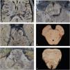
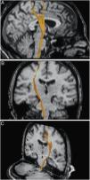
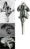
To demonstrate tridimensionally the anatomy of the cortico-spinal tract and the medial lemniscus, based on fibre microdissection and diffusion tensor tractography (DTT).
Material and methodsTen brain hemispheres and brain-stem human specimens were dissected and studied under the operating microscope with microsurgical instruments by applying the fibre microdissection technique. Brain magnetic resonance imaging was obtained from 15 healthy subjects using diffusion-weighted images, in order to reproduce the cortico-spinal tract and the lemniscal pathway on DTT images.
ResultsThe main bundles of the cortico-spinal tract and medial lemniscus were demonstrated and delineated throughout most of their trajectories, noticing their gross anatomical relation to one another and with other white matter tracts and grey matter nuclei the surround them, specially in the brain-stem; together with their corresponding representation on DTT images.
ConclusionsUsing the fibre microdissection technique we were able to distinguish the disposition, architecture and general topography of the cortico-spinal tract and medial lemniscus. This knowledge has provided a unique and profound anatomical perspective, supporting the correct representation and interpretation of DTT images. This information should be incorporated in the clinical scenario in order to assist surgeons in the detailed and critic analysis of lesions located inside the brain-stem, and therefore, improve the surgical indications and planning, including the preoperative selection of optimal surgical strategies and possible corridors to enter the brainstem, to achieve safer and more precise microsurgical technique.
Realizar un estudio anatómico de microdisección de fibras y radiológico mediante tractografía basada en tensor de difusión (DTT) para demostrar tridimensionalmente el tracto corticoespinal y el lemnisco medial.
Material y métodosBajo visión microscópica y con el uso de instrumental microquirúrgico se disecaron y estudiaron 10 hemisferios cerebrales y 15 troncoencéfalos humanos a través de la técnica de microdisección de fibras. Se obtuvieron imágenes de resonancia magnética cerebrales de 15 sujetos sanos, empleando secuencias potenciadas en difusión para el trazado y reproducción mediante DTT del tracto corticoespinal y la vía del lemnisco.
ResultadosSe demostraron y describieron anatómicamente el tracto corticoespinal y lemnisco medial en gran parte de sus trayectorias, identificando las relaciones entre sí y con otros haces de sustancia blanca y núcleos de sustancia gris cercanos, especialmente en el troncoencéfalo, con la correspondiente representación mediante DTT.
ConclusionesMediante la técnica de microdisección se apreció la disposición, arquitectura y organización topográfica general del tracto corticoespinal y lemnisco medial. Este conocimiento ha aportado una perspectiva anatómica única y profunda que ha favorecido la representación y la correcta interpretación de las imágenes de DTT. Esta información debe ser trasladada a la práctica clínica para favorecer el análisis crítico y exhaustivo por parte del cirujano ante posibles lesiones localizadas en el interior del troncoencéfalo y, en consecuencia, mejorar la indicación y planificación quirúrgica, incluyendo la selección preoperatoria de estrategias óptimas y posibles zonas de abordajes a su interior, alcanzando una técnica microquirúrgica más segura y precisa.
Article

If it is the first time you have accessed you can obtain your credentials by contacting Elsevier Spain in suscripciones@elsevier.com or by calling our Customer Service at902 88 87 40 if you are calling from Spain or at +34 932 418 800 (from 9 to 18h., GMT + 1) if you are calling outside of Spain.
If you already have your login data, please click here .
If you have forgotten your password you can you can recover it by clicking here and selecting the option ¿I have forgotten my password¿.


