

C0394 - CAN HEMODYNAMIC ALTERATIONS APPEAR FOLLOWING A CEREBELLAR ARTERIOVENOUS MALFORMATION RESECTION? CASE REPORT AND QUANTITATIVE ANALYSIS FROM CT IMAGING
Hospital 12 de Octubre, Madrid, España.
Objectives: Cerebellar arteriovenous malformations (CAVMs) are rare and challenging lesions with an aggressive natural history. They require prompt management of both the infratentorial bleeding and the vascular malformation. The mechanisms whereby a patient can worsen clinically after a supratentorial AVM resection include an acute alteration in cerebral hemodynamics, which is a known cause of postoperative hyperemia, edema and/or hemorrhage. While these phenomena have been largely documented for supratentorial AVMs, no case report supports these concepts to the posterior fossa AVMs.
Methods: We report a patient that presented an abrupt neurological deterioration after CAVM surgical resection.
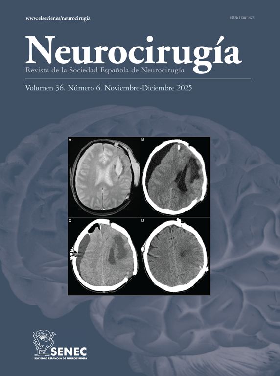
Results: Emergent external ventricular drainage to treat incipient hydrocephalus only partially reverted the patient’s deterioration. Consecutive post-surgery computed tomography (CT) images analysis revealed an increased cerebellum mean intensity and fourth ventricle compression that concurred with a new neurological deterioration. Importantly, our patient presented with venous drainage impairment given that ipsilateral transverse and sigmoid sinuses were absent. Venous congestion may effectively increase blood volume and so, lead to an increase in overall cerebellar CT intensity.
Conclusions: We postulate that venous outflow obstruction was responsible of cerebellar hyperemia, swelling and hemorrhage that conditioned overall neurological status. Our findings suggest that hemodynamic complications associated with supratentorial AVMs might also occur in the posterior fossa after CAVM resection.


